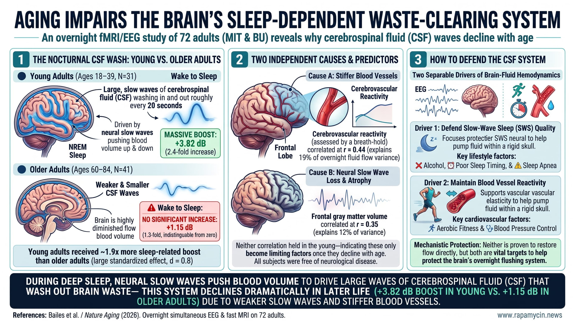
During deep non-REM sleep the young brain generates large, slow waves of cerebrospinal fluid that wash in and out roughly every twenty seconds, driven by neural slow waves pushing blood volume up and down inside a rigid skull. Researchers at MIT and Boston University scanned 31 young adults and 41 older adults overnight with simultaneous EEG and fast MRI to ask whether this still happens in later life. It largely does not. Young adults more than doubled their low-frequency fluid flow going from wake to sleep, while older adults showed no statistically significant increase at all. Daytime experiments using a flickering checkerboard and a breath-hold task separated two independent causes: weaker neural slow waves and stiffer, less reactive blood vessels. Reduced flow tracked frontal lobe shrinkage. All participants were free of neurological disease.
The brain has no lymphatic vessels in the ordinary sense. What it has instead is cerebrospinal fluid, and a growing body of work suggesting that fluid moves through brain tissue most vigorously during deep sleep, carrying metabolic waste out with it. The mechanism is mechanical and rather elegant. Your skull is a sealed box of fixed volume. When neurons across the cortex synchronize into the slow waves of deep sleep, blood vessels rhythmically dilate and constrict in response. Blood volume rises, fluid gets pushed out. Blood volume falls, fluid rushes back in. The result, in a young adult, is a slow tidal wave of fluid cycling roughly every twenty seconds.
Whether that pump keeps working into old age had never been directly measured. The studies that looked at fluid flow in older people were done during the day, awake, and came back contradictory. Some found slightly less flow, some slightly more. Nobody had put older adults in a scanner at midnight and watched what happens when they actually fall asleep.
The group at MIT and Boston University did exactly that. They recruited 31 young adults averaging 24 years old and 41 older adults averaging 69, screened to exclude neurological disease, psychiatric conditions, sleep disorders, cognitive impairment, and sleep medication. Everyone was scanned starting around midnight using a fast MRI sequence that simultaneously tracks blood oxygenation in gray matter and fluid velocity in the fourth ventricle, with EEG recording underneath to score sleep.
The young adults did what young adults do. Falling into deep sleep raised their low-frequency fluid flow power by about two and a half times. The older adults, going into the same sleep stages, showed a change so small it did not reach statistical significance. Whatever the overnight rinse cycle is, it was effectively absent in this older group.
The more interesting work was figuring out why, because there turned out to be two answers rather than one.
The first is neural. Older adults had less slow-wave power during deep sleep, concentrated in the frontal regions, and less slow-wave power predicted less fluid flow. A formal mediation analysis confirmed this pathway carried a significant share of the age effect.
The second is vascular, and it is independent of sleep entirely. The team brought participants back during the day for two tasks. In the first, everyone watched a flickering checkerboard. The EEG response to that stimulus was identical in young and old, meaning the neurons were firing fine. But the blood flow response in visual cortex was substantially weaker in the older group, and so was the resulting pulse of fluid. The neurons were shouting and the vessels were not listening. In the second task, participants held their breath for fifteen seconds at a time, a standard way to force vessels to dilate. Older vessels dilated less, and the fluid pulse was correspondingly smaller.
So one can have perfectly preserved slow-wave sleep and still fail to move fluid, if the vessels have stiffened. Or one can have supple vessels and fail to move fluid, if the slow waves are gone. The authors are explicit that this means different people will need different interventions, and that telling them apart will require measuring the individual rather than the group.
One finding will attract attention and deserves care. Among older adults, less fluid flow during sleep correlated with smaller frontal lobe gray matter volume, and not with volume in any other lobe. The authors lay out three possible readings and decline to pick one: atrophy causes slow-wave loss, atrophy weakens the mechanics, or poor fluid flow damages tissue over time. The data here cannot separate them.
What the study does not show is anything about waste clearance. It was not measured. The link from fluid waves to amyloid removal to dementia remains an inference, and the authors say so in plain language.
Actionable Insights
The central number: young adults increased low-frequency fluid flow power by 3.82 dB going from wake to sleep, which is about a 2.4-fold increase. Older adults managed 1.15 dB, roughly 1.3-fold, and that change was not statistically distinguishable from zero. Young adults got about 1.9 times more sleep-related boost than older adults. As a standardized effect this is around d = 0.8, a large difference by conventional benchmarks.
Two things predicted who in the older group did better, and both are measurable today. Cerebrovascular reactivity, assessed with a simple breath-hold in the scanner, correlated with nighttime fluid flow at r = 0.44, explaining about 19% of the variance. Frontal gray matter volume correlated at r = 0.35, about 12%. Neither correlation held in the young group, which is what you would expect if these only become limiting once they decline.
The practical implication is indirect but real. Two separable things drive this system: slow-wave sleep quality and blood vessel reactivity. Slow-wave sleep is affected by alcohol, sleep timing, and sleep apnea. Vascular reactivity is the standard cardiovascular target, responsive to aerobic fitness and blood pressure control. Neither is proven to restore fluid flow. Both are already worth doing on other evidence, and this paper gives a mechanistic reason to think the brain-fluid system may be one of the things they protect.
Context and Source
- Open Access Paper: The aging human brain exhibits reduced hemodynamic and cerebrospinal fluid flow waves during sleep., Published: 13 November 2026.
- Institution: Massachusetts Institute of Technology and Boston University.
- Country: United States of America.
- Journal: Nature Aging.
- Impact Evaluation: The impact score of this journal is 16.6, evaluated against a typical high-end range of 0-60 for top general science, therefore this is a High impact journal.
Related Reading
- Two Clocks, Two Windows: How Knowing When Sleep Fails Tells You When to Intervene
- Older Brains Still Need Their Sleep. They Have Lost the Machinery to Make It
- The Brain's Broken Plumbing: Why Diminishing Blood Flow Drives Dementia
- Protecting Arterial Elasticity by Targeting the Hidden Drivers of Vascular Aging
- New Proteomic Clocks Reveal How the Brain and Arteries Drive Human Longevity
- Estrogen Exposure Across the Lifespan Shields the Aging Female Brain from Vascular Damage
- Rapamycin induced Autophagy Ameliorates Age-Related Neurogenesis Decline and Neurodysfunction
- The Arginine Paradox Solved: Why Citrulline is the Superior Vascular Anti-Aging Agent
- The Vascular Pacemaker: How the Secretory Engine of Aging Arteries Synchronizes Whole-Body Decay