Protecting Arterial Elasticity by Targeting the Hidden Drivers of Vascular Aging

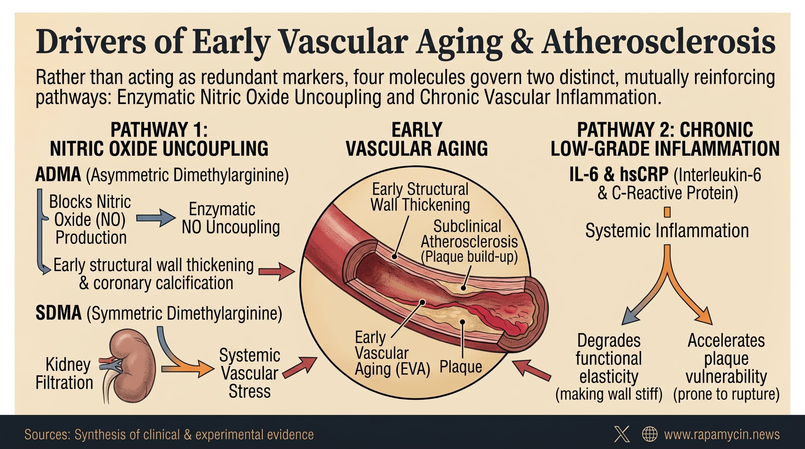
This narrative review synthesizes clinical and experimental evidence on how asymmetric dimethylarginine (ADMA),symmetric dimethylarginine (SDMA), interleukin-6 (IL-6), and high-sensitivity C-reactive protein (hsCRP) collectively drive early vascular aging and subclinical atherosclerosis. Rather than acting as redundant markers, these molecules govern two distinct yet mutually reinforcing pathways: the enzymatic uncoupling of nitric oxide production and chronic low-grade vascular inflammation. While elevated ADMA correlates independently with early structural wall thickening and coronary calcification, SDMA primarily reflects systemic vascular stress intertwined with renal filtration capacity.Simultaneously, systemic inflammation mediated by IL-6 and hsCRP degrades functional elasticity and accelerates plaque vulnerability.

Arteries do not all age at the same speed. Two 55-year-olds with identical cholesterol and blood pressure can carry arteries that behave like 45 or 70. Researchers call the fast track “early vascular aging” and the slow track “supernormal” aging. The problem is that standard risk calculators, built on cholesterol, blood pressure, smoking and diabetes, often miss who is on which track. Plenty of people labeled low or intermediate risk already have silent plaque.

This review asks whether a handful of blood tests could close that gap. The candidates fall into two camps.

The first camp concerns nitric oxide, the gas that keeps artery linings relaxed and healthy. ADMA, a molecule left over when cells break down methylated proteins, directly blocks the enzyme that makes nitric oxide. When ADMA builds up, that enzyme can misfire and generate damaging free radicals instead. SDMA, a close chemical cousin, works more indirectly by competing with arginine, the raw material for nitric oxide, at the cell’s entry gates.

The second camp is inflammation. IL-6 is a signaling molecule that activates blood vessel linings and recruits immune cells. It also tells the liver to make CRP, the protein measured by the familiar hsCRP test.

The evidence is uneven. ADMA most often turns up alongside thicker carotid artery walls and more coronary calcium. A pooled analysis of over 6,000 people found a modest link with carotid thickness, strongest in kidney disease. Yet the details wobble. In the Framingham study, ADMA tracked thickening at the carotid fork, where plaques form, but not in the straight segment below it. In a large German population study, ADMA showed no link to either carotid measure at all.

SDMA is cleared almost entirely by the kidneys, so high levels may simply reflect declining kidney function. Still, in the Dallas Heart Study and the German cohort, SDMA stayed linked to coronary calcium and carotid thickness even after accounting for kidney function.

For inflammation, genetics makes the strongest case. People born with variants that dampen IL-6 signaling have fewer heart attacks, which suggests causation rather than coincidence. But in the large US MESA cohort, IL-6 stopped predicting new coronary calcium once standard risk factors and statin use were considered.

hsCRP carries the most practical weight. In the Women’s Health Study, women in the top fifth of hsCRP had about 70 percent higher risk of cardiovascular events over 30 years than those in the bottom fifth. In a Chinese cohort of over 25,000 people, hsCRP above 3 mg/L went with faster arterial stiffening over about four and a half years. A Dutch analysis suggested inflammation may drive stiffening more than wall thickening, though it explained only a sliver of age-related change.

One small Greek trial offers a glimpse of modifiability. Six months of intensive diet and exercise coaching lowered ADMA and improved carotid stiffness and wall thickness in people with mild hypertension, and the more inflammatory the diet, the higher the ADMA.

The authors argue the four markers are complementary rather than interchangeable. That is plausible, but untested. Nobody has yet shown that adding them to a standard risk score changes who gets correctly flagged.

Actionable Insights

The evidence demonstrates that protecting long-term arterial youth requires two practical interventions: maintaining cellular nitric oxide bioavailability and aggressively suppressing systemic inflammation.

First, dietary adjustments produce measurable structural benefits. Clinical trial data cited in the review reveal that adopting an intensive lifestyle intervention consisting of an anti-inflammatory diet and aerobic exercise over six months drove a significant decline in asymmetric dimethylarginine. This biochemical reduction directly correlated with reduced arterial stiffness and decreased carotid wall thickness. For each one-unit decrease in the dietary inflammatory index score,circulating dimethylarginine concentrations declined predictably, verifying that dietary quality directly impacts endothelial enzymatic function.

Second, tracking high-sensitivity C-reactive protein (HS-CRP) provides a functional gauge of arterial stiffness. Prospective cohort data tracking over 25,000 adults indicate that maintaining baseline levels below 1 milligram per liter preserves arterial elasticity. Individuals with concentrations exceeding 3 milligrams per liter experience an additional stiffening rate of more than 40 centimeters per second over four to five years compared to those with low inflammation, an acceleration in mechanical vascular aging equivalent to several additional years of physical deterioration. In women followed for 30 years, the top fifth of hsCRP carried about 1.7 times the event risk of the bottom fifth, a small-to-moderate effect. Retest if a result looks high, since a cold or injury can spike it temporarily.

Third, preserving renal filtration capacity prevents symmetric dimethylarginine accumulation. Maintaining optimal glycemic control, sustaining insulin sensitivity, and engaging in habitual zone-2 aerobic conditioning prevent the lipotoxic and oxidative insults that lock endothelial enzymes into a permanent uncoupled state.

Protect kidney function. Both ADMA and SDMA rise as kidney filtration falls.

Do not expect ADMA, SDMA or IL-6 tests to guide decisions yet. Assays are not standardized, and ADMA explains only about 4 percent of the person-to-person variation in carotid wall thickness.

Context and Source

  • Open Access Paper: Vascular aging biomarkers: the role of ADMA, SDMA, and inflammation, Published 07 October 2026.
  • Authors and Affiliations: Aleksandra Melnitskaia, Liubov Machekhina, Valentina Pykhtina, Alexey Churov, Olga Maltseva, Wu Zhenghi, Olga Tkacheva, and Irina Strazhesko; Russian Gerontological Research and Clinical Center, Moscow, Russia, and Department of Neurology, Shenzhen Institute of Translational Medicine, The First Affiliated Hospital of Shenzhen University / Shenzhen Second People’s Hospital, Shenzhen, China.
  • Journal Name: Frontiers in Cardiovascular Medicine
  • Impact Evaluation: The impact score of this journal is 3.0 (CiteScore 6.1), evaluated against a typical high-end range of 0–60+ for top general science, therefore this is a Medium impact journal.

Hasn’t the recent failed trial with the IL-6 inhibitor thrown shade on IL-6/CRP as causal mediators of vascular disease? I thought the focus is now on inflammatory mediators higher up the chain.

Interesting question… there were some failures, but I was unsure if it was a target issue, or class of drug issue. There were also conflicting results with colchicine too, so it seems the jury it still undecided.

I asked Gemini for a summary of both positive and negative clinical outcomes:

Targeting the interleukin-6 pathway suppresses downstream inflammatory cascades, but clinical data on its ability to reverse atherosclerosis or prevent cardiovascular events is contradictory. A critical pharmacological distinction exists between drug classes: direct IL-6 ligand antibodies (such as ziltivekimab) lower measured free IL-6 and high-sensitivity C-reactive protein, whereas IL-6 receptor antagonists (such as tocilizumab) block receptor clearance, causing circulating total IL-6 levels to surge while shutting down downstream signaling and C-reactive protein production.

Clinical trials show a clear divergence between phase 2 trials measuring surrogate inflammatory or imaging markers, which generally report positive results, and hard-endpoint phase 3 trials, which have failed to show vascular protection.

Evidence Supporting Positive Impact on Vascular Markers and Surrogate Endpoints

  • The RESCUE Trial (Ziltivekimab): A randomized phase 2 trial evaluated ziltivekimab across three dose levels (7.5 mg, 15 mg, and 30 mg monthly) against placebo in 264 patients with advanced chronic kidney disease and elevated baseline hsCRP (2 mg/L or greater). Ziltivekimab produced dose-dependent reductions in hsCRP of up to 92 percent compared to placebo. It also suppressed other prothrombotic and inflammatory drivers, including fibrinogen, serum amyloid A, haptoglobin, and lipoprotein(a), without disrupting the apolipoprotein B to apolipoprotein A1 ratio. The trial proved systemic vascular inflammation can be largely turned off, though it was not powered to evaluate plaque regression or clinical events.
  • The ASSAIL-MI Trial (Tocilizumab): A randomized trial administered a single dose of tocilizumab or placebo to 199 patients presenting with acute ST-segment elevation myocardial infarction undergoing primary percutaneous coronary intervention. Tocilizumab cut C-reactive protein exposure by more than 50 percent and increased the myocardial salvage index measured by cardiac magnetic resonance imaging at 3 to 7 days (0.69 versus 0.64). It also reduced systemic markers of neutrophil extracellular trap formation, pointing to reduced acute microvascular inflammatory injury.
  • Surrogate Endothelial and Vascular Function Studies: Small clinical cohorts evaluating patients with rheumatoid arthritis treated with tocilizumab demonstrated improvements in flow-mediated dilation of the brachial artery and reductions in pulse wave velocity over 6 to 12 months. Vascular ultrasound evaluations in large-vessel vasculitis also showed rapid regression of vessel wall edema and reductions in arterial intima-media thickness.
  • Correlative Evidence from CANTOS: While the CANTOS trial tested an IL-1 beta inhibitor (canakinumab), on-treatment biomarker analyses demonstrated that the cardiovascular benefits were mediated through downstream IL-6 reduction. Patients who achieved on-treatment IL-6 levels below the study median of 1.65 ng/L experienced a 32 percent reduction in major adverse cardiovascular events and a 31 percent reduction in cardiovascular mortality, establishing circulating IL-6 as an independent epidemiological risk driver.

Studies Showing Null, Opposing, or Counter-Regulatory Vascular Effects

  • The ZEUS Trial (Ziltivekimab): The primary cardiovascular outcomes trial testing direct IL-6 inhibition evaluated once-monthly ziltivekimab against placebo in over 6,000 patients with established atherosclerotic cardiovascular disease, chronic kidney disease, and systemic inflammation. Despite achieving profound target engagement with steep drops in hsCRP and free IL-6, ziltivekimab failed to reduce the primary composite endpoint of major adverse cardiovascular events (cardiovascular death, myocardial infarction, or stroke). The hazard ratio remained essentially neutral at approximately 0.99, showing that aggressively shutting down the IL-6 pathway in established multi-morbid vascular disease did not alter macrovascular event rates.
  • The ENTRACTE Trial (Tocilizumab vs. Etanercept): A phase 4 cardiovascular safety trial randomized 3,080 rheumatoid arthritis patients with cardiovascular risk factors to either tocilizumab or the TNF inhibitor etanercept. Over a median follow-up of 3.2 years, tocilizumab failed to show any superiority in reducing major adverse cardiovascular events (hazard ratio: 1.05). Nonfatal stroke rates were numerically higher in the tocilizumab cohort.
  • Paradoxical Pro-Atherogenic Dyslipidemia: Nearly all trials targeting IL-6 or its receptor document adverse shifts in circulating lipid profiles. In ENTRACTE, MEASURE, and related rheumatoid arthritis cohorts, tocilizumab consistently raised low-density lipoprotein cholesterol by 10 to 22 percent and triglycerides by 10 to 15 percent, largely driven by alterations in hepatic lipid clearance and reduced LDL receptor density. In the RESCUE trial, ziltivekimab produced dose-dependent increases in triglycerides of up to 36 percent. This persistent elevation in circulating atherogenic particles provides a biologically plausible explanation for why anti-inflammatory gains fail to translate into reduced clinical events.
  • Post-NSTEMI Coronary Microvascular Flow Studies: A randomized trial testing tocilizumab in patients with non-ST-elevation myocardial infarction undergoing coronary angiography found that IL-6 receptor inhibition failed to improve coronary flow reserve or long-term index of microcirculatory resistance at 6 months compared to placebo. Treatment was also accompanied by a transient, paradoxical increase in circulating vascular cell adhesion molecule-1, indicating an incomplete or complex endothelial response.

Summary Comparison

Study Drug & Target Inflammatory Marker Effect Vascular / Clinical Outcome Result
RESCUE Ziltivekimab (Anti-IL-6) hsCRP reduced up to 92 percent Reduced Lp(a), fibrinogen, and thrombotic markers Positive (Surrogates)
ASSAIL-MI Tocilizumab (Anti-IL-6R) C-reactive protein reduced over 50 percent Increased myocardial salvage index on cardiac MRI Positive (Acute Injury)
ZEUS Ziltivekimab (Anti-IL-6) Marked hsCRP and free IL-6 suppression No reduction in primary MACE composite (HR approximately 0.99) Neutral / Negative (Hard Endpoints)
ENTRACTE Tocilizumab (Anti-IL-6R) Robust downstream signaling suppression No MACE reduction versus TNF inhibition; elevated LDL-C Neutral / Negative (Safety & Lipids)

References

  • Ridker PM, Devalaraja M, Baigent C, et al. Reduction of C-reactive protein and intermediate biomarkers of risk with the interleukin-6 inhibitor ziltivekimab in patients with chronic kidney disease (RESCUE): a randomized, double-blind, placebo-controlled, phase 2 trial. The Lancet. 2021;397(10289):2060-2069. https://pubmed.ncbi.nlm.nih.gov/34000257/
  • Broch K, Anstensrud AK, Woxholt S, et al. Randomized trial of interleukin-6 receptor inhibition in patients with acute ST-elevation myocardial infarction: The ASSAIL-MI study. Journal of the American College of Cardiology. 2021;77(15):1845-1855. https://pubmed.ncbi.nlm.nih.gov/33858620/
  • Giles JT, Sattar N, Gabriel S, et al. Cardiovascular safety of tocilizumab versus etanercept in rheumatoid arthritis: A randomized controlled trial (ENTRACTE). Arthritis & Rheumatology. 2020;72(1):31-40. https://pubmed.ncbi.nlm.nih.gov/31418520/
  • Ridker PM, Libby P, MacFadyen JG, et al. Modulation of the interleukin-6 signalling pathway and incidence of cardiovascular events in patients with prior myocardial infarction: An analysis from the CANTOS trial. European Heart Journal. 2018;39(38):3499-3507. https://pubmed.ncbi.nlm.nih.gov/30165610/
  • Protogerou AD, Zampeli E, Fragiadaki K, et al. A small single-center study demonstrates that IL-6 receptor inhibition improves large artery stiffness and endothelial function in rheumatoid arthritis. Atherosclerosis. 2011;219(2):734-736. https://pubmed.ncbi.nlm.nih.gov/21968316/
  • Kleveland O, Kunszt G, Bratlie M, et al. Effect of a single dose of the interleukin-6 receptor antagonist tocilizumab on myocardial injury in patients with non-ST-elevation myocardial infarction: A double-blind, randomized, placebo-controlled phase 2 trial. European Heart Journal. 2016;37(30):2406-2413. https://pubmed.ncbi.nlm.nih.gov/27099264/
  • ZEUS Clinical Trial Details: A Research Study to Look at How Ziltivekimab Works Compared to Placebo in People With Cardiovascular Disease, Chronic Kidney Disease and Inflammation. ClinicalTrials.gov Identifier: NCT05021835. https://clinicaltrials.gov/study/NCT05021835