Men Lose Thymic Output Faster: Sex Differences and Other Usable Findings From a Global Immune Atlas

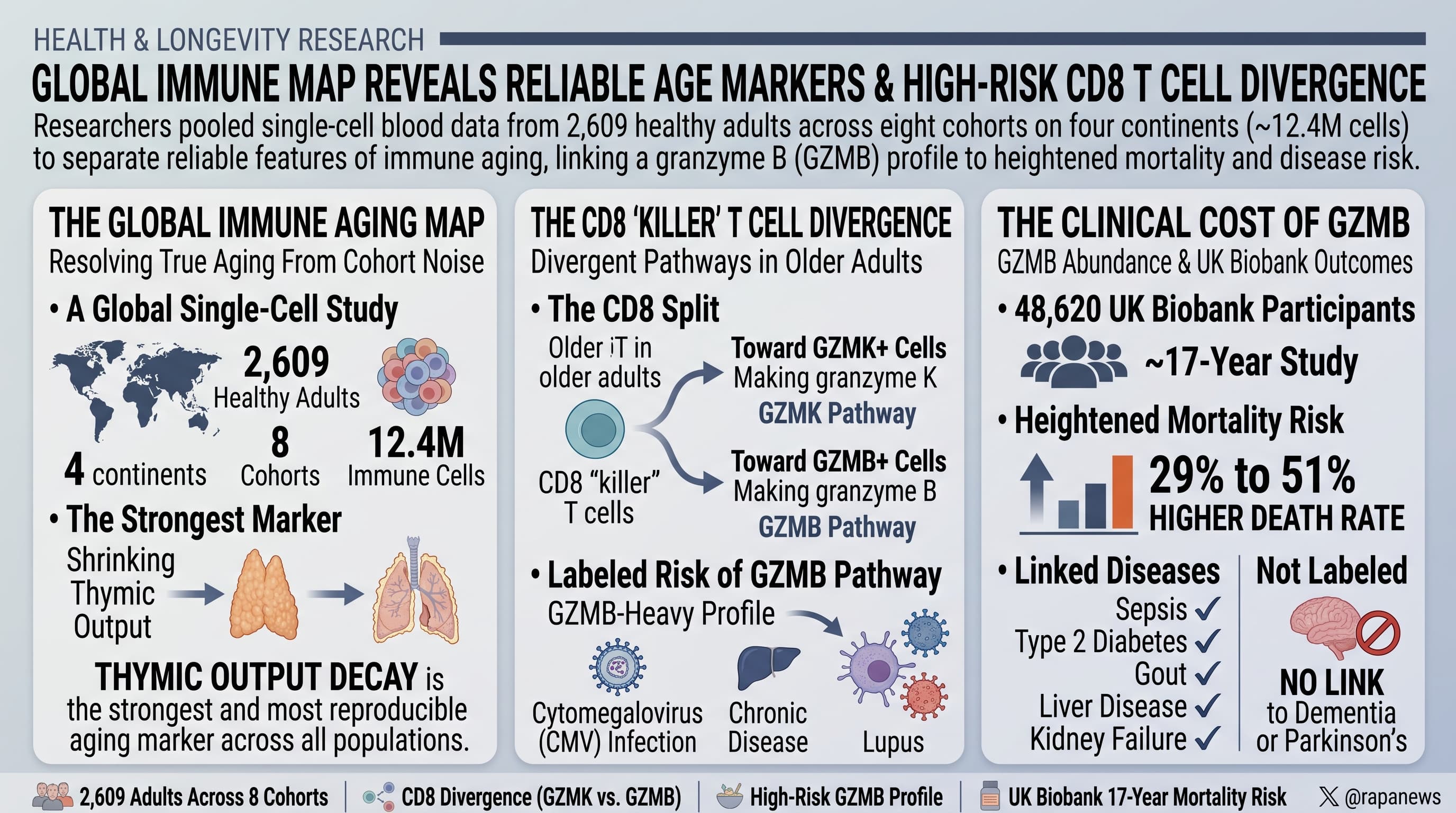
Researchers pooled single-cell blood data from 2,609 ostensibly healthy adults across eight cohorts on four continents (about 12.4 million immune cells) to separate reliable features of immune aging from cohort noise. Shrinking thymic output was the strongest and most reproducible marker of age. Among older adults, CD8 “killer” T cells diverged in two directions: toward cells making granzyme K (GZMK) or toward cells making granzyme B (GZMB). A GZMB-heavy profile was linked to cytomegalovirus (CMV) infection, chronic disease and lupus. A blood protein signature that estimates GZMB cell abundance, applied to 48,620 UK Biobank participants, was associated with a 29 to 51% higher rate of death over about 17 years, and with more sepsis, type 2 diabetes, gout, liver disease and kidney failure, but not dementia or Parkinson’s disease.

Narrative

Two people of the same age can have very different immune systems, and studies of immune aging have a habit of contradicting each other. A team led from Washington University in St. Louis, King’s College London and Nationwide Children’s Hospital tried to settle the matter with scale. They combined seven public single-cell datasets with a new cohort of 188 Londoners, re-labeled every cell using one shared scheme of 59 cell types, and asked which age-related changes show up in at least five of the eight cohorts.

Several did. Freshly minted T cells from the thymus, called recent thymic emigrants, fall steadily with age, as do naive CD8 T cells. These two populations carried the most weight in an “immune clock” the team built, although that clock is blunt: it guesses a person’s age to within about eight years. Type 2 immune cells and activated T cells rise with age across populations. Sex and ancestry mattered less than expected, and which study a sample came from explained more variation than any biological factor.

The more interesting result concerns what happens after the naive cells are gone. When the researchers mapped each person’s CD8 T cell makeup, older adults did not cluster together. They spread between two corners: one rich in effector memory cells carrying granzyme K, the other rich in cells carrying granzyme B. The ratio between the two did not track age itself or body mass index. It tracked CMV, a common lifelong herpesvirus, which pushes the balance toward granzyme B. It was also lower in a small group of older Londoners with mostly cardiovascular conditions and in lupus patients, even among those without CMV.

Counting these cells requires single-cell sequencing, so the team looked for a shortcut. They found that blood levels of a handful of proteins rise in step with granzyme B cells, trained a model to estimate the cells from proteins, and applied it to the UK Biobank. People in the top quarter of predicted granzyme B cells died at higher rates than those in the bottom quarter in every age band tested, and developed more infections, metabolic disease and organ failure. Neurodegenerative disease showed no link.

There is also a caution for anyone extrapolating from mice. In aged mice, granzyme K cells are the terminal, inflammatory population. In human blood they look like an intermediate state, and granzyme B cells are the endpoint.

The caveats are substantial. The mortality link rests on a protein-based estimate, not counted cells, and was adjusted only for age and sex. Five of the authors are named on a related provisional patent.

Actionable Insights

This paper describes a marker. It tests no intervention, so the practical takeaways are modest.

  1. Size of the risk signal. Among 60 to 70 year olds, the top quarter for predicted GZMB cells had a hazard ratio of 1.43 for death versus the bottom quarter. In plain terms, at any moment they were dying at a 43% higher rate. Read off the published curves, that is roughly 27 to 28 deaths per 100 people over 16 years versus about 20 per 100, an absolute gap of around 7 to 8 percentage points (approximate). For comparison, being male carried a larger hazard ratio (1.66) in the same model.
  2. CMV status is the most accessible lever of understanding. A standard CMV IgG blood test tells you whether you carry the virus that most strongly shifts this ratio. No evidence here supports antiviral treatment in healthy carriers.
  3. Inflammation tracks with it. C-reactive protein rose across GZMB groups, so a routine hs-CRP captures part of the same signal.
  4. You cannot order this test. Standard clinical T cell panels cannot separate these populations.
  5. Weight was not a factor. The ratio did not differ by BMI group.

Context/Source

Related Reading:

Another interesting Thymus Paper:

AI Reads Old Chest Scans and Finds the Thymus Tracks With Who Lives Longer

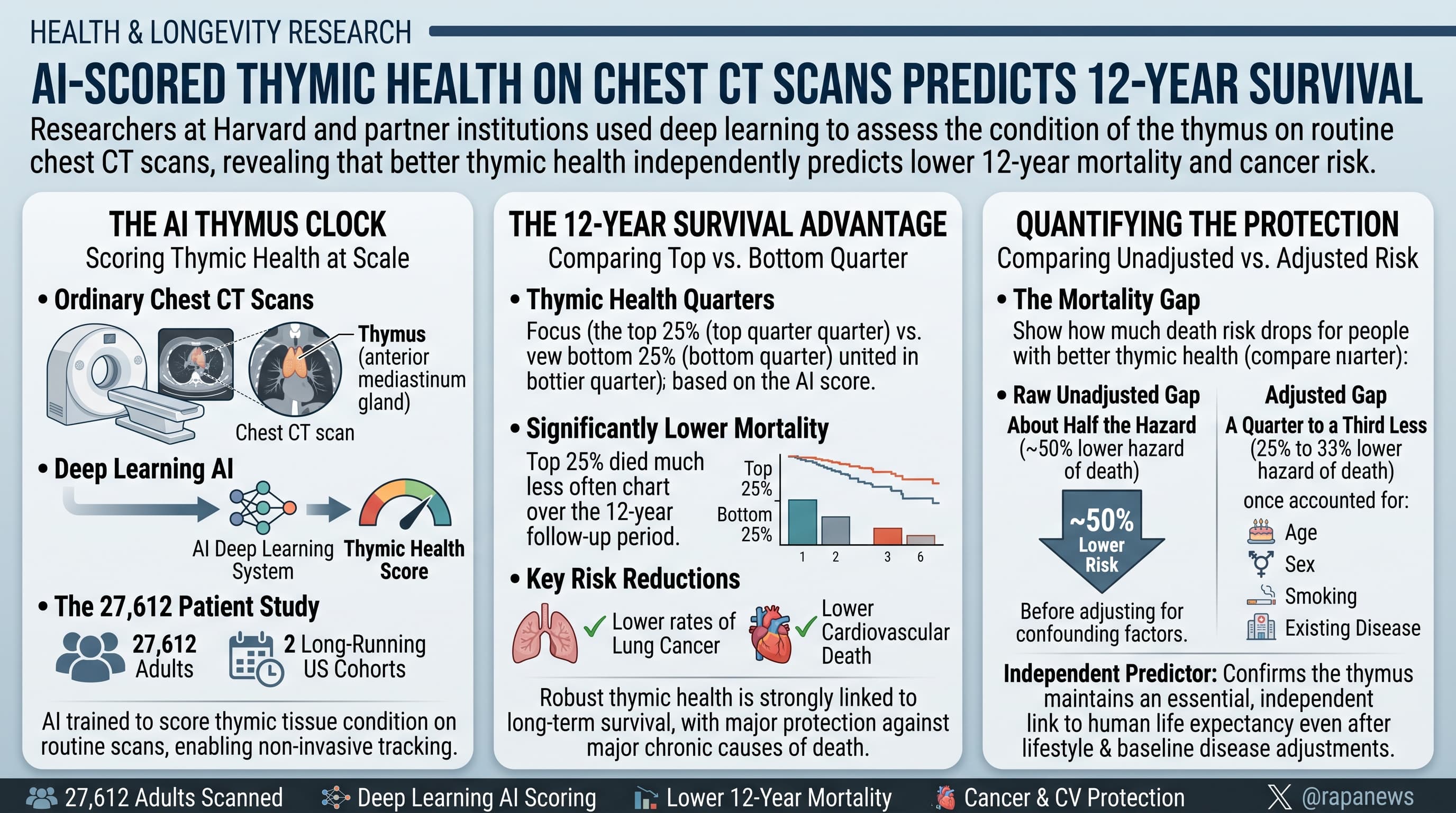
Researchers at Harvard and partner institutions trained a deep learning system to score the condition of the thymus on ordinary chest CT scans, then applied it to 27,612 adults from two long-running US cohorts. People in the top quarter of “thymic health” died less often over 12 years than those in the bottom quarter, with lower rates of lung cancer and cardiovascular death. The raw gap was large (about half the hazard of death), but it shrank to roughly a quarter to a third once age, sex, smoking and existing disease were accounted for.

For decades, medical students have been taught that the thymus does its work in childhood and then retires. The small gland behind the breastbone trains T cells, the immune system’s specialist defenders, and then slowly turns to fat. By middle age, the textbook view goes, it is a leftover.

A study in Nature challenges that view using scans that were sitting in archives. A team led by Hugo Aerts at Mass General Brigham and Harvard Medical School built an artificial intelligence system that finds the thymus on a chest CT scan and assigns it a score from 0 to 100, based on how much soft tissue remains rather than fat. They ran it on scans from 25,031 current and former heavy smokers in the National Lung Screening Trial and 2,581 participants in the Framingham Heart Study, all without symptoms when scanned.

The first finding is that adults of the same age differ widely. Women scored higher than men. Scores fell with age and with higher body weight. Smokers scored lower, as did people with high triglycerides, high blood sugar, high blood pressure and persistent inflammation. Alcohol intake made no measurable difference.

The second finding is the headline. In the lung screening group, 25.5 percent of people in the bottom quarter of scores died within 12 years, against 13.4 percent in the top quarter. Lung cancer, cardiovascular death, and deaths from lung, digestive and metabolic disease all followed the same pattern. In Framingham, the gap in cardiovascular deaths was wider still, though it rested on very few deaths.

Those raw numbers flatter the result. Age, sex and smoking drive both thymic decline and death, so part of the gap would exist even if the thymus did nothing. After the researchers adjusted for these factors, the difference in death risk fell from about 51 percent to about 33 percent, and to about 23 percent once existing illnesses such as diabetes, emphysema and heart disease were included. In Framingham, the adjusted link with overall mortality was not statistically distinguishable from zero.

The authors propose that a better-preserved thymus keeps supplying fresh T cells, which helps the body detect tumors and control the inflammation that damages arteries. That is plausible and fits a 2023 study showing that adults who had the thymus surgically removed fared worse years later. But this paper does not test it. No one measured T cell output. The score may partly be a readout of fat distribution and metabolic health in the chest, which already predict early death.

The authors state that their design cannot show cause and effect. A failing body may shrink the thymus, rather than the reverse.

What the work does offer is a cheap, automated measurement that can be pulled from millions of existing scans, and a strong argument that the adult thymus deserves study. Whether restoring it extends life is a question for trials that have not been run.

Actionable Insights

This study tested no treatment, so nothing here is proven to work through the thymus. It shows which habits travel with a better-preserved thymus on a scan.

The associated factors are familiar: not smoking, lower body weight, lower triglycerides, blood sugar and blood pressure, higher HDL cholesterol, more physical activity, and low chronic inflammation. Each link is small. A meaningful step up in HDL (one standard deviation, the gap between a typical person and someone at about the 84th percentile) went with about 5 points on the 100-point thymic scale. The same step up in triglycerides went with about 5 points lower. A comparable step up in smoking exposure went with about 1.5 points lower, in a group that were all heavy smokers already.

For outcomes, people in the top quarter had about a 23 to 33 percent lower rate of death than the bottom quarter after adjustment. In absolute terms that is roughly 5 to 8 fewer deaths per 100 people over 12 years (my estimate from the published figures), among older heavy smokers.

Practical points:

  • The score is not available clinically. Do not seek a CT scan for it.
  • The lifestyle measures above are already worth doing for independent reasons.

Context and Source

  • Paywalled Paper: Thymic health consequences in adults
  • Lead institution: Artificial Intelligence in Medicine Program, Mass General Brigham and Harvard Medical School, with Boston University and the Framingham Heart Study, Aarhus University, Maastricht University, University College London and the Francis Crick Institute
  • Country: USA (lead), with Denmark, the Netherlands, Germany and the UK
  • Journal: Nature, published online 18 March 2026
  • Impact evaluation: The impact score of this journal is 56.1 (2025 Journal Impact Factor), evaluated against a typical high-end range of 0 to 60+ for top general science, therefore this is an Elite impact journal.

Lifelong Cyclists Keep a Younger Immune System, But Exercise Does Not Stop Every Sign of Immune Aging

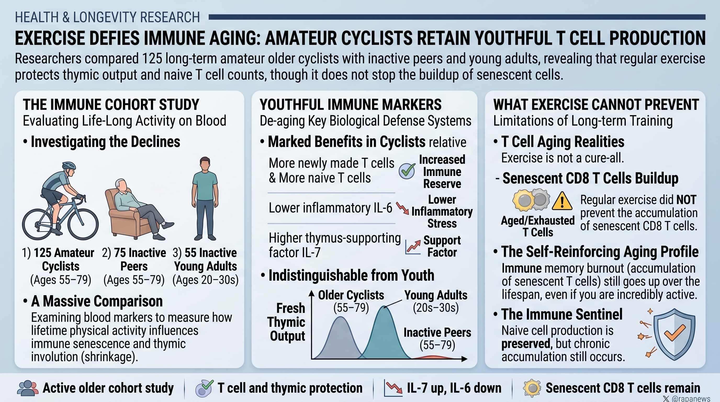
Researchers compared the blood of 125 long-term amateur cyclists aged 55 to 79 with 75 inactive people of the same age and 55 inactive young adults. The cyclists had more newly made T cells, more naive T cells, lower inflammatory IL-6, and higher levels of the thymus-supporting factor IL-7 than their inactive peers. On one marker of fresh thymic output they were indistinguishable from people in their 20s and 30s. Exercise did not prevent the buildup of senescent CD8 T cells.

For decades, immunologists have treated the shrinking of the thymus as one of the most dependable facts of human aging. The gland sits behind the breastbone and trains new T cells. It starts turning to fat in early adulthood, and by the 70s it is thought to release only a trickle of fresh cells. Fewer new T cells means a weaker response to unfamiliar infections and to vaccines.

A British team now argues that part of this decline may reflect how little older people move. Researchers at the University of Birmingham and King’s College London took blood from 125 amateur cyclists aged 55 to 79 who had ridden for most of their adult lives. These were not former professionals, but they were fit. The men had to be able to ride 100 km in under 6.5 hours and the women 60 km in under 5.5 hours. Their blood was compared with that of 75 healthy but inactive people of the same age and 55 inactive adults aged 20 to 36.

The cyclists’ blood carried considerably more recently made T cells than that of their sedentary peers. For one marker of newly released CD4 T cells, the cyclists matched the young adults. They also kept more naive T cells, the unprogrammed cells needed to meet new pathogens, although not as many as the young group.

The team also looked for a reason. Cyclists had about twice the blood level of interleukin-7, a growth factor that supports the thymus and that muscle can produce. They had roughly 70 percent less interleukin-6, an inflammatory signal linked to thymic shrinkage. Levels of interleukin-15, which helps naive T cells survive, were higher too. The suggestion is that well-used muscle sends chemical signals that keep the thymus working.

Exercise did not hold back everything. Worn-out, senescent CD8 T cells had built up just as much in the cyclists as in the inactive group. These cells accumulate mainly through decades of fighting persistent viruses such as cytomegalovirus, and about half the cyclists carried that virus. Cortisol, a stress hormone that can damage the thymus, was raised in both older groups.

There are important cautions. This was a single blood draw, not a trial. Nobody was assigned to cycle, so the study cannot show that exercise caused the difference. People who can still ride 100 km at 75 are an unusual, self-selected group, probably leaner and perhaps genetically luckier than average, and the analysis did not adjust for body fat, diet, or sex. The researchers measured the proportions of cell types in blood, not absolute cell counts and not the thymus itself. They did not test whether the cyclists fight infections or respond to vaccines any better.

Even so, the work challenges the idea that immune aging runs on a fixed internal clock. Much of what textbooks describe as normal immune aging comes from studies of mostly inactive people. If the finding holds up in controlled trials, part of that decline is a consequence of inactivity and may be preventable.

Actionable Insights

The practical message is that sustained endurance exercise through adult life is associated with a younger-looking immune profile in your 60s and 70s. The size of the differences, based on values I read from the paper’s figures, was as follows:

  • Newly made CD4 T cells: about 9.5 percent of CD4 cells in cyclists versus 4 percent in inactive elders, more than double, and equal to young adults.
  • Naive CD8 T cells: about 25 percent versus 18 percent. This closes only about a quarter of the gap to young adults, who sit near 48 percent.
  • Naive CD4 T cells: about 46 percent versus 39 percent, closing about half the age gap.
  • IL-6 (inflammation): about 1.5 versus 5.4 pg/ml, roughly 70 percent lower.
  • IL-7: about 42 versus 21 pg/ml, double.

In plain terms these are medium to large differences between groups, not marginal ones. The realistic framing is partial preservation, not reversal. Senescent CD8 T cells were unchanged, so exercise is not a complete answer.

Two cautions apply. The cyclists rode at high volume for decades, and nothing here shows that starting at 60, or doing 150 minutes a week, gives the same result. No infection or vaccine outcomes were measured, so the real-world health benefit is inferred.

Context and Source

1 Like