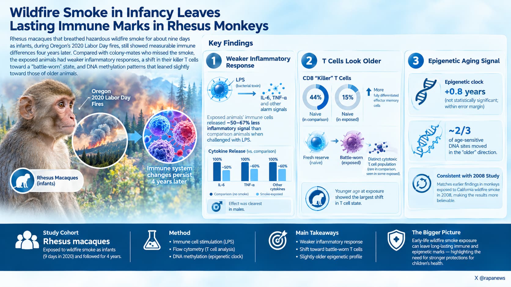
Rhesus macaques that breathed hazardous wildfire smoke for about nine days as infants, during Oregon’s 2020 Labor Day fires, still showed measurable immune differences four years later. Compared with colony-mates who missed the smoke, their blood cells produced roughly half to two-thirds less inflammatory signal when challenged with a bacterial toxin, their killer T cells had shifted from a “fresh reserve” state toward a “battle-worn” state, and their DNA methylation patterns leaned slightly toward those of older animals.
In September 2020, smoke from the Labor Day wildfires settled over the Oregon National Primate Research Center. Fine particle pollution peaked at around 273 micrograms per cubic meter, roughly twenty times the normal local maximum, and stayed in the “unhealthy” range for about a week. Fifteen infant macaques living in open-air enclosures, all under three months old, breathed it in. That age window matters: in rhesus monkeys it corresponds to about the first year of human life, when the immune system is still being assembled.
Four years later, researchers at Oregon Health and Science University drew blood from those animals, now late adolescents, and from eight comparison animals that were either born after the fires or kept in a filtered indoor facility. They then ran the cells through a battery of modern tests: cytokine panels, single-cell gene expression, and genome-wide DNA methylation.
Three findings stand out.
First, the exposed animals’ immune cells responded weakly to a mock bacterial infection. When stimulated with lipopolysaccharide, a bacterial wall component, their cells released markedly less IL-6, TNF-alpha and other alarm signals. The effect was clearest in males. This matches an earlier report from monkeys exposed to California wildfire smoke in 2008, which makes it more believable.
Second, the T cell compartment looked older than it should. In the comparison animals, roughly 44 percent of CD8 “killer” T cells were still naive, meaning uncommitted and available for new threats. In the smoke-exposed animals that figure was about 15 percent, with a matching rise in fully differentiated effector memory cells. The animals exposed at the youngest ages showed the largest shift. A distinct population of cytotoxic cells, almost absent in the comparison group, also appeared in some exposed animals.
Third, the epigenome carried a faint aging signature. An epigenetic clock estimated the exposed animals as 0.8 years older than expected, though that result did not reach statistical significance and sits within the clock’s own error margin. A second analysis found that about two-thirds of age-sensitive DNA sites had moved in the “older” direction.
What the study cannot tell us is whether any of this matters for health. No infections, vaccine responses, lung function or disease outcomes were measured. A dampened inflammatory response could mean vulnerability to infection, or it could be a harmless or even protective adaptation. The authors acknowledge they do not know which.
There are also reasons for caution about cause and effect. The exposed animals were about four months older at blood draw, were born in a different season, and half of the comparison group may have been exposed to the same smoke in the womb. The study does not report whether the groups differed in common viral infections that are known to reshape T cells.
Still, the central message is hard to dismiss: one short, severe smoke episode in infancy was followed, years later, by an immune system that looks different across several independent measurements.
Actionable Insights
This paper tested an exposure, not a treatment, so it offers nothing adults can take or do to reverse anything. Its practical message is about prevention for the youngest children.
The size of the differences was large by biological standards. Exposed animals released about 55 to 65 percent less IL-6 and TNF-alpha after a bacterial challenge. Their share of naive killer T cells was roughly one-third of the comparison group’s (about 15 percent versus 44 percent). In plain terms, a typical exposed animal fell outside the normal range of the unexposed group on that measure. The epigenetic age difference (0.8 years, roughly 19 percent of the animals’ age) was not statistically reliable.
Practical takeaways, all precautionary:
- Treat infants under one year as the highest-priority group during smoke events. The youngest animals showed the largest changes.
- During “unhealthy” air days (PM2.5 above about 55 ug/m3), keep infants indoors with windows closed and run a HEPA purifier sized for the room.
- Consider temporary relocation during multi-day severe smoke. Masks are not suitable for infants.
- Do not assume harm has occurred in an exposed child. No disease outcomes were shown.
Context/Source
- Open Access Paper: Early-Life Wildfire Smoke Exposure Is Associated with Long-Term Systemic Immune Remodeling and Epigenetic Reprogramming
- Lead institution: Oregon Health and Science University, including the Oregon National Primate Research Center, with collaborators at University of Oregon, Arizona State University and the US National Institute on Aging
- Country: USA
- Journal: bioRxiv, a preprint server.
Related Reading:
- Smoke Gets in Your Cells: How Air Pollution Accelerates Biological Aging
- Broccoli Sprout Drink Helped Chinese Villagers Clear a Polluted Air Carcinogens
- The Overlooked Variable in Heat Death Projections Was Everyone Over Sixty
- Egg to Exit: How Socio-economic Status Impacts Your Healthspan and Lifespan