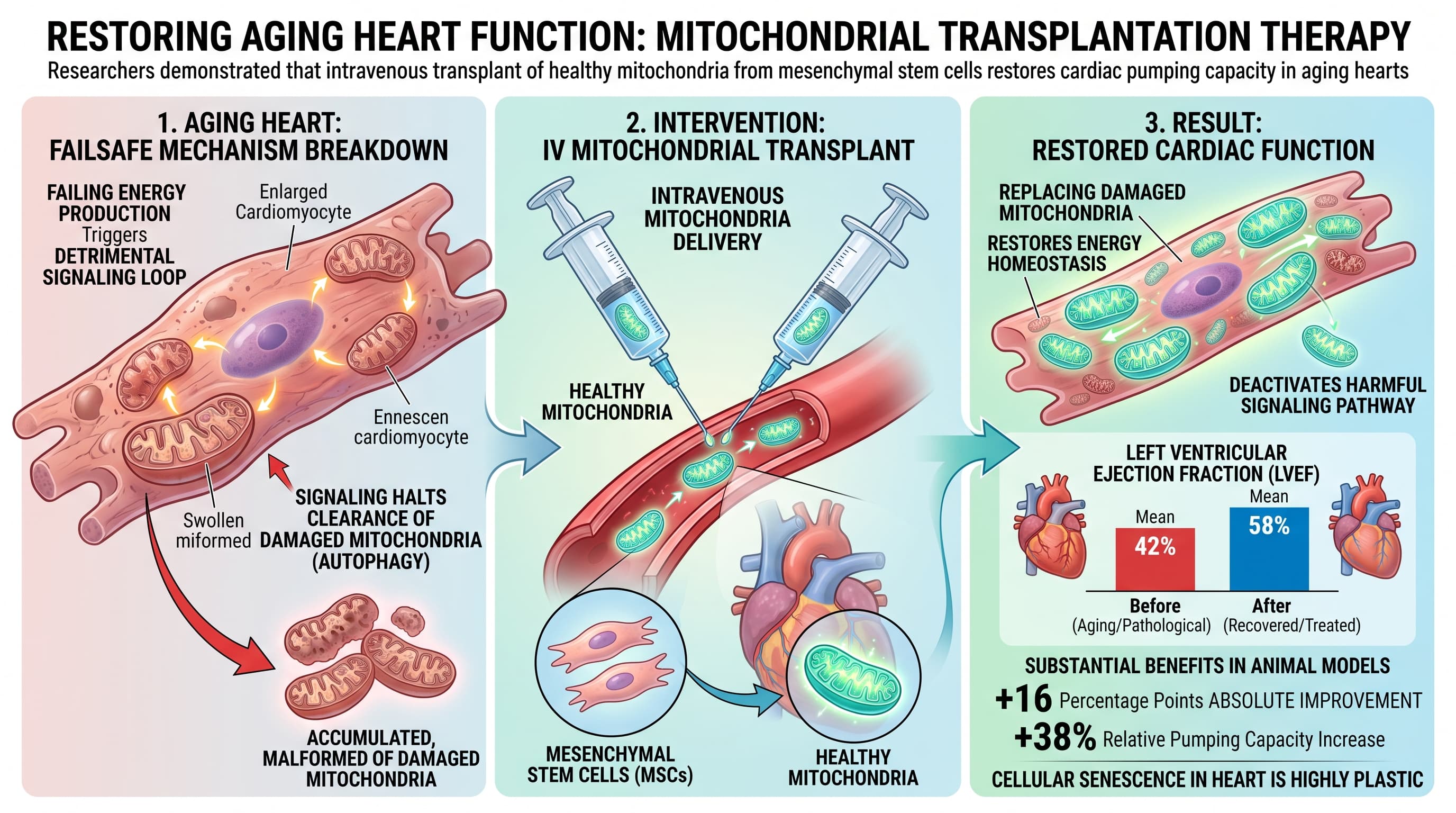
Researchers have demonstrated that failing energy production in aging heart tissue triggers a detrimental signaling loop that halts the clearance of damaged mitochondria. By intravenously transplanting healthy mitochondria derived from mesenchymal stem cells, the research team successfully restored cellular energy levels, deactivated the harmful signaling pathway, and improved overall cardiac pumping capacity in animal models.
Cardiac aging is characterized by a gradual failure of cellular energy systems and an accumulation of cellular damage. A central driver of this decline is the failure of mitophagy, the cellular recycling system responsible for breaking down defective mitochondria. When this process stalls, damaged mitochondria accumulate, spilling reactive oxygen species and accelerating tissue decay.
A newly identified mechanism explains this recycling failure. In senescent cardiomyocytes, low levels of adenosine triphosphate (ATP) trigger the abnormal accumulation of a protein called hypoxia-inducible factor 3 alpha (HIF-3a). While traditionally viewed as a genetic repressor, HIF-3a acts as an active driver in aging hearts, directly binding to and overexpressing the BNIP3 gene. BNIP3 is a receptor that initiates mitophagy by tagging mitochondria for disposal. However, when BNIP3 is persistently overexpressed, the initiation of mitophagy outpaces the cell’s degradation capacity. The recycling system becomes physically congested with unprocessed mitophagosomes.
To resolve this energy crisis and clear the congestion, researchers turned to mitochondrial transplantation. They isolated healthy mitochondria from bone marrow mesenchymal stem cells and injected them intravenously into mice suffering from accelerated cardiac aging. The exogenous mitochondria integrated into the cardiac tissue, providing an immediate boost to cellular ATP production.
This restoration of energy homeostasis created a profound downstream effect. The renewed ATP supply caused HIF-3a levels to drop, which subsequently normalized BNIP3 expression. With BNIP3 signaling quieted, the mitophagy traffic jam cleared. Cells resumed normal degradation of damaged components, and the structural and functional hallmarks of cardiac aging were reversed.
The findings shift the paradigm of cardiac aging from a model of simple wear and tear to one of reversible energy-sensing dysfunction. The data strongly suggests that metabolic collapse is a primary upstream driver of structural tissue decay, and that restoring bioenergetics can reset the biological age of the myocardium.
Actionable Insights
For individuals focused on health optimization, systemic mitochondrial transplantation is not yet a clinically available or safe intervention. However, the core mechanism provides a clear biological target. Preserving intracellular ATP levels is critical to prevent the pathological activation of the HIF-3a to BNIP3 axis.
Interventions that support continuous mitochondrial ATP generation such as endurance exercise, NAD+ precursor supplementation, or coenzyme Q10 optimization theoretically share the same downstream target as the experimental transplant. By keeping ATP levels high, the heart avoids the metabolic panic state that triggers HIF-3a accumulation.
The purported benefits demonstrated in the paper are substantial. The authors report that restoring energy homeostasis increased the left ventricular ejection fraction from a pathological mean of roughly 42 percent to a recovered mean of 58 percent. This represents an absolute improvement of 16 percentage points and a relative pumping capacity increase of 38 percent. This magnitude of functional recovery highlights that cellular senescence in the heart remains highly plastic and responsive to metabolic interventions.
Context/Source
- Open Access Paper: Mitochondrial Transplantation Rejuvenates Aging Heart by Restoring Mitophagy Flux via the HIF-3a-BNIP3 Axis
- Institution: Shanxi Medical University
- Country: China
- Journal Name: Aging Cell
- Impact Evaluation: The impact score of this journal is approximately 7.8, evaluated against a typical high-end range of 0 to 60+ for top general science, therefore this is a High impact journal.
Related Reading:
- Mitochondria transplants could cure diseases and lengthen lives (Economist)
- Mitochondria Transplant Improves Chemotherapy in Lung Cancer
- Mitochondrial Medicine, Podcast with Tom Benson of Mitrix Bio
- Longevity Treatments for a 91-Year-Old? A Bold Bet in Silicon Valley’s Immortality Race (Mitrix)
- The 130-Year-Old-Lifespan Trials - Mitrix Bio
- Mitochondrial transfer or “mitotherapy” for disease & longevity: how much real hope vs. hype?