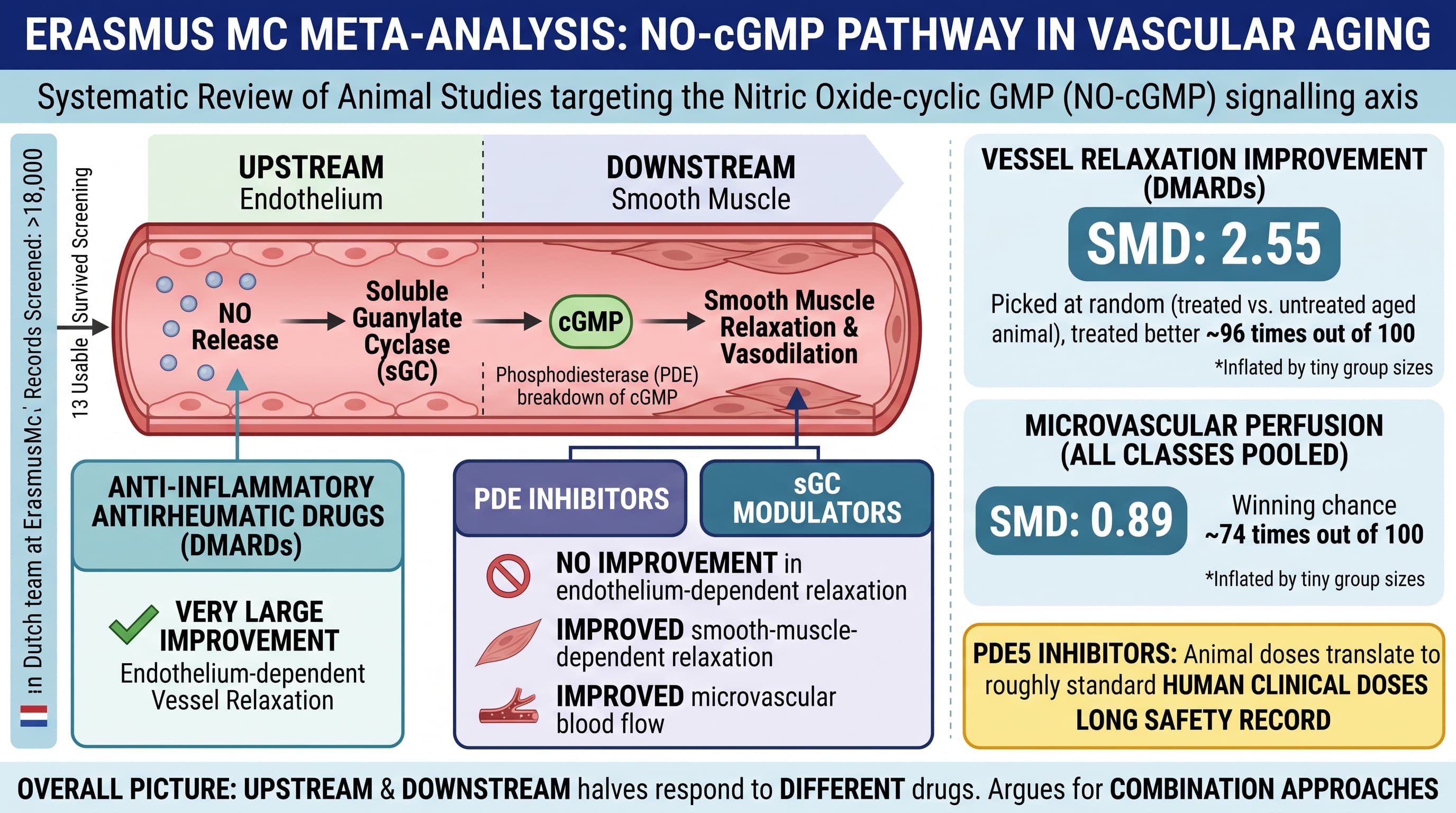
A Dutch team at Erasmus MC has pooled every usable animal study on drugs that target the nitric oxide to cyclic GMP (NO-cGMP) signalling axis in aged or prematurely aged animals. Thirteen studies survived screening out of more than 18,000 records. Three drug classes were tested: anti-inflammatory antirheumatic drugs (DMARDs), phosphodiesterase (PDE) inhibitors, and soluble guanylate cyclase (sGC) modulators. DMARDs produced a very large improvement in endothelium-dependent vessel relaxation. PDE inhibitors and sGC modulators did not improve endothelium-dependent relaxation but did improve smooth-muscle-dependent relaxation and microvascular blood flow. The overall picture is that upstream and downstream halves of the same pathway respond to different drugs, arguing for combination approaches.
Blood vessels age badly. They stiffen, they stop dilating properly, and they leak inflammation. Most of the clinical effort so far has gone into scraping cholesterol plaque out of the equation with statins, but a large share of cardiovascular risk in older people has nothing to do with plaque. It is the vessel wall itself losing the ability to relax on command.
The command signal in question is nitric oxide. The endothelial lining makes it, it activates an enzyme called soluble guanylate cyclase in the muscle layer beneath, that enzyme makes cyclic GMP, and cyclic GMP tells the muscle to let go. Aging degrades this chain at every link: less nitric oxide made, a damaged and unresponsive receptor enzyme, and more phosphodiesterase enzyme chewing up the cyclic GMP before it can act.
Jouabadi and colleagues asked a simple question of the animal literature. If you pharmacologically attack any one of those three links in an old animal, does the vessel work better? They ran three separate systematic searches, all preregistered, and found thirteen studies.
The results split along a mechanistic fault line that is the most interesting thing in the paper. Old anti-inflammatory drugs used for rheumatoid arthritis and lupus, methotrexate, hydroxychloroquine, gold and penicillamine, produced the single largest effect on endothelium-dependent relaxation, the standard readout of endothelial health. They work upstream, by lowering the inflammatory and oxidative burden that cripples nitric oxide production in the first place.
Drugs acting downstream did the opposite. Sildenafil and its relatives, and sGC activators, failed to improve endothelium-dependent relaxation, which makes sense because they bypass the endothelium entirely. But they did improve microvascular perfusion, actual blood flow through small vessels, and smooth-muscle relaxation.
So the aging vessel appears to have two separable lesions: a broken signal source and a leaky signal amplifier. Fixing one does not fix the other. The authors suggest that combining anti-inflammatory upstream treatment with downstream cyclic nucleotide support may be additive, though no included study actually tested that combination.
The honest verdict is that this is a mapping exercise, not a demonstration of efficacy. Thirteen studies is very few. Only two used naturally aged animals, both rats. The rest used progeroid or disease models. No study measured arterial stiffness by pulse wave velocity, the measure that actually predicts human outcomes.
Insights
First, the size of the reported benefits. The effect sizes are given as standardized mean differences, which express the gap between treated and control groups in units of the natural spread within each group. The DMARD figure of 2.55 means that if you picked one treated aged animal and one untreated aged animal at random, the treated one would have better vessel relaxation about 96 times out of 100. For microvascular perfusion pooled across all three drug classes, the figure of 0.89 corresponds to winning about 74 times out of 100. Those are the honest translations. They are also almost certainly inflated by tiny group sizes.
Second, the mechanistic lesson is more portable than the drugs. Downstream drugs did not repair endothelial function. If endothelial nitric oxide output is your target, upstream inflammation control is where the signal was, not cyclic GMP support.
Third, of the compounds tested, PDE5 inhibitors are the only class where the animal doses translate to roughly standard human clinical doses, and they have a long safety record.
Context and Source
- Open Access Paper: Targeting the NO-cGMP axis in age-related vascular dysfunction: a systematic review and meta-analysis of preclinical animal studies
- Institutions: Division of Pharmacology and Vascular Diseases, Department of Internal Medicine, Erasmus MC University Medical Center, Rotterdam, Netherlands; Erasmus MC Medical Library, Rotterdam; Department of Anesthesiology, Pain and Palliative Care, Radboud University Medical Centre, Nijmegen, Netherlands
- Country: The Netherlands
- Journal: GeroScience (Springer, official journal of the American Aging Association)
- Impact evaluation: The impact score of this journal is 6.0 (2025 Journal Impact Factor, up from 5.7 the prior year; CiteScore approximately 8.3), evaluated against a typical high-end range of 0 to 60+ for top general science, therefore this is a Medium impact journal.
Related Reading:
- How the Collapse of Nitric Oxide Signaling Accelerates Aging
- Eat Your Greens, Reboot Your Nitric Oxide: How a Backup Chemistry System Keeps Ageing Blood Vessels Alive
- The Arginine Paradox Solved: Why Citrulline is the Superior Vascular Anti-Aging Agent
- Nitric Oxide for improved bloodflow and healing -- WiseAthletes podcast
- The Brain's Broken Plumbing: Why Diminishing Blood Flow Drives Dementia
- Video Interview - Nitric Oxide and Functional Health - with Dr. Nathan Bryan