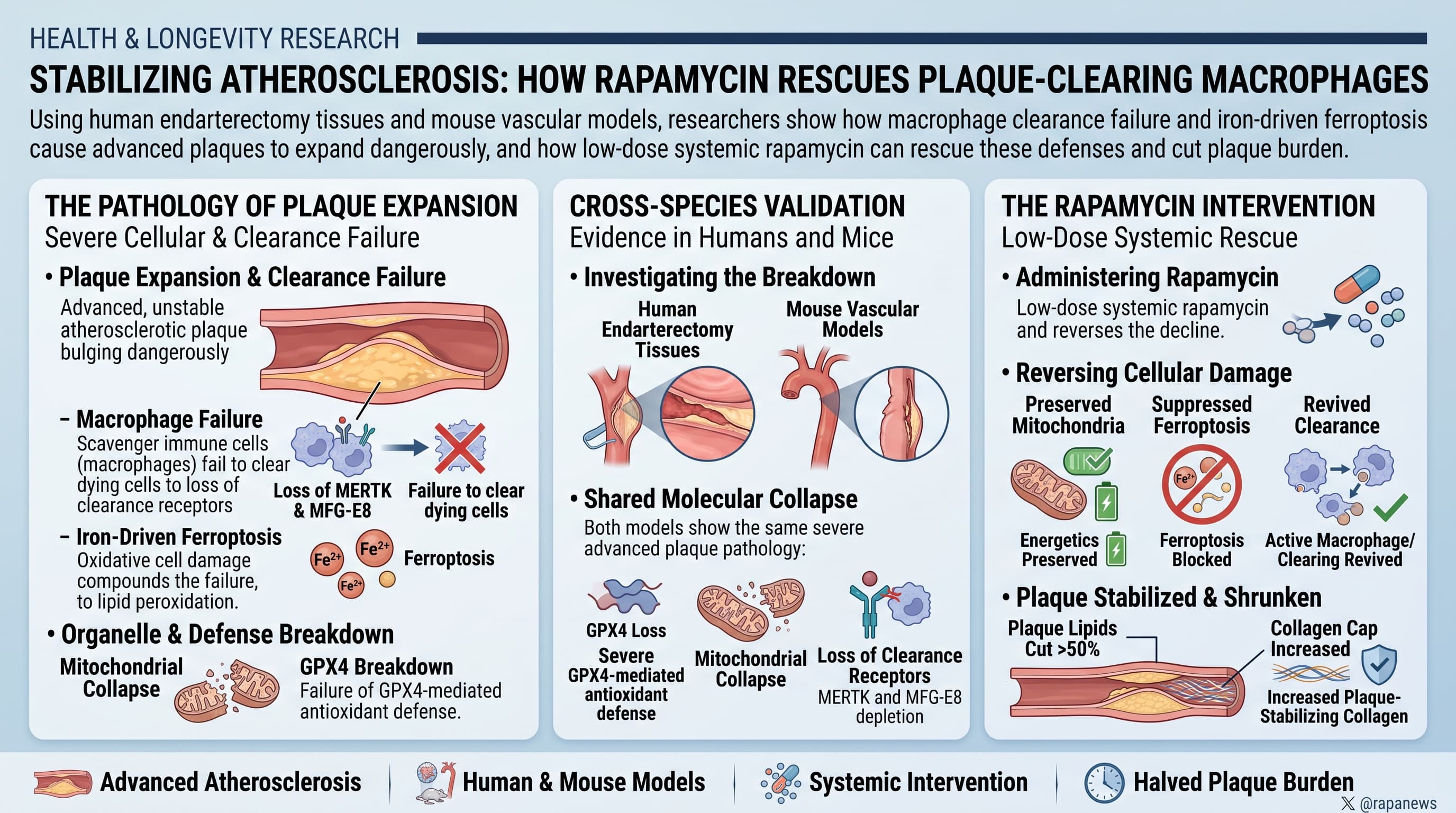
Atherosclerotic plaques expand dangerously when scavenger immune cells called macrophages fail to clear dying cells, a failure compounded by iron-driven oxidative cell damage known as ferroptosis. Examining both human endarterectomy tissues and mouse vascular models, researchers discovered that advanced plaques suffer from a severe breakdown in GPX4-mediated antioxidant defense, mitochondrial collapse, and loss of the clearance receptors MERTK and MFG-E8. Administering low-dose systemic rapamycin preserved mitochondrial energetics, suppressed iron-mediated lipid peroxidation, revived macrophage clearance capability, and cut whole-aorta plaque lipid burden by more than half while substantially increasing plaque-stabilizing collagen.
The defining crisis of advanced arterial disease is necrotic debris accumulation. As blood vessels accrue cholesterol, local macrophages ingest lipids until they become foam cells. In early stages, dying cells undergo efferocytosis, an orderly biological engulfment and waste-processing cycle that prevents intracellular contents from spilling into surrounding tissue. In mature lesions, this process falters. Uncleared apoptotic corpses undergo secondary necrosis, expelling inflammatory compounds, enzymatic debris, and crystalline lipids into the vessel wall. This cycle creates a growing, unstable necrotic core prone to catastrophic rupture.
A primary driver of this efferocytic collapse is ferroptosis, an iron-catalyzed cascade of lipid peroxidation that damages delicate subcellular membranes. Analyzing human carotid plaque samples harvested during vascular surgery, researchers observed that the primary lipid-peroxide neutralizer, glutathione peroxidase 4 (GPX4), was severely depleted in advanced fibrous lesions relative to adjacent early lesions. This deficiency leaves vascular macrophages defenseless against oxidative stress. Without functional GPX4, intracellular iron accelerates the peroxidation of polyunsaturated fatty acids, triggering mitochondrial calcium overload, membrane potential breakdown, and acute energy exhaustion. Because the mechanical engulfment of dying cells requires substantial cellular energy, energy-depleted macrophages shed their primary phagocytic tethering molecules, MERTK and MFG-E8.
The investigative team modeled this pathway in human macrophages using the chemical compound FIN-56, reproducing the cellular damage, glycolytic shift, and clearance deficits seen in diseased vessels. Co-treatment with rapamycin rescued these cellular cohorts. By dampening baseline mTOR activity, rapamycin restored GPX4 expression, lowered intracellular free iron and toxic lipid aldehydes, and preserved oxygen consumption and cellular energy production. Macrophages regained their physical capacity to bind and internalize dying cells. Crucially, the compound also stopped surrounding vascular smooth muscle cells from adopting a harmful synthetic phenotype that accelerates arterial disease.
In an animal model using genetically susceptible mice fed an atherogenic high-fat diet, a four-week regimen of twice-weekly rapamycin reversed established vascular decay. The treatment restored the ratio of active clearance events relative to free-floating dying cells from 0.37 to 1.37, while dropping lipid deposition across the entire length of the aorta from 25.32 percent down to 12.28 percent. Plaque coverage at the aortic root dropped from 35.56 percent to 16.71 percent, while protective fibrous collagen caps expanded from 22.42 percent to 39.73 percent. These findings indicate that rapamycin works in part by rescuing macrophage vitality and restoring cellular housekeeping.
Actionable Insights
This paper highlights that arterial plaque stability depends heavily on the energy status of tissue-resident macrophages and their capacity to clear cellular waste. The practical takeaway centers on the potential of targeted mTOR modulation and lipid antioxidant support to resolve vascular inflammation.
In mice, low-dose rapamycin produced an absolute reduction of 13.04 percentage points in aortic lipid accumulation (falling from 25.32% to 12.28%, a relative reduction of 51.5%), while reducing the aortic root plaque footprint by an absolute 18.85 percentage points (falling from 35.56% to 16.71%, a relative reduction of 53.0%). Concurrently, it produced an absolute increase of 17.31 percentage points in structural collagen (rising from 22.42% to 39.73%, a relative improvement of 77.2%), and improved the cellular clearance index nearly fourfold (increasing from 0.37 to 1.37).
These findings suggest that strategies preserving mitochondrial glutathione pools, minimizing systemic iron overload, and periodically attenuating hyperactive mTOR signaling may help stabilize existing plaques. The data also support the clinical development of drug-coated balloons that deliver rapamycin locally to damaged blood vessels, securing these cellular benefits without causing systemic harm.
Context and Source
- Paywalled Paper: Rapamycin attenuates ferroptotic stress and improves macrophage efferocytosis in experimental atherosclerosis, 2026 Oct 2.
- Primary Institutions: Department of Cerebrovascular Diseases, People’s Hospital of Zhengzhou University (Henan Provincial People’s Hospital); Henan International Joint Laboratory of Cerebrovascular Diseases, Zhengzhou, China
- Journal Name: International Immunopharmacology
- Impact Evaluation: The impact score of this journal is 5.6 (Clarivate Web of Science Journal Impact Factor), evaluated against a typical high-end range of 0 to 60+ for top general science, therefore this is a Medium impact journal.
Related Reading:
- Rapamycin Quiets the Aging Immune System Around Arterial Plaques
- Repurposing Rapamycin to Reverse Brain Aging in APOE4 Carriers
- Rapamycin exerts geroprotective effects in the ageing human immune system by enhancing resilience against DNA damage
- Rapamycin Tames Inflammaging Without Compromising Immunity
- Low-Dose Rapamycin Suppresses Senescence Biomarkers in Frail Heart Failure Patients