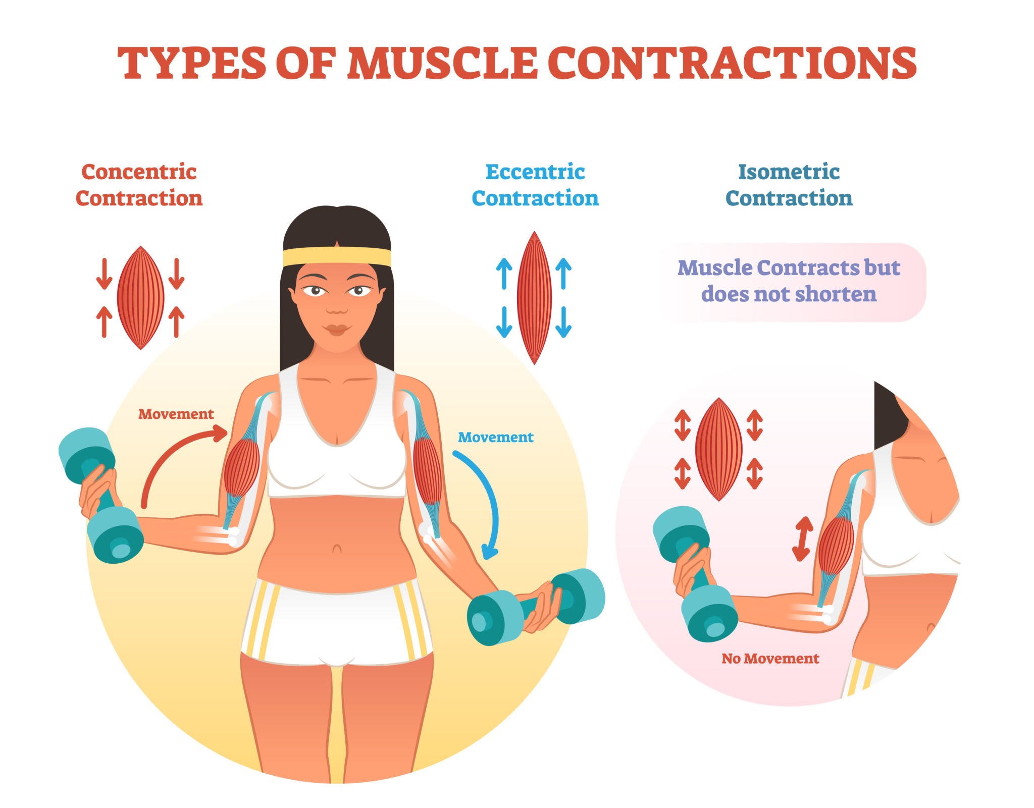
Danish researchers stretched isolated leg muscles from young and old mice in a dish and watched which internal growth signals fired. The main growth-and-build pathway (mTORC1) responded just as strongly in old muscle as in young. But a second pathway that helps switch on muscle-remodeling genes (JNK feeding into SMAD2) was roughly cut in half in the old muscle. Because the muscles were tested outside the body, with no blood, nerves, or hormones involved, the result argues that a good chunk of the “anabolic resistance” seen in aging bodies is not baked into the muscle fiber itself. Instead it likely comes from the aging environment around the muscle. One repair-signaling pathway, though, does appear to weaken from the inside.
For decades the accepted story of muscle aging has been that old muscle simply stops listening to exercise. Load it, feed it protein, and it responds more weakly than young muscle. This blunted response, called anabolic resistance, is one of the mechanistic engines behind sarcopenia, the age-related loss of muscle mass and strength that erodes independence in later life. The open question has always been where the fault lies. Is the muscle fiber itself broken, or is the problem the aged body it lives in, with its poorer blood flow, altered hormones, and simmering low-grade inflammation?
A group at Aarhus University in Denmark went after that question by taking the body out of the equation. They isolated the extensor digitorum longus, a fast-twitch lower-leg muscle, from young adult mice (16 weeks) and old mice (24 months), then stretched each muscle mechanically in an oxygenated bath with no nerves, no circulation, and no hormones. Each animal donated one muscle for stretching and the opposite muscle as an untouched control, a clean within-animal comparison.
The headline result splits in two. The mTORC1 pathway, which controls the protein-building machinery, lit up just as strongly in old muscle as in young after stretch. The molecular readouts (mTOR, p70S6K, rpS6, 4E-BP1) rose to similar levels regardless of age. If old muscle were intrinsically deaf to mechanical load, this pathway should have been quieter. It was not.
The second pathway told a different story. JNK, a strain-sensitive kinase, and its downstream target SMAD2 at its linker region, drive the gene-level remodeling that lets muscle adapt. Here the old muscle responded at roughly half the magnitude of young muscle, and this age gap was statistically robust.
The interpretation is that aging does not flip one master switch. It selectively dampens the branch tied to turning genes on and off, while leaving the branch that builds protein largely intact. The practical implication is provocative. If translational signaling is preserved inside the fiber, then much of the anabolic resistance observed in living older people may be imposed from outside the muscle, by the systemic environment, rather than being an irreversible defect of the muscle itself. That is a more hopeful framing, because systemic factors are more modifiable than hardwired cellular decline.
Actionable Insights
Be careful here, because this is a mouse study done on muscles in a dish, not a human trial. It tests mechanism, not a supplement or a workout you can copy. That said, a few useful take-home messages emerge.
First, the growth-signaling machinery in aging muscle is not broken. Mechanical tension alone was enough to fully activate the main build pathway in old muscle. The practical translation is that resistance and stretch-loading exercise remains a legitimate anabolic stimulus into old age, and the “you are too old to build muscle” narrative is not supported at the level of the muscle fiber.
To put a number on the stimulus itself, stretching produced large signaling jumps. In old muscle the build-pathway markers rose roughly 1.7-fold (mTOR) up to 3.9-fold (p70S6K) over the unstretched control. Expressed as a standardized effect size, which measures how big a change is relative to its own variability, most of these responses land at a Cohen’s d well above 0.8, the conventional threshold for a large effect. In plain terms, the signal clearly rises above the noise.
Second, the aging deficit was specific to the remodeling pathway, which fell to about half of the young response (a roughly 45 to 51 percent reduction, effect size Cohen’s d near 1.0). This is where age genuinely bites at the cellular level.
Third, and most actionable at the whole-body level, the preserved internal machinery points the finger at systemic factors you can influence: circulating inflammation, protein and amino acid delivery, insulin sensitivity, and blood flow. Managing those may matter as much as the exercise itself.
Context and Source
- Open Access Paper: Aging preserves mTORC1 but attenuates JNK-SMAD2L signaling sensitivity to passive stretch-induced tension development in isolated mouse skeletal muscle.
- Institution and Country: Aarhus University, Aarhus, Denmark (Exercise Biology, Department of Public Health), with one co-author affiliated with NMD Pharma A/S, Aarhus, Denmark.
- Journal: Experimental Gerontology (Elsevier).
- Impact evaluation: The most recent metrics place the Journal Impact Factor around 5.1 (2025 release, June 2026) and the CiteScore at 6.7. Taking the impact factor: the impact score of this journal is 5.1, evaluated against a typical high-end range of 0 to 60+ for top general science journals, therefore this is a Low-to-Medium impact journal. Within its own specialist field of aging and gerontology it is a respectable, established mid-tier venue