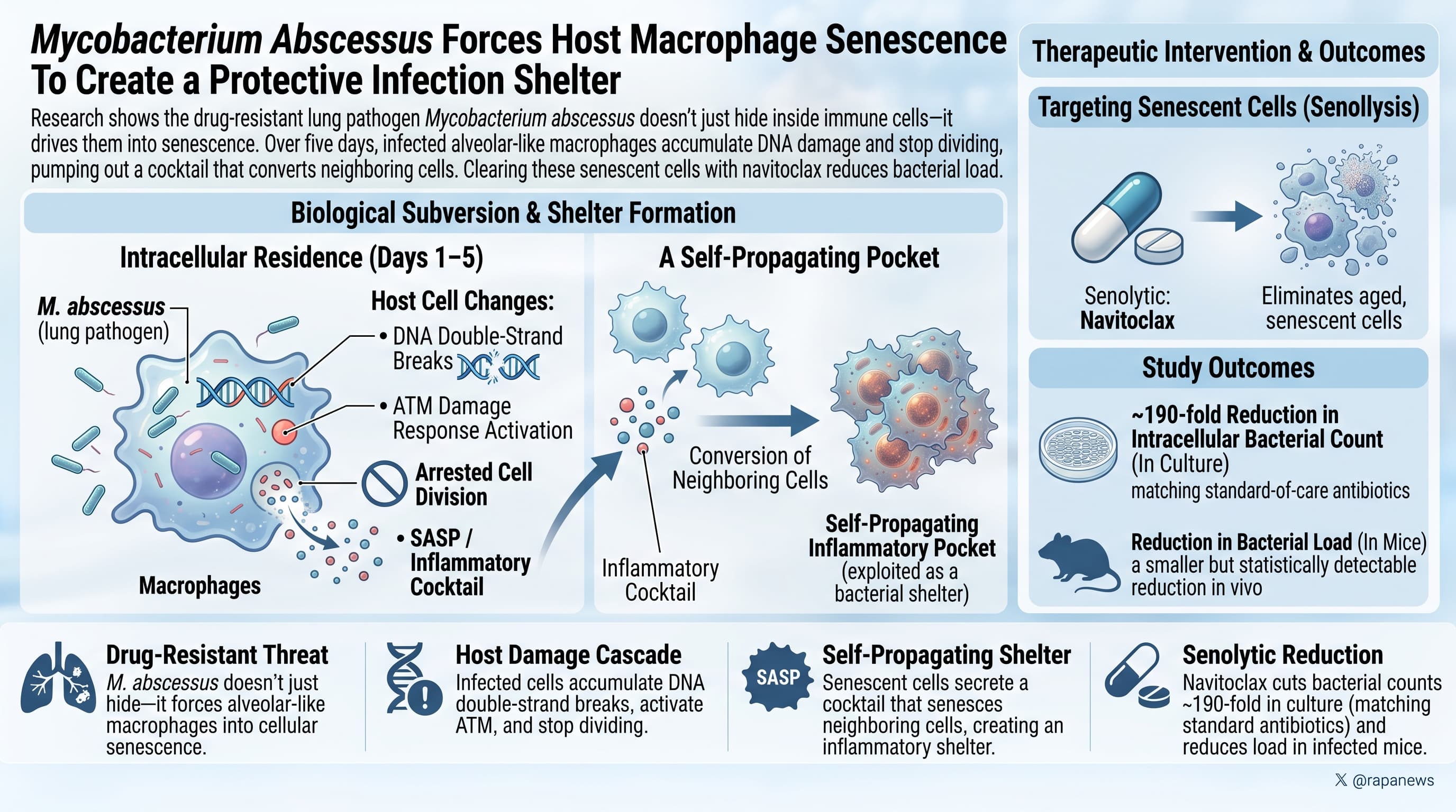
An Italian group has shown that Mycobacterium abscessus, a drug-resistant lung pathogen, does not simply hide inside immune cells; it pushes them into cellular senescence. Over five days of intracellular residence, infected alveolar-like macrophages accumulate DNA double-strand breaks, activate the ATM damage response, stop dividing, and begin pumping out an inflammatory secretory cocktail. That cocktail then converts neighboring uninfected cells into senescent cells as well, creating a self-propagating inflammatory pocket that the bacterium appears to exploit as a shelter. Clearing those senescent cells with the senolytic navitoclax cut intracellular bacterial counts by roughly 190-fold in culture, matching the standard-of-care antibiotic, and produced a smaller but statistically detectable reduction in bacterial load in infected mice.
The hallmarks-of-aging framework treats senescent cells as a byproduct of time, oxidative wear, and replication limits. This paper argues for a different entry point into the same pathway: an infection that sits inside your cells and damages their DNA on purpose.
The team built a chronic infection model using murine alveolar-like macrophages, the cell type that M. abscessus actually colonizes in human lungs. They used a deliberately low bacterial dose, a two-hour exposure window, and daily washing to keep the infection intracellular rather than letting bacteria overgrow in the dish. By day five, about two thirds of cells carried bacteria, and those cells looked wrong: larger, less circular, with irregular nuclei, and almost completely stalled in the cell cycle. Only around two percent were still synthesizing DNA.
What followed is the standard senescence checklist, ticked off one item at a time. RNA sequencing showed a coordinated shift in the SenMayo senescence gene panel, with the usual inflammatory suspects, including IL-1 beta, IL-6 and TNF, climbing to the top. Protein markers confirmed it: p21 rose, lamin B1 fell, and senescence-associated beta-galactosidase activity went from roughly a quarter of cells to roughly sixty percent.
The mechanistic claim is the strongest part of the work. The authors found DNA damage foci, confirmed real strand breaks with comet assays, and showed the damage only appears when the bacteria are alive. Killed bacteria produced nothing. Blocking the ATM kinase with a small molecule cut p21-positive cells by more than half, placing the damage response upstream of the senescence program rather than alongside it.
Then comes the part with the broader implications. Senescence did not stay with the infected cells. Culture medium taken from infected dishes, filtered to remove bacteria, turned healthy macrophages senescent on its own, reaching about seventy-five percent at the highest dose. Cells sitting within two cell widths of an infected cell were overwhelmingly senescent even when they carried no bacteria. Worse, cells pre-treated with that medium were roughly fifty percent more likely to become infected when challenged. The infection appears to build its own hospitable neighborhood.
The therapeutic test follows logically. Kill the senescent cells, and you remove the reservoir. Navitoclax, a Bcl-2 family inhibitor used in senolytic research, did exactly that in culture, and critically it did not spill live bacteria into the surrounding medium. In mice, the effect was real but modest, and smaller than amikacin. Combining the two added nothing.
The conceptual contribution outlives the drug result: chronic infection may be a source of senescent cell burden, which would place infectious history inside the aging equation rather than beside it.
Actionable Insights
There is no supplement, dose, or protocol to take from this paper. What it offers is a revised model of where senescent cells come from.
The magnitudes are worth knowing. Chronically infected macrophage cultures went from roughly 25 percent senescent cells to roughly 60 percent, an absolute increase of about 35 percentage points. Among the cells actually carrying bacteria, approximately 75 percent were senescent and approximately 80 percent expressed p21. Uninfected cells sitting near an infected cell were senescent 85.7 percent of the time. These are not subtle shifts; they are the kind of effect size where you can see the difference by eye in a microscope field.
The senolytic result was a 2.3 log10 reduction in intracellular bacteria, which means roughly 190 bacteria killed for every one that survived, or about 99.5 percent clearance. In live mice the same drug produced closer to a tenfold reduction, which is a real effect but an order of magnitude weaker.
The practical takeaway for anyone tracking inflammatory markers: a persistent, unresolved infection may be contributing to senescent cell burden in a way that supplements aimed at “inflammaging” do not address. Navitoclax itself causes dose-limiting thrombocytopenia and is not a self-experimentation candidate.
Context and Source
- Open Access Paper: Cell senescence emerges as a hallmark and therapeutic target of chronic intracellular infection
- Institutions: National Institute of Molecular Genetics (INGM), Milan; University of Milan; IRCCS San Raffaele Scientific Institute, Milan; Istituto Italiano di Tecnologia, Genova and Rome; Sapienza University of Rome; University School of Advanced Studies IUSS, Pavia
- Country: Italy
- Journal: Nature Communications, published online 18 August 2026
- Impact evaluation: The impact score of this journal is 18.1 (2025 Journal Citation Report data year, released June 2026), evaluated against a typical high-end range of 0 to 60+ for top general science, therefore this is a High impact journal.
Related Reading:
- Death by a Thousand Colds: The Hidden Aging Tax of Infections You Never Noticed
- Analysis: How Stem Cells Respond to Infection, Inflammation, and Ageing
- Infection with cat parasite Toxoplasma may drive 'inflammation aging' in older adults
- Repeated doses of BCG vaccine reduces risk of Alzheimer’s and all cause mortality
- Vaccines for longevity