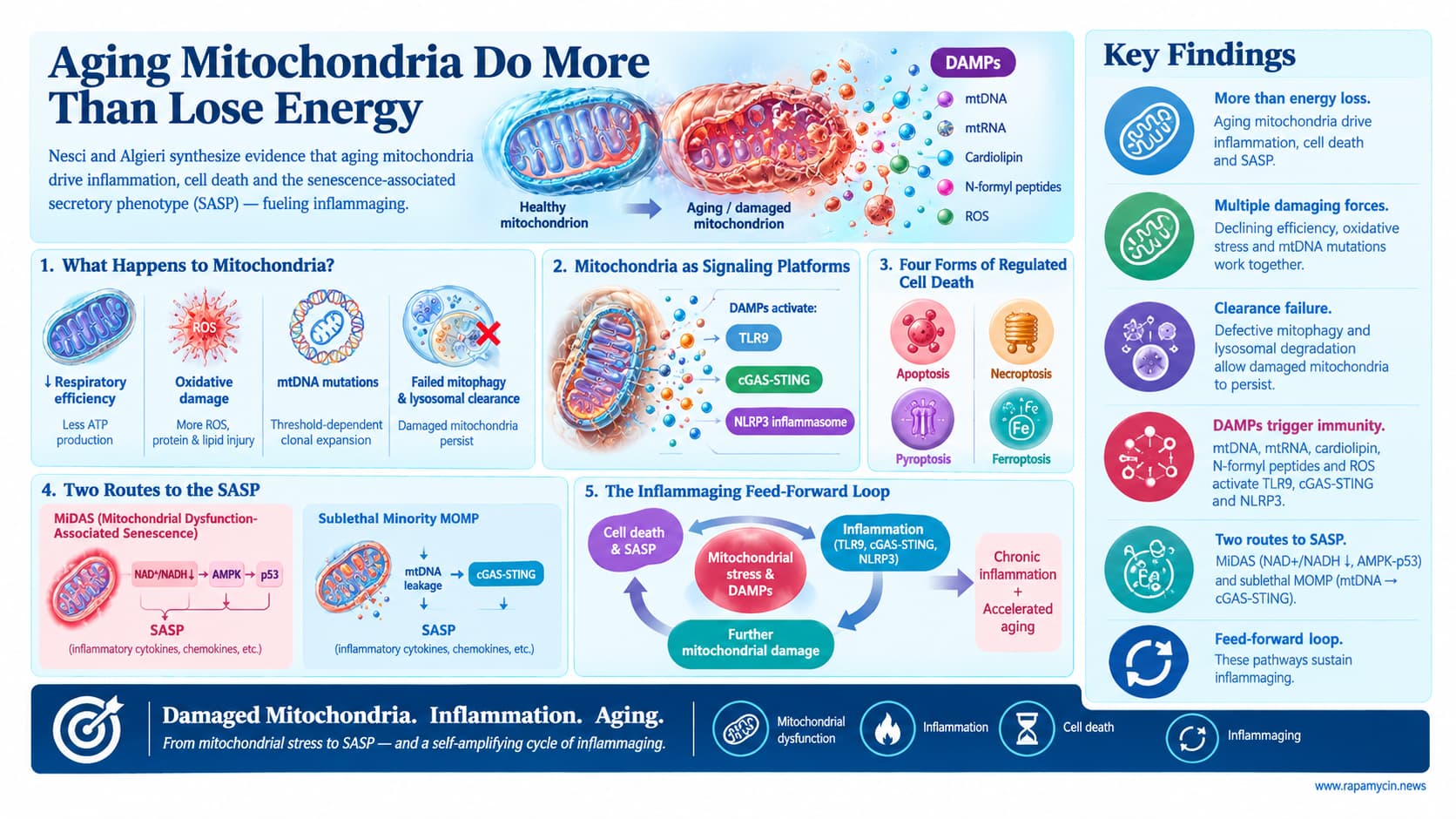
Nesci and Algieri synthesize evidence that aging mitochondria do far more than lose energy output. Declining respiratory efficiency, oxidative damage and the threshold-dependent clonal expansion of mtDNA mutations progressively compromise the organelle, while failing mitophagy and lysosomal clearance let damaged mitochondria persist. These organelles then behave as signaling platforms. They release damage-associated molecular patterns (mtDNA, mtRNA, cardiolipin, N-formyl peptides, ROS) that activate TLR9, cGAS-STING and the NLRP3 inflammasome, and they shape four forms of regulated cell death: apoptosis, necroptosis, pyroptosis and ferroptosis. The review highlights two distinguishable routes from mitochondrial stress to the senescence-associated secretory phenotype (SASP): mitochondrial dysfunction-associated senescence (MiDAS), driven by a falling NAD+/NADH ratio and AMPK-p53 signaling, and sublethal minority MOMP, which leaks mtDNA to cGAS-STING without killing the cell. Together these form a feed-forward loop the authors propose sustains inflammaging.
For decades, the textbook picture of aging mitochondria was a power plant slowly running down. A new review in GeroScience argues that picture is incomplete. Damaged mitochondria, say Salvatore Nesci of the University of Bologna and Cristina Algieri of Link University in Rome, do not simply fail quietly. They send out alarm signals that the immune system mistakes for a bacterial infection, and those false alarms may help keep the aging body in a state of slow-burning inflammation.
The idea rests on mitochondrial ancestry. These organelles descend from bacteria engulfed by our ancestral cells more than a billion years ago, and they still carry bacterial fingerprints: their own circular DNA, rich in a sequence pattern our immune sensors associate with microbes, and proteins that begin with a chemical tag bacteria also use. In healthy mitochondria these molecules stay locked inside. In damaged ones, pieces leak out.
The review traces how the damage builds. As the energy machinery loses efficiency, more electrons escape and form reactive oxygen species, which further harm mitochondrial DNA, proteins and fats. Mutations in mitochondrial DNA can accumulate silently for years, because each cell holds hundreds to thousands of copies. Only when mutant copies pass a tissue-specific threshold does energy production falter. In the human colon, for instance, the share of energy-deficient crypts rises with age as individual mutations slowly take over.
Once mitochondrial DNA escapes into the cell’s interior, a sensor called cGAS detects it and switches on an antiviral-style alarm through a partner protein called STING. Other mitochondrial fragments trigger the NLRP3 inflammasome, which releases the potent inflammatory messengers IL-1 beta and IL-18. Strikingly, the cell does not have to die for this to happen. Experiments in human cells show that when only a handful of mitochondria in a cell rupture, enough DNA spills out to drive inflammation while the cell survives.
The second half of the story concerns senescent cells, the so-called zombie cells that stop dividing but refuse to die. Mitochondrial failure alone can push a cell into this state. These cells secrete their own cocktail of inflammatory signals, but it differs from the cocktail produced by cells pushed into senescence by DNA damage. A drop in a key energy ratio, NAD+ to NADH, activates a stress pathway that locks the cell in arrest while muting part of its inflammatory output.
Put together, the authors describe a loop. Damaged mitochondria create senescent cells and leak danger signals; those signals inflame neighboring tissue; inflammation stresses mitochondria further. In principle the loop could be broken from several directions: better mitochondrial cleanup, clearing senescent cells, or blocking the alarm sensors themselves.
The authors are clear that human evidence lags well behind. A trial of urolithin A, a compound that promotes mitochondrial recycling, improved muscle endurance but missed its main goals. A senolytic drug combination failed its primary bone endpoint in postmenopausal women. Drugs that block NLRP3 or STING are in clinical testing today, but remain early-stage or untested for aging.
Context and Source
- Open Access Paper: Mitochondrial dysfunction and inflammaging: a mechanistic bridge between bioenergetic failure and senescence
- Institutions: Department of Veterinary Medical Sciences, University of Bologna (Ozzano Emilia); Department of Life Sciences, Health and Health Professions, Link University (Rome)
- Country: Italy
-
Journal: GeroScience (Springer; official journal of the American Aging Association).
Impact evaluation: The impact score of this journal is 4.9 (JIF), evaluated against a typical high-end range of 0 to 60+ for top general science (roughly 5 to 20 among leading aging-specialty journals), therefore this is a Medium impact journal.
Related Reading:
- BioAge Presentation (Longevity Summit, 2025): NLRP3, Apelin Targets
- The Pilot Light of Aging: How Your "Healthy" Diet May Be Fueling Silent Inflammation
- The Eye's Inflammatory Alarm Bell: Can Silencing NLRP3 Save Aging Vision?
- Running Down the NLRP3 Alarm: How Exercise Quiets the Brain's Inflammatory Trigger
- Quenching the Vascular Fire (NLRP3): How Phytochemicals Combat the Residual Inflammatory Risk of Heart Disease