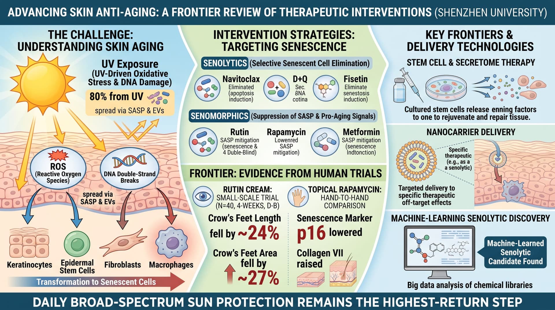
This narrative review from Shenzhen University argues that senescent cells are central to skin aging. It covers four skin cell types (fibroblasts, macrophages, keratinocytes and epidermal stem cells), how damage from UV-driven oxidative stress and DNA damage spreads between cells through the SASP and extracellular vesicles, and how histone modifications keep aged skin macrophages in an inflammatory state. It then surveys interventions: senolytics (navitoclax, dasatinib plus quercetin, fisetin), senomorphics (rutin, rapamycin, metformin), stem cell and secretome therapies, nanocarrier delivery, and machine-learning senolytic discovery. It contains no new data. Most of the evidence it summarizes comes from cell culture, rodents and human skin grafted onto mice. The human trials it cites are small, short, and mostly split-face cosmetic studies.
Skin shows age sooner than any other organ, and the authors argue it shows it for a specific reason: old, damaged cells that refuse to die.
These are senescent cells. They stop dividing but stay metabolically active, and they release a cocktail of inflammatory signals, enzymes and small vesicles known as the senescence-associated secretory phenotype, or SASP. In skin, the review says, the main senescent populations are fibroblasts in the dermis, keratinocytes in the outer layer, immune macrophages, and the stem cells that normally replenish the epidermis and hair follicles.
Each population fails differently. Senescent fibroblasts make less collagen and more of the matrix metalloproteinases (MMPs) that break collagen down. The authors describe a feedback loop: fragmented collagen reduces the mechanical tension fibroblasts feel, which raises their internal oxidative stress, which drives further MMP production. Senescent keratinocytes renew the epidermis more slowly, thinning the barrier and letting moisture out and bacteria such as Staphylococcus aureus in. Aged macrophages get stuck in an inflammatory mode and fail to shift into the repair mode that heals wounds.
The review’s more unusual contribution is its attention to how the damage spreads. Senescent cells release more extracellular vesicles than young cells. UV-damaged cells may export fragments of their own DNA in these vesicles, and neighboring cells detect the foreign DNA through the cGAS-STING immune sensing pathway and become senescent in turn. Vesicles also carry microRNAs, such as miR-30a, that suppress renewal in young keratinocytes. The authors also compile evidence that epigenetic regulators, including the enzymes JMJD3, MOF and SETDB2, lock macrophages into a chronic inflammatory state. Much of that evidence, however, comes from diabetic wounds rather than aged skin.
On treatments, the authors cover the familiar longevity pharmacopeia. Navitoclax clears senescent dermal fibroblasts in mouse and human-skin-graft models but causes low platelet and neutrophil counts when taken systemically. Dasatinib plus quercetin, and fisetin alone, reduce senescent fibroblast counts in similar models. Rutin, a quercetin glycoside, acts more as a SASP dampener than a cell killer. Topical rapamycin gets a mention for one small human trial that found fewer p16-positive cells and more collagen VII. Metformin is included for AMPK activation, mostly on the strength of rodent and worm data.
Stem cell therapy receives equal emphasis. The authors conclude that cell-free products, meaning conditioned media and vesicles from mesenchymal stem cells, are more practical than transplanted cells, which survive poorly after injection. Seven clinical studies are tabulated, and nearly all delivered the product by microneedling.
The review ends by pointing to machine-learning screens that have identified new senolytics, including ginkgetin, periplocin and oleandrin, from compound libraries.
The big idea is coherent: skin aging is a senescence problem that can be spread from cell to cell, and so it should be treatable by removing or silencing senescent cells locally. The weakness is that this remains a hypothesis supported mainly by mechanistic and animal work. The human data are sparse, small and unblinded or confounded more often than not.
Actionable Insights
The most reliable intervention remains the least exotic one. The review repeats a common estimate that UV exposure causes about 80% of visible skin aging. That figure is widely cited but loosely sourced, and the true share is hard to measure. Daily broad-spectrum sun protection is still the highest-return step.
Among topicals discussed, two have small human trials:
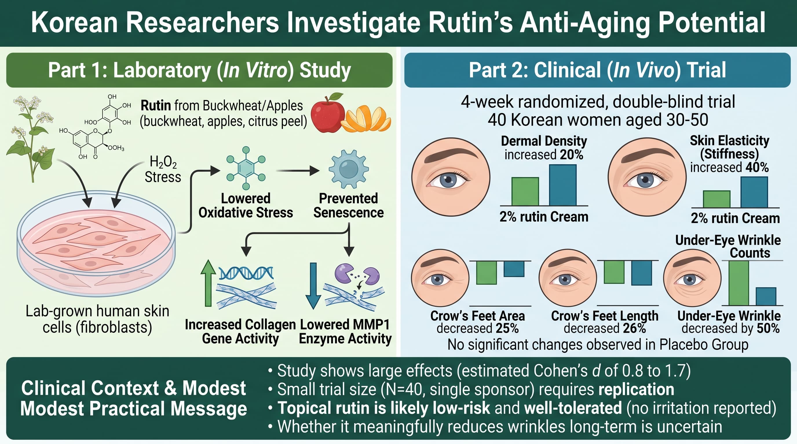
- Rutin cream: In a 40-person, 4-week double-blind study, crow’s feet length fell by about 24% and crow’s feet area by about 27% in the rutin group. These are improvements from each person’s own baseline. The paper did not report a standardized effect size, and 4 weeks is too short to judge durability.
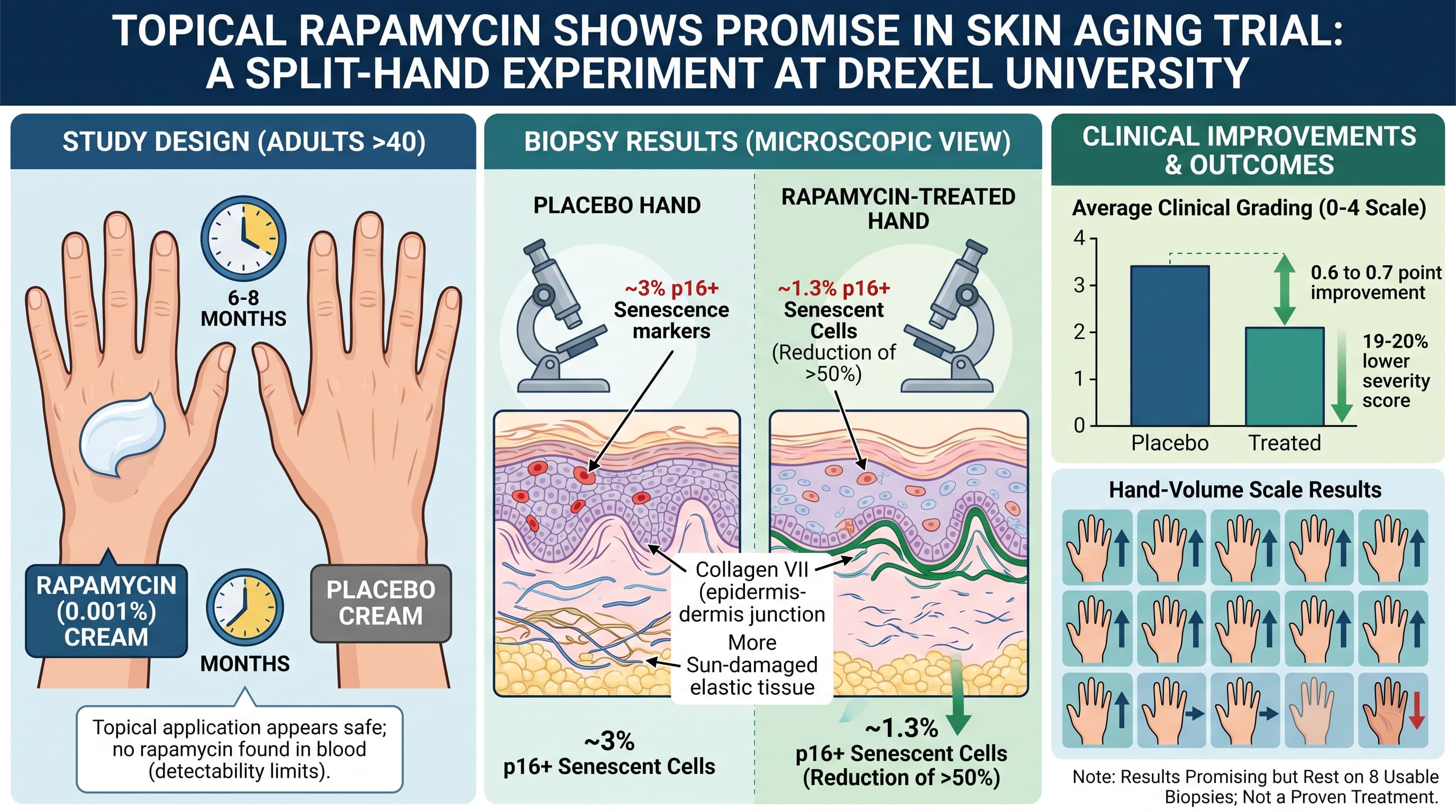
- Topical rapamycin: In a hand-to-hand comparison, rapamycin cream lowered the senescence marker p16 and raised collagen VII.
Stem cell conditioned media trials all used microneedling, which stimulates collagen on its own. The one trial that compared delivery methods found no difference between groups. Any added benefit from the stem cell product itself remains unproven.
Oral senolytics (fisetin, dasatinib plus quercetin) have no controlled human skin-aging outcome data in this review.
Context and Source
- Full title: Skin Aging: From Molecular Mechanisms to Therapeutic and Technological Innovations
- Institution: Medical School, Shenzhen University; Laboratory of Regenerative Medicine, The 2nd Affiliated Hospital of Shenzhen University
- Country: China
- Journal: Journal of Cellular and Molecular Medicine
- Impact evaluation: The impact score of this journal is 4.7, evaluated against a typical high-end range of 0–60+ for top general science, therefore this is a Medium impact journal.
Related Reading:
- Senolytics Topically Administered to Skin for Antiaging Effects
- Topical "Zombie Cell" Killer (Navitoclax) Primes Aged Skin for Rapid Healing, Bypassing Systemic Toxicity
- Cellular Senescence in Human Skin Aging: Leveraging Senotherapeutics (Rapamycin)
- Beyond senolytics: senoadaptive drugs & clinical data on GPX4 modulation (Dr. Marco Quarta, Rubedo) - Skin Aging
- Breakthrough drug reverses skin aging and dramatically speeds healing