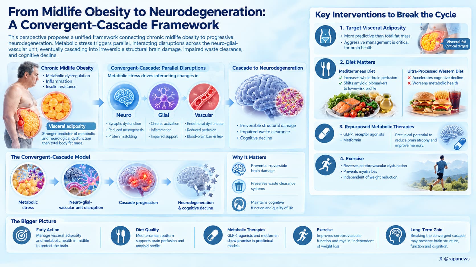
This perspective proposes a unified framework connecting chronic midlife obesity to progressive neurodegeneration. The authors outline a “convergent-cascade” model where metabolic stress induces parallel, interacting disruptions across the neuro-glial-vascular unit, eventually cascading into irreversible structural brain damage, impaired waste clearance, and cognitive decline.
For decades, obesity was treated as a problem of the body. Cardiologists cared about it and endocrinologists managed it. Neurologists mostly watched from a distance. This Perspective from researchers at UC San Diego, Yale and Scripps argues that the separation no longer makes sense.
Their central claim is that chronic overeating, and especially the deep belly fat that wraps around internal organs, puts the brain under slow, compounding stress. The damage does not come from one cause. The authors describe a sequence of overlapping failures.
It starts with the brain’s plumbing. Active neurons normally signal nearby blood vessels to deliver more blood, a process called neurovascular coupling. In obesity this signaling weakens. Blood flow drops, particularly in the hippocampus and other regions involved in memory and in Alzheimer’s disease. The same vessel movements that deliver blood also help pump cerebrospinal fluid through the brain to wash out waste. When the pump weakens, so does waste removal.
The brain then tries to compensate. It grows new vessels and loosens the BBB to let in more nutrients and immune cells. In mice, BBB leakiness can be measured within an hour of a high-fat, high-sugar meal and becomes structural after several months of high-fat feeding. In the short term these adaptations may help. Over time they backfire. New vessels are immature and leaky, immune cells enter the brain tissue, and inflammation becomes chronic.
In the final stage, inflammation breaks down the scaffolding between brain cells, called the extracellular matrix, and the insulation around nerve fibers, called myelin. The authors highlight that tracts myelinated late in development, which are also the first to deteriorate with normal aging, appear most vulnerable. Human imaging shows lower myelin content in these regions in people with higher BMI and more central fat.
A recurring theme is regional difference. The hypothalamus, which senses nutrients and controls appetite, appears able to adapt by growing vessels and increasing fluid clearance. Higher cognitive regions such as the cortex and hippocampus seem less flexible and therefore more fragile.
There are reasons for caution. Most of the mechanistic evidence comes from mice fed high-fat diets, and human data are largely correlational. The authors acknowledge that most people with obesity never develop dementia, and many people with Alzheimer’s were never obese. Their framework describes increased probability, not destiny.
The most practically interesting thread is that some of this damage looks reversible in animals. Voluntary running in obese mice restored myelin and vascular function without weight loss. Surgically removing visceral fat in aged mice reduced brain iron and myelin loss. The authors also point to metabolic drugs as possible brain protectors. However, they omit a major recent setback. The large EVOKE trials of oral semaglutide in early Alzheimer’s disease reported null results on clinical progression before this paper was accepted.
The big idea is that metabolic health and brain health share one system. If that holds, the strongest brain-protective strategy may be to prevent visceral fat buildup decades before memory problems start, rather than to treat dementia once it appears.
Actionable Insights
The paper itself provides no intervention data, so these takeaways come from the studies it cites, and their strength varies.
First, target visceral fat specifically, not just weight. The authors identify belly fat around the organs as the strongest driver of the brain changes they describe.
Second, exercise regardless of whether the scale moves. In obese mice, running reversed demyelination and blood vessel damage even without weight loss. Human data show physical activity moderates the link between obesity and reduced brain blood flow. No human effect size on brain structure is available from this paper.
Third, eat less ultra-processed food and more whole foods. These associations come from observational studies, so the true causal size is uncertain.
Fourth, if you have type 2 diabetes, newer drugs may lower dementia risk. In one large US records study, people starting GLP-1 drugs had a 33% lower relative risk of dementia than those on other diabetes drugs, and SGLT2 inhibitor users had a 43% lower risk. In absolute terms, that is about 2 to 3 fewer dementia cases per 1,000 person-years. This is observational evidence and cannot prove cause.
Context and Source
- Paywalled Paper: Obesity as a catalyst for neurodegeneration., Published 27 February 2026.
- Institution: UC San Diego School of Medicine, Yale University School of Medicine, Scripps Research Translational Institute.
- Country: USA.
- Journal: Nature Metabolism.
- Impact Evaluation: The impact score of this journal is 20, evaluated against a typical high-end range of 10 to 40 for top metabolic and general science journals, therefore this is a High impact journal.