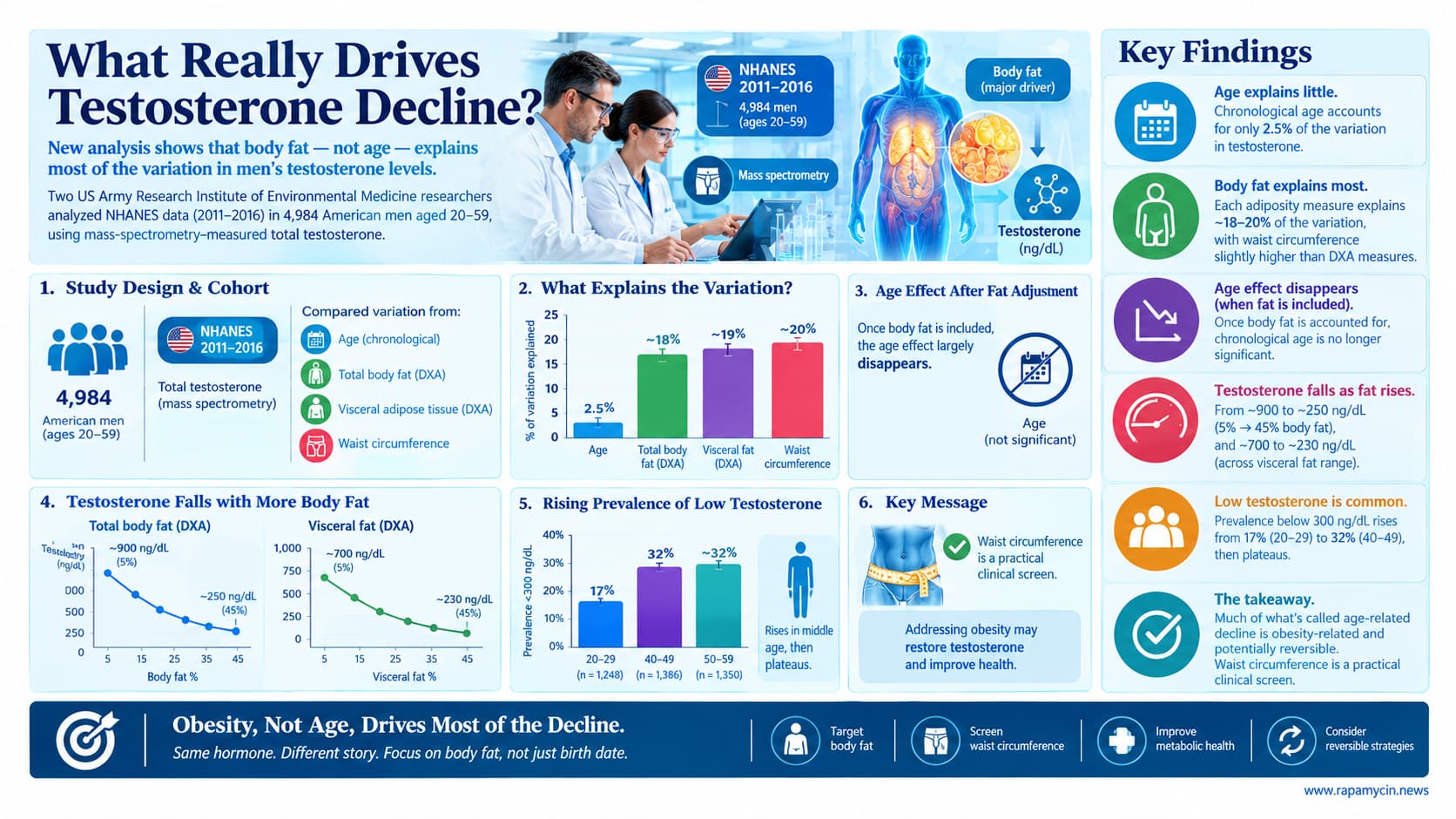
Two researchers at the US Army Research Institute of Environmental Medicine analyzed NHANES data from 2011 to 2016, covering 4,984 American men aged 20 to 59 with mass-spectrometry-measured total testosterone. They compared how much variation in testosterone was explained by chronological age versus by three measures of body fat: DXA-derived total percent body fat, DXA-derived visceral adipose tissue, and simple waist circumference. Age explained 2.5 percent. Each adiposity measure explained roughly 18 to 20 percent, with waist circumference slightly edging out the DXA scan. Once fat was accounted for, the age effect largely disappeared. Predicted testosterone fell from roughly 900 ng/dL at 5 percent body fat to about 250 ng/dL at 45 percent, and from about 700 to 230 ng/dL across the visceral fat range. The prevalence of testosterone below 300 ng/dL rose from 17 percent in men aged 20 to 29 to 32 percent at 40 to 49, then plateaued. The authors argue that much of what is called age-related testosterone decline is actually obesity-related and potentially reversible, and that waist circumference is a practical clinical screen.
The standard story about male aging is that testosterone falls roughly 1 percent a year after 30, and that this is simply what happens to men. That story has supported a very large testosterone prescribing industry, now increasingly delivered by online platforms that ask few questions.
A new analysis from the US Army Research Institute of Environmental Medicine puts a number on how much of that story is actually about age. Adam Potter and Karl Friedl pulled six years of NHANES data, the federal survey that combines physical examination with laboratory testing on a nationally representative sample. They had 4,984 men aged 20 to 59 with testosterone measured by mass spectrometry, the reference method, and most of them had whole-body DXA scans giving both total fat and visceral fat.
Age by itself accounted for 2.5 percent of the variation in testosterone. Body fat accounted for about 18 percent. Visceral fat, the metabolically active fat packed around the organs, accounted for about 18 percent. Waist circumference, which costs nothing and takes fifteen seconds, accounted for 20 percent.
That last result is the one clinicians should notice. A tape measure slightly outperformed a dual-energy X-ray scanner at explaining testosterone. The authors are appropriately careful about overinterpreting a small difference, but the practical message holds: you do not need imaging to identify the men whose testosterone is being suppressed by fat.
The magnitude is substantial. Predicted testosterone ran from about 900 ng/dL at 5 percent body fat down to about 250 ng/dL at 45 percent. Across the visceral fat range it fell from roughly 700 to 230. For comparison, the full span of the age curve from 20 to 59 was about 110 ng/dL, and the curve actually turned upward after 50.
Why would fat do this? Adipose tissue contains aromatase, which converts testosterone into estradiol. Estradiol feeds back on the hypothalamus and pituitary to reduce the signal telling the testes to produce testosterone. Insulin resistance suppresses the liver’s production of sex hormone binding globulin, which lowers total testosterone independently. Inflammatory signals from visceral fat add further suppression. The loop runs both ways, since low testosterone promotes fat accumulation.
The authors give context from military physiology: in lean men under severe caloric restriction and physical stress, testosterone can drop to near-castrate levels and then recover fully on refeeding. Testosterone suppression can be a reversible adaptation to metabolic circumstances rather than organ failure.
One finding deserves attention from anyone weighing TRT. Men with low testosterone had higher fat-free mass index than men with normal testosterone, in every age decade. They were not sarcopenic. They were simply larger, carrying more of everything, with a worse fat-to-lean ratio. The intuition that low testosterone means wasted muscle does not hold at the population level.
The caveats are real. This is a single snapshot, so it cannot prove that losing fat raises testosterone. NHANES took one blood draw with no repeat confirmation and no symptom assessment, so “low testosterone” here is a lab value, not a diagnosis. And the study stops at 59, so men in their seventies, where genuine testicular failure becomes more common, are outside its scope.
Still, for the large population of men in their thirties, forties and fifties being told their numbers are low, the sequence this paper implies is sensible: measure the waist first.
Actionable Insights
Measure your waist circumference. It explained about 20 percent of testosterone variation, slightly more than a DXA scan. This is free and repeatable.
The gradient is steep in the range most men occupy. Going from roughly 15 percent to 25 percent body fat was associated with about 100 ng/dL lower predicted testosterone. By 30 percent body fat, predicted testosterone was approaching 400 ng/dL. These are not extreme numbers; they describe a large share of American men.
A visceral fat target of under 100 cm squared corresponds to a waist of roughly 95 cm (about 37 inches) in men. Above about 110 to 115 cm (43 to 45 inches), the authors argue low testosterone is far more likely to be driven by visceral fat than by age.
Translating the effect size for a non-statistician: age explained 2.5 percent of the variation in testosterone, meaning 97.5 percent came from something else. Adiposity explained roughly eight times as much. The model including both fat and sex hormone binding globulin explained about 47 percent. That is unusually high for a biological variable in a population survey.
If your testosterone comes back low, the evidence-aligned sequence is: confirm with a second morning fasted measurement, check whether symptoms are actually present, assess waist and metabolic markers, and address adiposity before starting replacement. Weight loss via surgery or GLP-1 receptor agonists has been associated with rising testosterone in published studies. [Confidence: Medium]
One caution against over-reading: this study cannot show that losing fat raises testosterone, only that the two track together.
Context and Source
- Open Access Paper: Adiposity, not chronological aging, is the dominant determinant of circulating testosterone in US adult men
- Institution: US Army Research Institute of Environmental Medicine, Natick, Massachusetts
- Country: United States
-
Journal: Journal of the Endocrine Society, published 20 August 2026
Impact evaluation: The impact score of this journal is 4.4 (2025 Journal Impact Factor, Clarivate; five-year JIF 4.2; ranked 59 of 198 in Endocrinology and Metabolism; CiteScore 5.2), evaluated against a typical high-end range of 0-60+ for top general science, therefore this is a Medium impact journal.
Related Reading:
- Testosterone Replacement for Older Men
- The Testosterone Trade-Off: Optimizing Anabolism Without the Needle
- The Testosterone Trade-Off: Optimizing Anabolism Without the Needle
- Rapamycin used with Testosterone replacement
- New Testosterone Study FINALLY Explains Why Levels Are Falling - YouTube (Stanfield)
- ‘I’m on Fire’: Testosterone Is Giving Women Back Their Sex Drive — and Then Some (NYT)