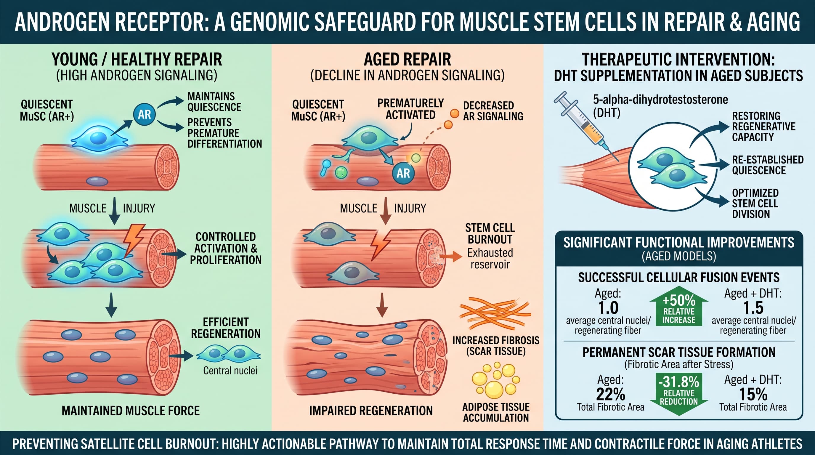
French researchers deleted the androgen receptor (AR) only in the muscle stem cells (satellite cells) of adult male mice. The muscles still healed after injury, but they healed worse. Fibers came out smaller and disorganized, there was more scar tissue and fat, and force was about 13% lower. The stem cells also stopped renewing themselves properly. Too many of them committed to becoming muscle at once, and after a second injury the reserve pool was about three times smaller than normal. In male mice, testosterone falls sharply by one year of age, and muscle AR protein falls with it. Older mice given a DHT implant repaired injured muscle with about a third less fibrosis. The stem cell pool itself did not recover.
Muscle carries its own repair crew. Satellite cells sit dormant under the surface of each muscle fiber. When the muscle is damaged, some of them wake up and rebuild it, and the rest go back to sleep so they are available next time. That balance between spending and saving stem cells is what makes muscle repair last a lifetime. It is also a big part of why older muscle heals badly.
This study, from the IGBMC institute near Strasbourg, points to testosterone signaling as one of the things that holds the balance. The team removed the androgen receptor, the protein that lets cells respond to testosterone and DHT, from satellite cells alone. They did it in young adult male mice, so every other tissue could still sense hormones normally.
Without injury, nothing obvious happened. After the team damaged a leg muscle with snake toxin, the knockout mice repaired it poorly. New fibers were smaller and structurally messy, inflammation lasted longer, scar tissue roughly doubled, and fat cells showed up where muscle should have been. Stem cell numbers were about half of normal one week after injury. After a second injury, they were about a third of normal.
The mechanism looks like poor rationing. AR is concentrated in the most dormant stem cells. When it is missing, those cells leave dormancy early and too many of them become committed muscle cells. The reserve gets used up. Molecular maps show AR moving around the genome during repair. It first sits near genes that keep stem cells dormant, then moves to genes for activation, and later to genes for maturation. The knockout cells also switched early to mitochondrial energy production, a metabolic sign that they had left the resting state.
The link to aging matters most here. Testosterone in these male mice fell from about 25 to about 3 to 5 nmol/L by one year of age, and AR protein in muscle dropped by nearly 80%. Aged normal muscle healed in a way that looked partly like the young knockouts. Aged knockouts did worse still. When 21-month-old mice received DHT implants, their repaired muscle had fewer scar-tissue regions and more nuclei per fiber. Stem cell counts did not change, and no one measured strength.
Human cells gave a similar picture. A muscle-precursor line from an older man had about half the AR of a line from a younger man and differentiated less well. Knocking AR down stopped differentiation in both lines. Adding extra AR sped up differentiation in the older line.
The big idea is that testosterone does more than build muscle mass. Acting on stem cells, it may help decide whether muscle can keep repairing itself over decades. That shifts the discussion of male hormonal decline from how big the muscles are to how well they can repair. The evidence comes from a small number of mice, all male, and one injury model, so it is a strong hypothesis rather than a settled mechanism.
Actionable Insights
This is a mechanistic mouse study, not a treatment trial. Its lessons are directional.
- Male hormone decline may matter most for repair. In these mice, testosterone fell by roughly 80 to 90% with age. When androgen signaling in stem cells was removed, recovery after injury was worse: about 13% less force, twice the scar tissue and three times fewer stem cells after re-injury. For older men, it is reasonable to track total and free testosterone and not only muscle size. [Confidence: Medium]
- Restoring hormones only partly fixed the problem. In old mice, DHT cut scar tissue in repaired muscle from about 22% to about 15% of the area, a relative reduction of about 36%. It did not restore the stem cell pool, and no strength data were collected. That is a modest, partial repair signal. It is not evidence that TRT or DHT reverses muscle aging. [Confidence: Low for translation]
- Do not self-prescribe androgens because of this paper. Outside androgens shut down the body’s own testosterone production and carry blood, fertility and prostate risks. Decisions about testosterone therapy belong with a clinician.
- Men on anti-androgen drugs, such as prostate cancer therapy, may have slower muscle repair. This is a hypothesis only.
- Women were not studied.
Context and Source
- Open Access Paper Androgen receptor imprints satellite cell stemness and preserves their reservoir for lifelong regeneration and optimal repair
- Institution: IGBMC (Université de Strasbourg, CNRS, Inserm), Illkirch, working with Institut de Myologie/Sorbonne (Paris), CRBS Strasbourg and I2MC Toulouse
- Country: France
- Journal: Science Advances, published 9 September 2026
- Impact evaluation: The impact score of this journal is about 12.5 (Journal Impact Factor; some trackers list up to 13.9), evaluated against a typical high-end range of 0 to 60+ for top general science, therefore this is a High impact journal.