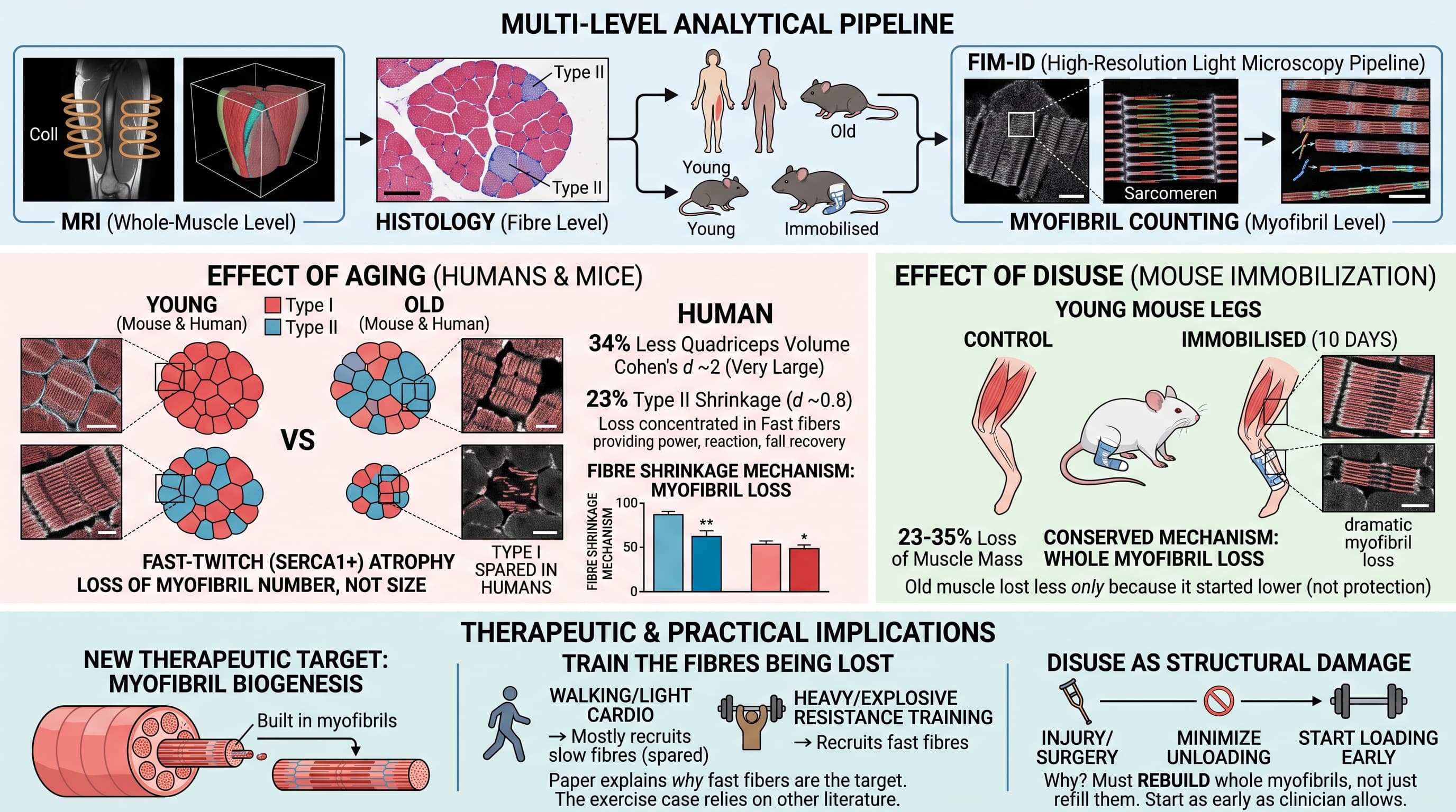
Using MRI, standard histology and a high-resolution light microscopy pipeline (FIM-ID), this study measured skeletal muscle at the whole-muscle, fibre and myofibril levels in young and older adults and in young and old mice, including mice with one hindlimb immobilised for 10 days. In both species, age-related shrinkage of fast-twitch (SERCA1-positive, Type II) fibres came mainly from fibres having fewer myofibrils, not from individual myofibrils getting thinner. Disuse in mice worked the same way. Slow (Type I) fibres in humans were spared. The authors conclude that loss of myofibril number is a conserved mechanism of fibre atrophy and a possible therapeutic target.
Every muscle fibre is packed with hundreds to thousands of myofibrils, the protein cables that actually generate force. When a fibre shrinks with age or bed rest, there are two ways it could happen. Each cable could get thinner, or the fibre could carry fewer cables. The field has known for about fifty years that fibres shrink. It has not known which of these two things is going on, mostly because counting myofibrils by electron microscopy is extremely slow work.
A team led by Troy Hornberger at the University of Wisconsin-Madison, working with Christopher Sundberg’s group at Marquette University, now has an answer. They used a light microscopy method they built, called FIM-ID. It images myofibrils with enough contrast for software to trace them automatically. With it they measured muscle at three scales in 34 young and older adults, and in young and old mice. Some of the mice had one hind leg immobilised for ten days.
In people, the top-line result was familiar. Adults aged 65 to 84 had about a third less quadriceps volume on MRI than adults aged 19 to 40. Under the microscope, the fast-twitch Type II fibres were about 23% smaller in older people. Slow Type I fibres were untouched. The new finding came from the smallest scale. Individual myofibrils in the older fast fibres were the same size as in young fibres. What had changed was how many there were, down by roughly a quarter. Missing myofibrils accounted for almost all of the fibre shrinkage.
Mice showed much the same pattern. Old mice had fewer myofibrils per fast fibre. They also had some myofibril thinning, mostly in a muscle rich in a highly glycolytic fibre type (IIb) that humans do not have. Ten days of immobilisation in young mice cut soleus fibre area by about a third. Again, most of that came from losing myofibrils, with only slight thinning of the ones that remained. Old mice lost less muscle when immobilised. The authors think their muscles were already partly deconditioned by low daily activity.
Why does the distinction matter? If ageing muscle thinned every cable evenly, that would point to a general imbalance between protein building and breakdown. Losing whole cables points to something more specific: a failure in myofibril turnover. The muscle may be removing myofibrils whole, failing to build or split new ones, or both. That narrows the target list. It includes the machinery that assembles new myofibrils in response to mechanical load, which this lab has previously shown drives muscle growth.
The caveats are real. The human data are a snapshot comparing different people, not the same people followed over time, so they cannot show the order of events. Only one thigh muscle was biopsied, and about 30 fibres per person were analysed in detail. The myofibril “count” is calculated from areas, not tallied one by one. The study measured structure, not the molecular signals behind it, and it tested no treatment. What it gives is a clean map of where the muscle goes. That map is needed before anyone can design a way to stop the loss.
Actionable Insights
This paper tested no intervention, so everything below is an inference, not a demonstrated benefit.
- The loss is large and concentrated. Older adults had about 34% less quadriceps volume. Within each sex, that is a Cohen’s d of about 2, which is very large: the typical older person fell below nearly every young person of the same sex. The damage sat in fast-twitch fibres, which shrank about 23% (d about 0.8, large). Slow fibres were unchanged. Fast fibres supply power, quick reactions and fall recovery.
- Train the fibres that are being lost. Walking and light cardio mostly use slow fibres. Heavy or explosive resistance training recruits fast fibres. The case for this comes from the broader exercise literature, not this study. This paper explains why fast fibres are the ones to target.
- Treat disuse as structural damage. In young mice, 10 days of immobilisation removed 23% to 35% of mass in several leg muscles. Most of that loss was whole myofibrils, which have to be rebuilt rather than simply refilled. After injury or surgery, keep unloading periods short and start loading as early as your clinician allows.
- Old muscle lost less during immobilisation only because it started lower. That is not protection.
Context and Source
- Open Access Paper: Macroscopic to ultrastructural analyses identify the loss of myofibrils as the primary mediator of ageing- and disuse-induced muscle fibre atrophy
- Institutions: University of Wisconsin-Madison (Department of Comparative Biosciences; Division of Geriatrics and Gerontology) and Marquette University (Exercise and Rehabilitation Sciences), with an affiliation at Sohag University, Egypt
- Country: USA
- Journal: The Journal of Physiology (published online 25 September 2026)
- Impact evaluation: The journal’s publisher page currently lists a CiteScore of 7.2 and a Journal Impact Factor of 4.6. The impact score of this journal is 4.6 (JIF), evaluated against a typical high-end range of 0-60+ for top general science, therefore this is a Medium impact journal.
Related Reading:
- Muscle Is the New Longevity Drug Target, and the Pipeline Just Got Crowded
- Why Muscles Heal Slower With Age, and How Hormones Might Help
- Genetic Jackpot: Rare Myostatin Mutations Unlock Increased Muscle and Reduced Adiposity in Humans
- HIV Drug (Maraviroc) Reverses Muscle Aging by purging “Zombie Cell” Signals
- Botanical Compound Peucedanum japonicum (PJ) Reverses Age-Related Muscle Atrophy by Forcing Mitochondrial Biogenesis
- The Calcium Leak: Why Your Muscles Age Even If You Train (and how to stop it)