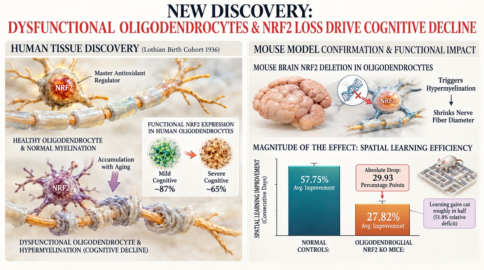
Researchers tracking brain tissue from the decades-long Lothian Birth Cohort 1936 discovered that human cognitive decline is unexpectedly driven by an accumulation of dysfunctional oligodendrocytes, which wrap nerve fibers in excessively thick, disordered myelin sheaths. These abnormal cells exhibit a marked loss of the master antioxidant regulator NRF2. Modeling this cell-specific defect in mice confirmed that deleting oligodendroglial NRF2 directly triggers hypermyelination, shrinks nerve fiber diameter, and blunts cognitive learning over time.
For decades, neuroscientists viewed age-related cognitive decline primarily through the lens of dying neurons, toxic protein aggregates, or the progressive stripping away of myelin—the specialized fatty insulation that coats central nervous system axons. Without sufficient myelin, electrical transmission slows and circuits degrade. A study published in Nature Medicine turns this assumption on its head, demonstrating that an overabundance of poorly regulated myelin can be just as destructive as demyelination.
Using post-mortem tissue from the Lothian Birth Cohort 1936, a landmark Scottish study tracking mental ability from age 11 into the ninth decade of life, investigators probed the corpus callosum to evaluate structural differences between individuals with preserved cognition and those suffering steep decline. High-resolution electron microscopy revealed that brains with severe mental decline did not lack myelin. Instead, they displayed an unexpected accumulation of oligodendrocytes that produced pathologically thick myelin sheaths around large nerve fibers. This structural distortion went hand-in-hand with an increased prevalence of empty, degenerating axons and shrunken fiber calibers, indicating that excess, abnormal insulation actively strangles or starves the underlying neuronal wiring.
Single-nucleus transcriptomic profiling revealed the driver behind this pathology. The accumulated oligodendrocytes suffered from a severe deficit of NRF2, a transcription factor that orchestrates cellular defenses against oxidative damage, maintains mitochondrial homeostasis, and governs macroautophagy. Rather than dying, these defective glial cells persisted and downregulated critical autophagic turnover genes, causing abnormal membrane build-up.
To determine whether oligodendroglial NRF2 deficiency is a causal driver rather than a passive byproduct of brain aging, the team engineered a conditional mouse model in which NRF2 was selectively deleted from oligodendrocytes in middle age. The rodents recapitulated the exact structural defects seen in human brains: thickened myelin coats, shrunken axon diameters, and impaired spatial learning in water maze tests. These findings firmly reclassify the oligodendrocyte from a benign support cell into an active mediator of age-related cognitive deterioration.
Actionable Insights While this research relied on post-mortem human tissue and genetic mouse models rather than clinical lifestyle interventions, it identifies a clear target for preserving white matter health: maintaining active NRF2 signaling in the aging brain.
-
Upregulate Endogenous NRF2 Activity: Because the pathological cascade begins when oligodendrocytes downregulate NRF2, maintaining this pathway via dietary electrophilic compounds (such as sulforaphane from broccoli sprouts) or therapeutic activators (such as dimethyl fumarate) represents a logical preventative strategy.
-
Support Autophagic Clearance: The downstream consequence of low NRF2 in these cells is suppressed autophagy (noted by reduced SQSTM1/p62 expression), which prevents normal turnover of bulky myelin membrane. Interventions that stimulate cellular recycling pathways may help curb aberrant membrane deposition.
-
Magnitude of the Effect: The functional impact of losing oligodendroglial NRF2 is substantial. In aged mice, normal controls improved their spatial learning efficiency by an average of 57.75% across consecutive training days, whereas mice lacking oligodendroglial NRF2 improved by only 27.82%. This represents an absolute drop in learning improvement of 29.93 percentage points, cutting their learning gains roughly in half (a relative deficit of 51.8%). In human tissue, the proportion of oligodendrocytes expressing functional NRF2 dropped from approximately 87% in individuals with mild cognitive decline down to roughly 65% in severe decline.
Context/Source
- Full Title: Oligodendrocyte dysfunction in human age-related cognitive decline
- Lead Institutions: Keenan Research Centre for Biomedical Science, Unity Health Toronto, and The University of Edinburgh
- Countries: Canada, United Kingdom, and United States
- Journal Name: Nature Medicine
- Impact Evaluation: The journal impact factor is 58.7 (Journal Citation Reports, 2024–2025; CiteScore 72.8). The impact score of this journal is 58.7, evaluated against a typical high-end range of 0–60+ for top general science, therefore this is an Elite impact journal.
Related Reading:
- Role of the Transcription Factor NRF2 in Neurodegenerative Disorders
- Sulforaphane inhibits mTOR in an NRF2-independent manner
- Does Nrf2 Play a Role of a Master Regulator of Mammalian Aging?
- Sulforaphane: how to supplement? Forms, dose, brands, timing
- The Newest Endurance Supplement Is a Broccoli Shot. Does It Work? (Outside)
- Can't Exercise? Sulforaphane from Broccoli Sprouts Reverses Frailty in Aging Mice
- How to Increase Sulforaphane in your Diet, Cancer Prevention, etc