Top causes of reduced HRV after age 60
Tier 1: Largely non-modifiable
1. Chronological age and intrinsic sinoatrial remodeling
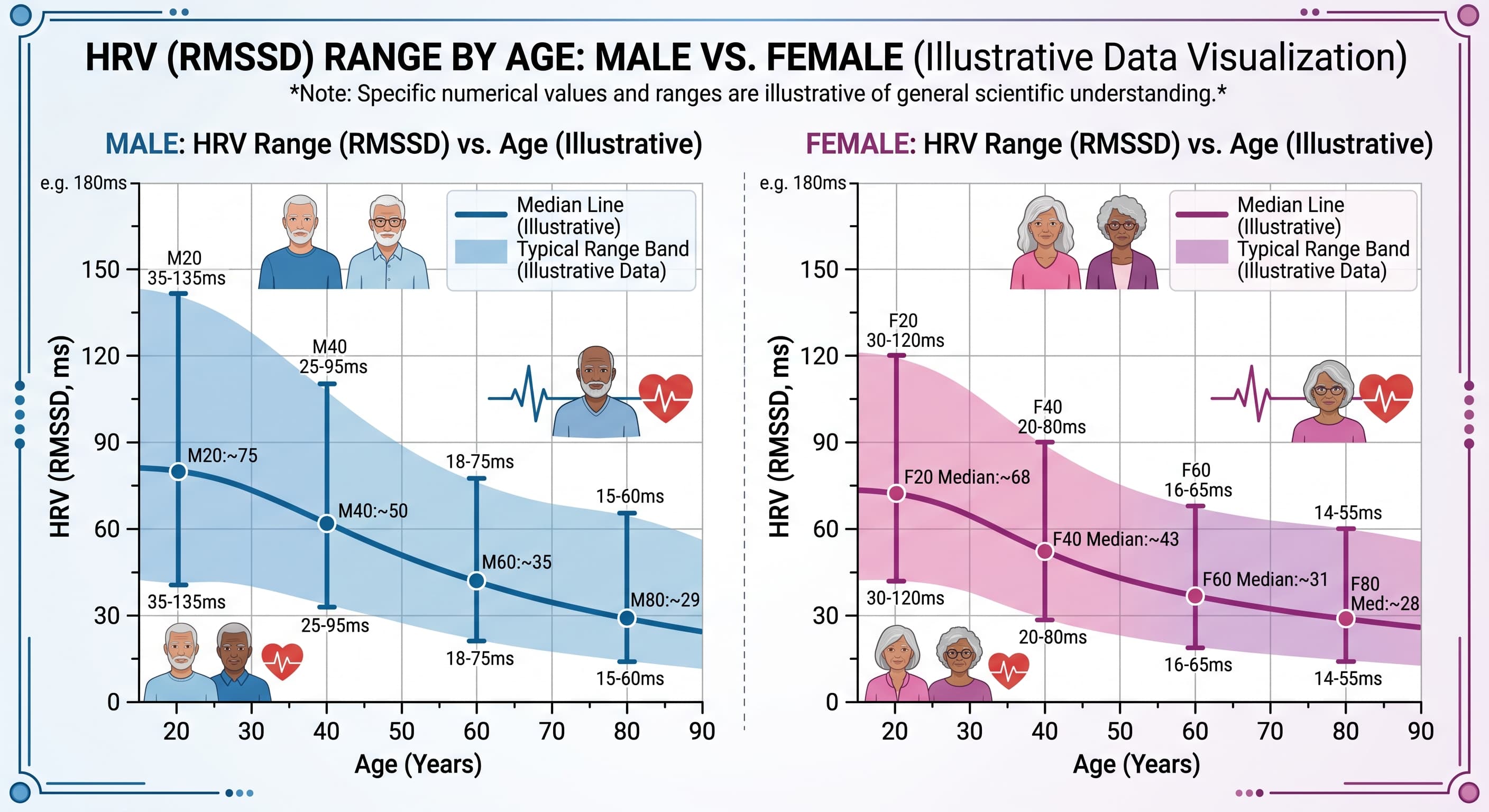
The single largest determinant, and the one biohackers most consistently underestimate. Twenty-four hour HRV declines steeply and roughly monotonically across the lifespan, with the sharpest fall between the third and sixth decades and continued decline thereafter (Umetani et al., JACC 1998). A systematic review of reference values in older adults confirms that published norms for this age band are both lower and highly heterogeneous (Rocha et al., Psychophysiology 2024).
Critically, a substantial fraction of this is structural, not autonomic tone. The sinoatrial node loses pacemaker cells and gains fibrosis with age, and intrinsic heart rate measured under full autonomic blockade declines roughly linearly with age. You cannot train that component back. [Confidence: High that age dominates, Medium on the precise structural versus neural split]
Tier 2: High magnitude, modifiable in principle
2. Type 2 diabetes and cardiac autonomic neuropathy
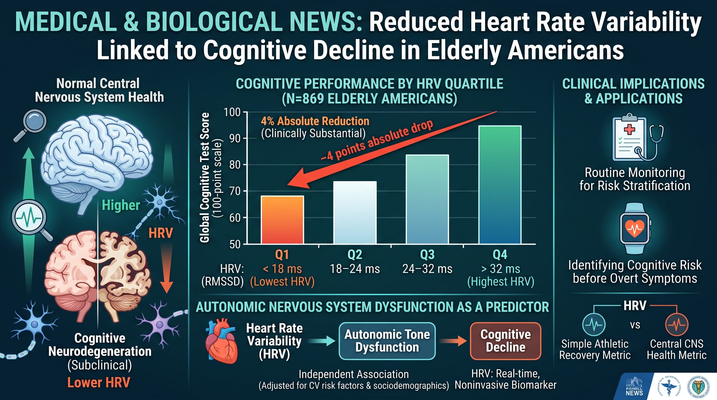
Diabetes reduces HRV across essentially every index, and cardiovascular autonomic neuropathy is common, underdiagnosed and progressive. Prevalence estimates run roughly 20 percent in unselected type 2 diabetes and substantially higher with longer duration (Pop-Busui, Diabetes Care 2010; Spallone, Diabetologia 2024). In the paper you just had me analyse, fasting insulin was 39 percent higher in the lowest HRV quartile. [Confidence: High]
3. Obesity and visceral adiposity
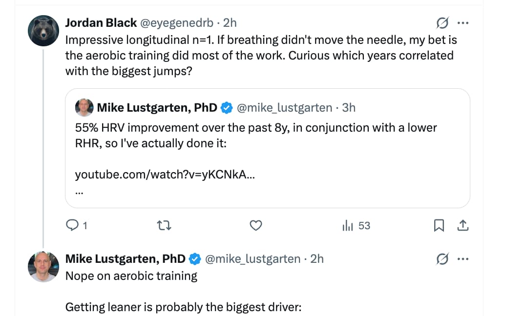
Consistent inverse relationship with vagal indices. The clearest evidence of reversibility comes from surgical weight loss: pooled HRV improvement after metabolic and bariatric surgery was a weighted mean difference of 12.0 (95 percent CI 6.98 to 17.04), with a dose relationship between BMI reduction and HRV gain (Updates in Surgery 2026). Only 11 studies and 322 patients, so precision is limited. [Confidence: Medium-High]
4. Medication burden
Routinely ignored in consumer HRV discussion and arguably the largest single correctable artifact in this age group. Tricyclic antidepressants produce a large HRV reduction (g = 1.24, 3 studies, 32 participants), while SSRIs showed no significant effect (g = 0.09) (Kemp et al., Biological Psychiatry 2010). Anticholinergic drugs suppress HRV directly and dose-dependently (Eur J Clin Pharmacol 2005).
Beta blockers are the counterintuitive case. They generally increase time-domain and vagal HRV indices while lowering heart rate, which means a person starting a beta blocker may see their wearable HRV rise for reasons unrelated to health improvement (Nature Sci Rep 2023). Do not read that number as progress. [Confidence: High]
Tier 3: Moderate magnitude, highly modifiable
5. Physical inactivity and low cardiorespiratory fitness
Inferred largely from the reverse direction, that is, from training trials showing HRV gain (see Part B). Cross-sectional fitness associations are consistent. [Confidence: High for direction, Medium for magnitude of the deconditioning contribution specifically]
6. Sleep loss, fragmentation and obstructive sleep apnea
Pooled across 11 RCTs and 549 participants, sleep deprivation reduced RMSSD (SMD −0.24, 95 percent CI −0.47 to −0.00) and raised LF/HF (SMD 1.47, 95 percent CI 0.62 to 2.33) (Frontiers in Neurology 2025). The RMSSD confidence interval touches zero, so acute sleep loss is a real but modest vagal suppressor in the short term. Untreated OSA is the more serious chronic case, and CPAP partially reverses it (Heart and Lung 2018). OSA prevalence rises sharply after 60 and is heavily underdiagnosed. [Confidence: High]
7. Depression and chronic psychological stress
Depression is associated with reduced HRV at g = −0.29 to −0.30 for time-domain and high-frequency measures across 18 studies (673 depressed, 407 controls), with a dose relationship to severity (r = −0.36) (Kemp et al. 2010). Note the effect size is small, and in older adults the antidepressant medication may contribute more than the depression. [Confidence: Medium-High]
8. Alcohol
Acute intake reduces short-term HRV in a dose-related fashion, demonstrated in controlled dosing studies (Am J Physiol Heart Circ Physiol 2010; Sci Rep 2021). This is the most reliably detectable single-night effect on a wearable in most people. Chronic heavy use produces sustained autonomic impairment. [Confidence: High for acute, Medium for chronic dose-response]
9. Smoking
In 4,751 adults, every 10 grams of daily tobacco was associated with 9.8 percent lower SDNN and 8.9 percent lower RMSSD (CHRIS study, PLOS ONE 2019). Oddly, that study found former smokers had higher HRV than never-smokers, which is biologically implausible as a causal effect and probably reflects selection or confounding. I flag it rather than smoothing it over. [Confidence: High that current smoking lowers HRV, Low on the former-smoker finding]
Tier 4: Disease endpoints and artifacts
10. Established cardiovascular disease. Heart failure, prior myocardial infarction and atrial fibrillation all reduce or invalidate HRV. AF in particular makes standard HRV indices uninterpretable rather than merely low, and AF prevalence climbs steeply after 65. [Confidence: High]
11. Measurement artifact. Respiratory rate is the dominant non-physiological driver of short-term HRV, since HRV rises mechanically as breathing slows. Posture, recording length, time of day, ectopic beats and device algorithm all move the number. Much of what people interpret as day-to-day biological variation is measurement variance. [Confidence: High]
PART B: Approaches most likely to raise HRV in middle age
Tier 1: Strong randomized evidence, moderate to large effects
1. Structured aerobic and high-intensity interval training
The best-supported intervention by a clear margin.
| Source |
Population |
Result |
|
Cureus 2024, 16 RCTs, 623 healthy adults |
Healthy adults |
SDNN SMD 0.58 (0.16 to 1.00); RMSSD SMD 0.84 (0.36 to 1.31); HF SMD 0.89 (0.27 to 1.51) |
|
Rev Cardiovasc Med 2024, network meta-analysis, 29 RCTs, 1,317 participants |
Adults |
HIIT ranked first for SDNN (SUCRA 98.7 percent), RMSSD (84.9 percent) and LF/HF (99.8 percent); resistance training first for HF |
|
PLOS ONE 2024, 19 studies |
Cardiovascular disease |
Aerobic superior to resistance alone; largest gains in heart failure |
Note the wide confidence intervals in the healthy-adult pooling, which signal heterogeneity and small-study effects. The point estimates are moderate to large; the true effects are probably smaller. HIIT’s top ranking comes from a network meta-analysis with only 29 trials spread across five modalities, so modality ranking is much less certain than the overall benefit of training. [Confidence: High that aerobic training raises HRV, Medium on magnitude, Low-Medium on HIIT specifically being superior]
Practical translation: the intervention with the best evidence is simply becoming more aerobically fit. Zone 2 volume plus one or two hard interval sessions per week is a defensible reading of these data.
2. Slow-paced breathing and HRV biofeedback, with an important asterisk
Breathing at roughly 5 to 6 breaths per minute produces large, immediate increases in HRV (Laborde et al., Neurosci Biobehav Rev 2022). This is the highest-magnitude acute effect available.
The asterisk matters. Much of the during-practice increase is a mechanical resonance phenomenon. Breathing slowly synchronizes the baroreflex and respiratory sinus arrhythmia, so HRV rises whether or not anything about your autonomic health has changed. That is the same maneuver the Zeki Al Hazzouri study used as a provocation test. Evidence that regular practice raises resting, non-practice HRV is considerably weaker than evidence that it raises HRV during practice, and biofeedback meta-analyses consistently report better evidence for symptom outcomes than for durable HRV change (Sci Rep 2021; Appl Psychophysiol Biofeedback 2025).
So: high value as a state-regulation tool, uncertain value as a trait-HRV intervention. [Confidence: High for acute effect, Low-Medium for durable resting HRV change]
Tier 2: Strong evidence, effect conditional on baseline
3. Weight loss if overweight or obese. Weighted mean difference 12.0 (95 percent CI 6.98 to 17.04) after bariatric surgery, with a dose relationship to BMI change (Updates in Surgery 2026). Evidence for non-surgical weight loss is thinner but directionally consistent. No expected benefit if you are already lean. [Confidence: Medium-High]
4. Diagnose and treat sleep apnea. CPAP improves HRV in OSA patients (Heart and Lung 2018). Middle age is exactly when undiagnosed OSA becomes common, and it is the most commonly missed explanation for a persistently low nocturnal HRV in an otherwise healthy-looking person. If your overnight HRV is low and your resting heart rate is high despite good fitness, this is the first thing to rule out. [Confidence: High]
5. Glycemic and insulin-sensitivity control. Follows directly from the diabetes and CAN literature (Spallone, Diabetologia 2024). Evidence that improving glycemia reverses established autonomic neuropathy is much stronger for prevention than for reversal, which argues for acting in middle age rather than after 65. [Confidence: Medium-High for prevention, Medium for reversal]
Tier 3: Reliable but smaller, or lower-quality evidence
6. Reduce alcohol. Among the fastest-acting changes, with clearly demonstrated acute dose-response (Am J Physiol 2010). Nightly drinking is a common and easily reversed cause of chronically depressed overnight HRV. No RCT has tested sustained abstinence against a control for resting HRV outcomes in middle age, so the long-term magnitude is inferred. [Confidence: High for acute, Medium for chronic]
7. Smoking cessation. Dose-response evidence is strong (CHRIS study 2019); cessation studies are small and short. [Confidence: Medium-High]
8. Sleep extension and consistency. Supported indirectly by deprivation trials (Frontiers in Neurology 2025). The intervention direction has not been tested as rigorously as the deprivation direction. [Confidence: Medium]
9. Yoga and tai chi. 17 RCTs: nHF g = 0.37, nLF g = −0.39, LF/HF g = −0.58, perceived stress g = −0.80. Yoga evidence stronger than tai chi; roughly 60 to 90 minutes per week needed. Only 6 of 17 trials had allocation concealment (J Clin Med 2018; Tai chi specific, J Integr Complement Med 2023). The reliance on normalized units rather than absolute RMSSD or HF weakens interpretation, since normalized indices can move without absolute vagal change. [Confidence: Medium]
10. Mindfulness meditation. Reviewed effects on vagally mediated HRV are inconsistent and generally small, with brief interventions showing little (Appl Psychophysiol Biofeedback 2025). Worth doing for other reasons; weak as an HRV lever. [Confidence: Medium that the effect is small]
Tier 4: Weak, null or overclaimed
11. Omega-3 fatty acids. The consistent, replicated effect is a small reduction in resting heart rate rather than a clear increase in vagal HRV (Eur J Clin Nutr 2018). Short-term fish oil meta-analyses show inconsistent HRV effects (Am J Clin Nutr 2013). Treat as marginal. [Confidence: Medium that the HRV effect is small or absent]
12. Sauna. A multi-arm RCT of regular post-exercise sauna bathing found no improvement in HRV (Physiological Reports 2025). Acute post-sauna vagal rebound is real but transient (Complement Ther Med 2019). Do not expect chronic HRV gain. [Confidence: Medium-High that chronic effect is null]
13. Cold exposure. Acute effects only, direction depends on timing relative to measurement, no evidence for durable resting HRV improvement. [Confidence: Low, evidence base is thin]
14. Most supplements marketed for HRV. No meta-analytic support I could locate. [Confidence: Medium, absence of evidence rather than evidence of absence]
Three things that will make your own tracking less misleading
Within-person trend, not cross-person comparison. Between-person HRV variance is enormous and heavily genetic. Your absolute number against a population norm tells you almost nothing. An eight to twelve week trend in your own data is the only signal worth acting on. [Confidence: High]
Hold the measurement conditions fixed. Same device, same posture, same time window, ideally overnight or on waking. Breathing rate alone can move short-term HRV more than a year of training will. [Confidence: High]
Watch for HRV going up for bad reasons. Beta blockade, bradycardia from overtraining or illness, and some arrhythmias all inflate common HRV indices. A rising number alongside a rising resting heart rate, worsening sleep or falling performance is a signal to investigate, not to celebrate. [Confidence: Medium-High]
Bottom line
If you want one intervention with real randomized evidence behind it, it is aerobic training, at SMD 0.58 to 0.89 across HRV indices. Everything in Tier 2 is essentially “remove a specific injury”: excess adiposity, untreated apnea, dysglycemia, alcohol, tobacco, a drug with anticholinergic load. For most middle-aged people, the ceiling on trainable HRV is set by how many of those injuries are present, not by breathing protocols. Slow-paced breathing is the highest-leverage acute tool and the most overclaimed chronic one.
This is a research summary and not medical advice. Any change to prescribed medication, including anything in the anticholinergic or beta blocker discussion above, belongs with your physician.
Sources:
- Umetani et al., 24-hour HRV across nine decades, JACC 1998
- Rocha et al., HRV reference values in older adults, Psychophysiology 2024
- Pop-Busui, Cardiac autonomic neuropathy in diabetes, Diabetes Care 2010
- Spallone, Cardiovascular autonomic neuropathy in diabetes, Diabetologia 2024
- Metabolic and bariatric surgery and HRV, Updates in Surgery 2026
- Kemp et al., Depression, antidepressants and HRV, Biological Psychiatry 2010
- Anticholinergic drug effects on HRV, Eur J Clin Pharmacol 2005
- Beta blockers and circadian HRV, Scientific Reports 2023
- Exercise training and HRV in healthy adults, Cureus 2024
- Exercise modality network meta-analysis, Reviews in Cardiovascular Medicine 2024
- Physical activity and HRV, PLOS ONE 2024
- Laborde et al., Voluntary slow breathing and HRV, Neurosci Biobehav Rev 2022
- HRV biofeedback and depressive symptoms, Scientific Reports 2021
- Remote HRV biofeedback for mental health, Appl Psychophysiol Biofeedback 2025
- Brief mindfulness interventions and HRV, Appl Psychophysiol Biofeedback 2025
- Sleep deprivation and HRV, Frontiers in Neurology 2025
- CPAP and HRV in OSA, Heart and Lung 2018
- Dose-related effects of alcohol on HRV, Am J Physiol Heart Circ Physiol 2010
- Acute ethanol and cardiac autonomic regulation, Scientific Reports 2021
- Smoking status, history and intensity and HRV, CHRIS study, PLOS ONE 2019
- Mind-body exercise (tai chi and yoga) and HRV, J Clin Med 2018
- Tai chi and HRV, J Integr Complement Med 2023
- Omega-3 and heart rate, Eur J Clin Nutr 2018
- Short-term fish oil and HRV, Am J Clin Nutr 2013
- Post-exercise sauna bathing and HRV RCT, Physiological Reports 2025
- Recovery from sauna bathing and cardiac autonomic function, Complement Ther Med 2019