RNA Is Rotting as You Age, Harvard's New Damage Clock Measures it, and Rapamycin Helps Prevent it

A Gladyshev-lab computational study has built the first aging clock derived from RNA-level damage signatures — not gene expression — showing that transcriptomic deterioration accumulates predictably with age, is partially reversible by rapamycin and caloric restriction, and is measurably accelerated in Alzheimer’s disease blood at a signal strength that conventional expression-based clocks completely miss.

Aging researchers have argued for decades that molecular damage — not just regulatory drift — is a root cause of biological decline. The evidence base for DNA mutations and protein oxidation is well established. But one entire layer of molecular deterioration has escaped systematic quantification: the damage accumulating in RNA itself.

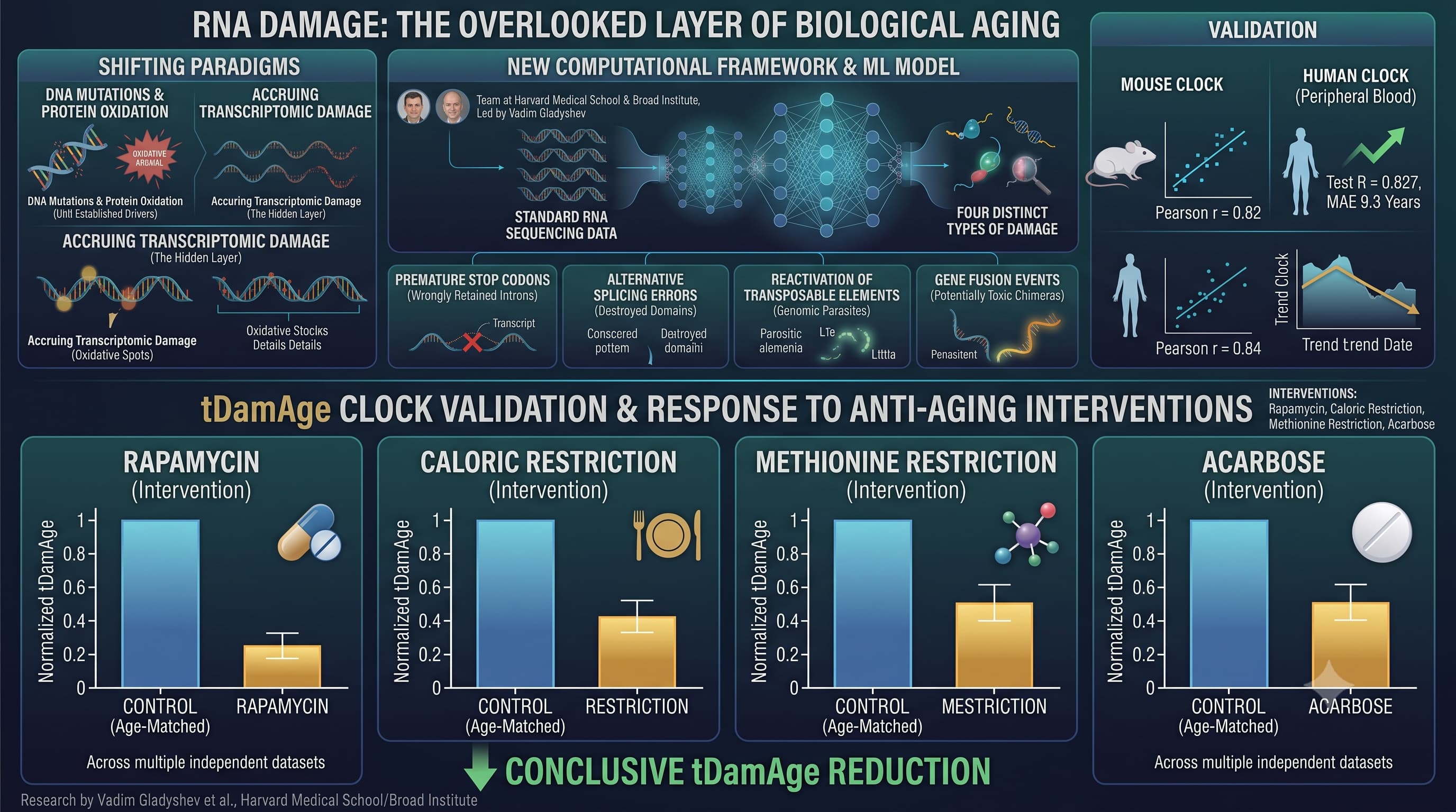
A team at Harvard Medical School and the Broad Institute, led by Vadim Gladyshev, has now built a computational framework that extracts four distinct types of transcriptomic damage from standard RNA sequencing data. These are not soft correlations. The damage types are structurally concrete: premature stop codons generated when introns are wrongly retained in transcripts (producing truncated, non-functional proteins or triggering degradation); alternative splicing errors that physically destroy conserved protein domains; the reactivation of transposable elements — genomic parasites that are normally suppressed — across multiple repeat classes; and gene fusion events, where unrelated RNA strands are incorrectly joined into potentially toxic chimeric molecules. Each of these increases with age. All four damage types positively correlate with chronological age across the majority of human and mouse tissues tested.

Using the GTEx dataset — over 16,000 human tissue samples from 26 tissue types — the team confirmed consistent age-related accumulation across nearly every tissue. The cerebellum carries the highest damage burden of any brain region, consistent with its documented vulnerability to aging-related neurodegeneration. Pancreas, muscle, and blood show the least damage, potentially reflecting higher cell turnover that clears damaged transcriptome-burdened cells before accumulation peaks.

From these signals, the team trained a machine learning aging clock called tDamAge — first in mice (Pearson r = 0.82) and then in human peripheral blood (test R = 0.827, mean absolute error 9.3 years). The clock responds to interventions in the expected direction across multiple independent datasets: rapamycin, caloric restriction, methionine restriction, and acarbose each reduce tDamAge relative to age-matched controls; SARS-CoV-2 lung infection, BubR1 progeroid mutation, and Klotho heterozygous knockout elevate it.

The most clinically striking finding is in Alzheimer’s disease. When the human blood tDamAge clock was applied to AD patient samples, it detected significantly elevated biological age acceleration compared to cognitively normal controls (p = 0.0099). A standard gene expression clock — built from the same datasets using the same pipeline — failed to detect any significant difference. Transcriptomic damage picks up a signal that expression levels obscure.

The paper also captures a transient rejuvenation window during mouse embryogenesis — a pronounced drop in tDamAge from approximately embryonic day E10/11 through E16 — with the damage clock’s nadir arriving slightly later than expression-based clocks, suggesting damage clearance lags transcriptional reset during development.

The authors frame this within Gladyshev’s deleteriome theory: that aging is defined by the totality of accumulated molecular damage across biological layers. tDamAge provides the first scalable, RNA-layer readout of that entropy. That framing is theoretically sound, though the paper stops short of formal causal proof — a distinction worth keeping in mind.


Actionable Insights

For the clinician and advanced biohacker, the intervention-response data is the most practically relevant output. Anti-aging interventions already in widespread use — rapamycin, caloric restriction, acarbose, and methionine restriction — each produced measurable reductions in transcriptomic damage age in mice, with fold-change reductions estimated at approximately 5 to 30 percent relative to age-matched controls depending on tissue and age stratum (from Figure 4H). Methionine restriction and dietary restriction showed the most consistent reductions across tissue types; rapamycin’s effects were meaningful but more tissue-specific.

Convergence analysis across 22 longevity intervention datasets identified RNA splicing fidelity, chromatin organization, and RNA catabolism as the central shared targets — suggesting these aren’t incidental effects but core mechanistic pathways through which diverse anti-aging strategies operate.

The AD blood signal (approximately 2 to 4 years of tDamAge acceleration relative to controls, estimated from Figure 6F) positions peripheral blood tDamAge as a potential early neurodegeneration marker. At current accuracy (MAE approximately 9.3 years), this clock is unsuitable for individual-level biological age tracking but may have utility as a population-level stratification tool.


Source:

  • Open Access Paper: Causally measuring aging and rejuvenation through transcriptomic damage
  • Institution: Division of Genetics, Department of Medicine, Brigham and Women’s Hospital, Harvard Medical School, Broad Institute of MIT and Harvard, Cambridge, MA, USA
  • Country: USA
  • Journal: bioRxiv preprint; posted June 29, 2026. Not peer-reviewed at time of analysis.
  • Impact Evaluation: This work is a preprint and carries no journal impact factor.

Related Reading:

Intervention Effect Sizes (estimated from Figure 4H, fold change in tDamAge vs. controls):

Explicit Cohen’s d values are not reported in the paper. The following are estimated from figure visualization:

Intervention Direction Estimated fold change in tDamAge
Methionine restriction Anti-aging 0.70 to 0.85 (15 to 30% reduction)
Dietary restriction Anti-aging 0.75 to 0.90 (10 to 25% reduction)
Caloric restriction Anti-aging 0.80 to 0.95 (5 to 20% reduction)
Rapamycin Anti-aging 0.85 to 0.95 (5 to 15% reduction)
Acarbose Anti-aging 0.85 to 0.95 (5 to 15% reduction)
Klotho heterozygous KO Pro-aging 1.40 to 1.60 (40 to 60% elevation)
SARS-CoV-2 (lung) Pro-aging 1.25 to 1.45 (25 to 45% elevation)
BubR1 MVA (muscle) Pro-aging 1.20 to 1.40 (20 to 40% elevation)

Mechanistic Deep Dive

mTOR Pathway

Rapamycin’s reduction of tDamAge is mechanistically plausible through at least two routes beyond canonical autophagy induction. First, mTORC1 directly phosphorylates and activates the splicing factor SRSF1, promoting alternative splicing patterns associated with oncogenic and age-related isoforms. Rapamycin-mediated mTORC1 inhibition may restore splicing fidelity and reduce domain-disrupting splice variants. Second, mTORC1 suppresses nonsense-mediated decay (NMD), the cellular quality control pathway that degrades transcripts containing premature stop codons. By relieving this suppression, rapamycin may enhance clearance of intron-retaining, truncated transcripts — one of the four damage classes directly measured here. [Confidence: Medium — mechanistically coherent, not directly tested in this paper]

AMPK and Metabolic Restriction

Caloric restriction, dietary restriction, and methionine restriction all activate AMPK and suppress mTORC1, which partially overlaps with rapamycin’s mechanism. Methionine restriction additionally depletes S-adenosylmethionine (SAM), the universal methyl donor including for RNA methylation at N6-methyladenosine (m6A) and 5-methylcytosine (m5C) positions. m6A modifications are recognized by YTHDF proteins that regulate alternative splicing, NMD efficiency, and translation. Reduced SAM availability under methionine restriction may therefore directly reshape the RNA epitranscriptome in ways that suppress the damage classes measured here. This could partially explain why methionine restriction shows among the most consistent tDamAge reductions in the dataset. [Confidence: Medium-low — speculative mechanistic bridge, requires experimental validation]

Transposable Elements and cGAS-STING

The repeat element damage class — particularly LINE-1 elements, endogenous retroviral LTRs, and DNA transposons — showed the strongest and most consistent right-shifted age correlation of all damage categories. Transposable element reactivation with age is well established, driven by heterochromatin loss, DNMT1 decline, and H3K9me3 erosion. Active LINE-1 elements generate cytosolic cDNA intermediates through their reverse transcriptase activity; these activate cGAS-STING, driving the interferon response and feeding the SASP. This paper provides a transcriptome-level, population-scale quantification of transposon reactivation across aging tissues — a readout that had not been systematically computed from standard RNA-seq at this scale. The convergence of anti-aging interventions on suppressing repeat element expression (implied by their tDamAge reduction) positions LINE-1/LTR silencing as a mechanistic link between these interventions and cGAS-STING attenuation. [Confidence: High for the association; Medium for the causal mechanistic link to cGAS-STING]

RNA Processing as the Central Convergence Hub

The functional enrichment network across 22 anti-aging intervention datasets (Figure 5D) identifies RNA splicing, snRNA/sncRNA processing, and RNA catabolism as the most consistently upregulated pathways. This is a biologically meaningful finding: it implies that interventions as mechanistically distinct as mTOR inhibition, insulin/IGF-1 pathway reduction (GHRKO, Snell dwarf), and methionine availability restriction all restore RNA processing fidelity as a shared downstream effect. This elevates the RNA surveillance machinery — the spliceosome, the exosome complex, the NMD pathway, and RNA helicase activity — to the status of potential first-line therapeutic targets rather than passive downstream read-outs. [Confidence: Medium — GO enrichment is correlational; causal experimental evidence in humans is absent]

Organ-Specific Aging Priorities

  • Cerebellum: highest transcriptomic damage burden across all brain subregions, consistent with documented cerebellar vulnerability to aging, motor decline, and neurodegeneration. Cerebellar Purkinje neurons are post-mitotic and do not regenerate, meaning damage cannot be diluted by cell division.
  • Blood (immune compartment): B cells positively correlate with transcriptomic damage in whole blood; T cell pathways show negative correlation. This maps onto classical immunosenescence — B cell expansion and T cell contraction with age — and suggests that B cell-associated transcriptomic instability (including immunoglobulin gene rearrangement-derived fusion events, which may register as age-associated gene fusions in blood) contributes substantially to the blood tDamAge signal.
  • Pancreas: lowest overall damage despite high secretory burden. Pancreatic acinar cell abundance negatively correlates with damage, suggesting that preserved acinar identity is protective. Loss of acinar cell identity during pancreatic aging (transdifferentiation or senescence) may appear as rising damage levels as cell composition shifts rather than intrinsic transcriptomic deterioration.

Novelty Assessment

What this paper adds that was not previously established:

  1. The first systematic, multi-tissue, multi-species computational pipeline to extract RNA-level damage from standard RNA-seq data. The gap this fills is not conceptual (damage causing aging has been theorized for decades) but operational — there was no scalable method to measure it.
  2. The first aging clock derived from damage metrics rather than expression levels or epigenetic marks, providing a mechanistically orthogonal biomarker layer with distinct information content.
  3. The AD blood finding is the single most clinically significant result: tDamAge in peripheral blood detects Alzheimer’s-associated biological age acceleration (p = 0.0099) in a context where a standard expression-based clock applied to the same data shows no significant signal. If replicated in larger, better-characterized cohorts with neuropathological confirmation, this could become a blood-based biomarker of neurological aging distinct from amyloid or tau.
  4. Quantitative confirmation that diverse anti-aging interventions converge specifically on RNA processing restoration — not just as a consequence of their primary mechanism but as a central shared pathway — strengthens the case for directly targeting spliceosomes and RNA quality control machinery.
  5. The embryonic tDamAge rejuvenation window (E10 to E16) arrives slightly later than expression-based age clock minima, indicating that transcriptomic damage clearance lags transcriptional reset. This temporal distinction has implications for understanding what a biological age reset actually entails at the molecular level.
  6. Empirical operationalization of Gladyshev’s deleteriome concept: biological entropy is no longer purely theoretical but is now a computed, dimensioned quantity measurable from RNA-seq data.

Its not the NMD pathway. That is working properly.You would not want these isoforms translated.