Old Cells, Thin Skin: A New Review Makes the Case for Senolytic Skincare

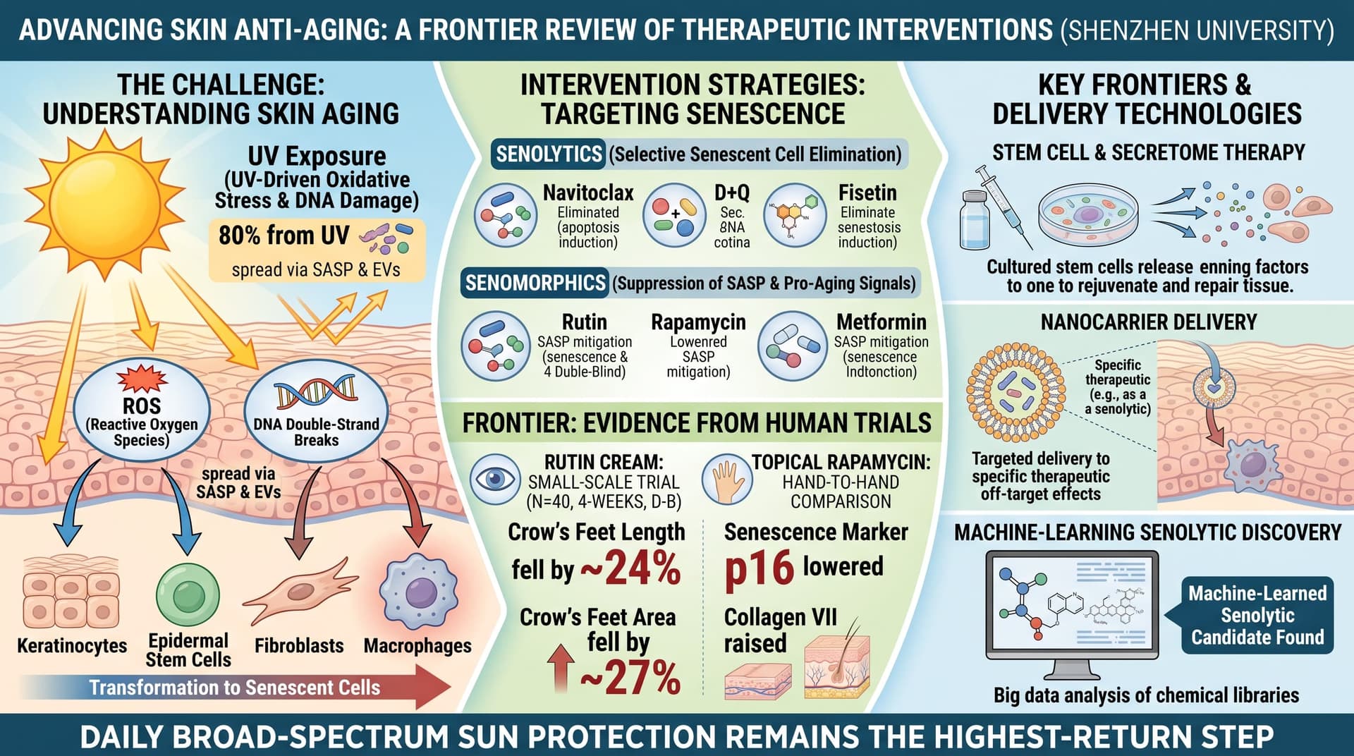
This narrative review from Shenzhen University argues that senescent cells are central to skin aging. It covers four skin cell types (fibroblasts, macrophages, keratinocytes and epidermal stem cells), how damage from UV-driven oxidative stress and DNA damage spreads between cells through the SASP and extracellular vesicles, and how histone modifications keep aged skin macrophages in an inflammatory state. It then surveys interventions: senolytics (navitoclax, dasatinib plus quercetin, fisetin), senomorphics (rutin, rapamycin, metformin), stem cell and secretome therapies, nanocarrier delivery, and machine-learning senolytic discovery. It contains no new data. Most of the evidence it summarizes comes from cell culture, rodents and human skin grafted onto mice. The human trials it cites are small, short, and mostly split-face cosmetic studies.

Skin shows age sooner than any other organ, and the authors argue it shows it for a specific reason: old, damaged cells that refuse to die.

These are senescent cells. They stop dividing but stay metabolically active, and they release a cocktail of inflammatory signals, enzymes and small vesicles known as the senescence-associated secretory phenotype, or SASP. In skin, the review says, the main senescent populations are fibroblasts in the dermis, keratinocytes in the outer layer, immune macrophages, and the stem cells that normally replenish the epidermis and hair follicles.

Each population fails differently. Senescent fibroblasts make less collagen and more of the matrix metalloproteinases (MMPs) that break collagen down. The authors describe a feedback loop: fragmented collagen reduces the mechanical tension fibroblasts feel, which raises their internal oxidative stress, which drives further MMP production. Senescent keratinocytes renew the epidermis more slowly, thinning the barrier and letting moisture out and bacteria such as Staphylococcus aureus in. Aged macrophages get stuck in an inflammatory mode and fail to shift into the repair mode that heals wounds.

The review’s more unusual contribution is its attention to how the damage spreads. Senescent cells release more extracellular vesicles than young cells. UV-damaged cells may export fragments of their own DNA in these vesicles, and neighboring cells detect the foreign DNA through the cGAS-STING immune sensing pathway and become senescent in turn. Vesicles also carry microRNAs, such as miR-30a, that suppress renewal in young keratinocytes. The authors also compile evidence that epigenetic regulators, including the enzymes JMJD3, MOF and SETDB2, lock macrophages into a chronic inflammatory state. Much of that evidence, however, comes from diabetic wounds rather than aged skin.

On treatments, the authors cover the familiar longevity pharmacopeia. Navitoclax clears senescent dermal fibroblasts in mouse and human-skin-graft models but causes low platelet and neutrophil counts when taken systemically. Dasatinib plus quercetin, and fisetin alone, reduce senescent fibroblast counts in similar models. Rutin, a quercetin glycoside, acts more as a SASP dampener than a cell killer. Topical rapamycin gets a mention for one small human trial that found fewer p16-positive cells and more collagen VII. Metformin is included for AMPK activation, mostly on the strength of rodent and worm data.

Stem cell therapy receives equal emphasis. The authors conclude that cell-free products, meaning conditioned media and vesicles from mesenchymal stem cells, are more practical than transplanted cells, which survive poorly after injection. Seven clinical studies are tabulated, and nearly all delivered the product by microneedling.

The review ends by pointing to machine-learning screens that have identified new senolytics, including ginkgetin, periplocin and oleandrin, from compound libraries.

The big idea is coherent: skin aging is a senescence problem that can be spread from cell to cell, and so it should be treatable by removing or silencing senescent cells locally. The weakness is that this remains a hypothesis supported mainly by mechanistic and animal work. The human data are sparse, small and unblinded or confounded more often than not.

Actionable Insights

The most reliable intervention remains the least exotic one. The review repeats a common estimate that UV exposure causes about 80% of visible skin aging. That figure is widely cited but loosely sourced, and the true share is hard to measure. Daily broad-spectrum sun protection is still the highest-return step.

Among topicals discussed, two have small human trials:

  • Rutin cream: In a 40-person, 4-week double-blind study, crow’s feet length fell by about 24% and crow’s feet area by about 27% in the rutin group. These are improvements from each person’s own baseline. The paper did not report a standardized effect size, and 4 weeks is too short to judge durability.
  • Topical rapamycin: In a hand-to-hand comparison, rapamycin cream lowered the senescence marker p16 and raised collagen VII.

Stem cell conditioned media trials all used microneedling, which stimulates collagen on its own. The one trial that compared delivery methods found no difference between groups. Any added benefit from the stem cell product itself remains unproven.

Oral senolytics (fisetin, dasatinib plus quercetin) have no controlled human skin-aging outcome data in this review.

Context and Source

  • Full title: Skin Aging: From Molecular Mechanisms to Therapeutic and Technological Innovations
  • Institution: Medical School, Shenzhen University; Laboratory of Regenerative Medicine, The 2nd Affiliated Hospital of Shenzhen University
  • Country: China
  • Journal: Journal of Cellular and Molecular Medicine
  • Impact evaluation: The impact score of this journal is 4.7, evaluated against a typical high-end range of 0–60+ for top general science, therefore this is a Medium impact journal.

Related Reading:

3 Likes

Home made 3% Rutin Cream Using Vanicream Moisturizing Lotion

Batch size: 50 g (enough for 3–4 weeks of use)

Ingredients

• Rutin powder — 1.5 g (Bulk Supplements, 100 g, $20)
• Distilled water — 10–15 g (hot)
• Vanicream Moisturizing Lotion — 35–40 g (Amazon, $13)
• Optional: Glycerin — 1 g (helps dissolve rutin, improves texture)
• Optional: Preservative — 0.5 g (Phenoxyethanol + Ethylhexylglycerin)

Vanicream lotion is stable enough that you can skip the preservative if you make small batches and use within 4–6 weeks.

Step‑by‑Step Instructions

  1. Dissolve the rutin

Rutin must be dissolved before mixing into lotion.

  1. Heat 10–15 g distilled water until very hot (not boiling).

  2. Add 1.5 g rutin powder.

  3. Stir until the water turns yellow and the powder dissolves.• A tiny bit of cloudiness is normal.

  4. Optional: Add 1 g glycerin to help solubilize and smooth texture.

Let the solution cool to room temperature.

  1. Prepare the lotion base

Place 35–40 g Vanicream Moisturizing Lotion into a clean bowl.

• If you want a thicker final cream → use 40 g lotion
• If you want a lighter cream → use 35 g lotion

  1. Combine

Slowly pour the cooled rutin solution into the lotion while stirring continuously.

Mix for 1–2 minutes until fully uniform.

The lotion will:

• become slightly thinner
• turn pale yellow
• remain smooth and stable

  1. Add preservative (optional)

  2. Package and store

Transfer to a clean, airtight container.

• Shelf life without preservative: 4–6 weeks
• Shelf life with preservative: 8–12 weeks
• Store at room temperature
• Avoid contamination (don’t dip fingers directly)

4 Likes

I’m tempted to make this and add some vitamin R to it as well! :slight_smile:

I would research it first before adding vit R to the mix. For example, I wanted to use Cerave cream instead of Vanicream lotion and my friend AI explained why it’s a bad idea.

1 Like

Here is the Claude Opus 5.5 summary of the Rutin Paper that came out in 2016:

Biological effects of rutin on skin aging

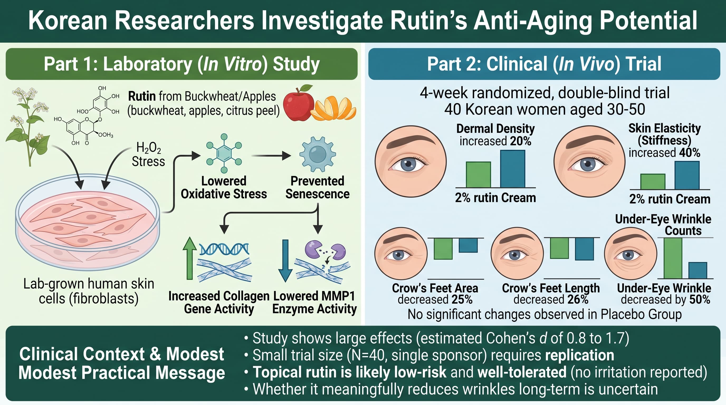
Korean researchers tested rutin, a plant flavonoid found in buckwheat, apples and citrus peel, in two ways. In lab-grown human skin cells stressed with hydrogen peroxide, rutin lowered oxidative stress, prevented cells from taking on a senescence-like state, raised collagen gene activity and lowered activity of the collagen-degrading enzyme MMP1. In a 4-week randomized, double-blind trial of 40 Korean women aged 30 to 50, a 2% rutin cream was reported to increase dermal density by 20%, raise skin stiffness (reported as “elasticity”) by 40%, cut crow’s feet length and area by roughly a quarter, and reduce under-eye wrinkle counts by half, with no change in the placebo group. The direction of the findings is plausible. The size of the effects is not.

Rutin is one of the most common flavonoids in the human diet. It is a sugar-bound form of quercetin, the compound many longevity enthusiasts know as half of the dasatinib plus quercetin senolytic combination. Rutin has a long history as a “vitamin P” supplement for fragile capillaries, and it already turns up in cosmetic ingredient lists. This study set out to give that use a scientific basis.

The laboratory half of the work followed a familiar script. The team grew human dermal fibroblasts, the cells that build and maintain the collagen scaffold of the skin, and hit them with hydrogen peroxide to mimic oxidative damage. Pretreating the cells with rutin kept more of them alive, cut the fluorescent signal used to track reactive oxygen species roughly in half, and reduced the share of cells staining positive for a common senescence marker from about 60% to under 20%. Rutin also nudged collagen gene expression back toward normal and pulled down expression of MMP1, the enzyme that chops up collagen in aging and sun-damaged skin.

The human trial is where the paper makes its headline claims. Twenty women applied a cream containing 2% rutin to their faces twice a day for four weeks, and twenty used an identical cream without it. Neither the women nor the assessors were told who got what. By week four, the rutin group showed a 20% rise in ultrasound-measured dermal density, a 40% rise in a suction-based skin mechanics score, a 24% reduction in crow’s feet length, a 27% reduction in crow’s feet area, and a 49% drop in the number of visible under-eye wrinkles. The placebo group barely moved. No one reported irritation.

Taken at face value, that would make rutin one of the most potent topical anti-aging agents ever tested, outperforming prescription retinoids in a fraction of the time. That is precisely why the results need skepticism. Dermal collagen turns over very slowly, over years rather than weeks, so a genuine 20% structural change in a month is hard to explain biologically. Rutin is also a large, water-loving molecule that penetrates intact skin poorly, and the study never measured whether it reached the dermis at all.

The design leaves several doors open for bias. The trial was small, short, unregistered, and approved by the ethics board of a company whose staff co-authored the paper. Rutin is bright yellow, so a 2% cream is likely visibly different from its placebo, which can quietly undo blinding. The statistics compared groups at individual time points without the analyses normally used for trials like this, and without adjusting for the many outcomes tested. One key measure, Young’s modulus, rises when skin gets stiffer, which is not obviously an improvement.

None of this means rutin does nothing. It is a competent antioxidant in a dish, and a mild topical benefit, perhaps via hydration or reduced inflammation, is believable. But this paper should be read as a promising pilot, not proof, and the effect sizes are best treated as an upper bound that independent trials have yet to confirm.

Actionable Insights

The practical message is modest. A topical rutin cream is probably low risk; no irritation was seen in 20 users over 4 weeks. Whether it meaningfully reduces wrinkles is uncertain.

On paper, the effects look large. Compared with placebo, the rutin group ended with about 18 mm less crow’s feet length (about a 26% relative difference), about 2.5 fewer under-eye wrinkles out of roughly 4 to 5, and about 10 points higher ultrasound dermal density. In standardized terms, these are “large” effects (estimated Cohen’s d of roughly 0.8 to 1.7, where 0.8 already counts as large). But with only 20 people per group, the true effect could plausibly be anywhere from small to very large, and effects from small, single-sponsor trials usually shrink when others repeat them.

For context, prescription tretinoin, the best-validated topical, typically needs 3 to 6 months to produce more modest measurable wrinkle change. Daily broad-spectrum sunscreen remains the intervention with the strongest evidence.

If you want to try rutin, treat it as an experiment: use it alongside, not instead of, sunscreen and a retinoid, and judge results over months with consistent photos. Oral rutin supplements were not tested here, and rutin has antiplatelet activity, so people on blood thinners should consult a clinician before supplementing.

Context and Source

  • Open Access Paper: Biological effects of rutin on skin aging, Published in 2016.
  • Institutions: Korea Institute for Skin and Clinical Sciences, Konkuk University; GeneCellPharm Incorporated (commercial); Department of Dermatology, Konkuk University School of Medicine
  • Country: Republic of Korea
  • Journal: International Journal of Molecular Medicine (Spandidos Publications)
  • Impact evaluation: The impact score of this journal is 8.5 (2025 JIF, released June 2026), evaluated against a typical high-end range of 0-60+ for top general science, therefore this is a Medium impact journal.

Here is the Claude Opus 5.5 summary of the Rapamycin on skin Paper that came out in 2019:

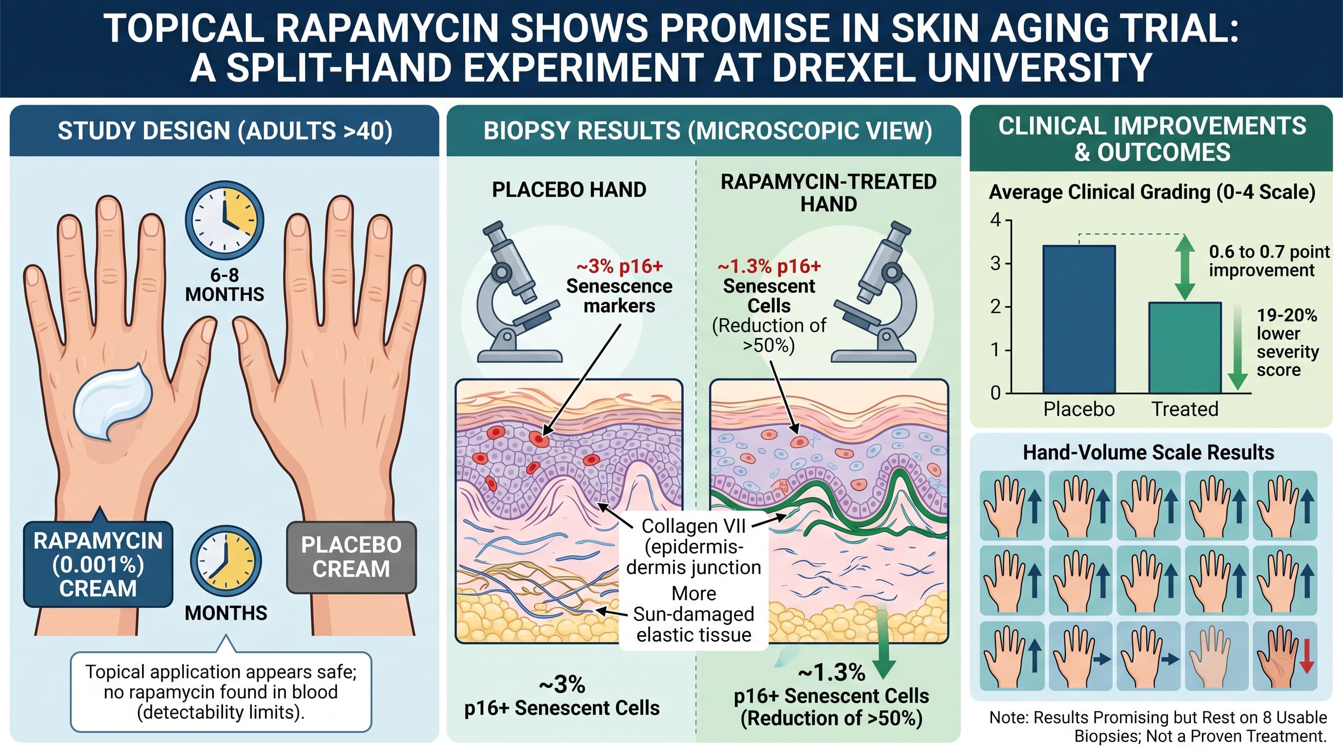
In a small split-hand trial at Drexel University, adults over 40 put a very dilute rapamycin cream (0.001 percent) on the back of one hand and a placebo cream on the other for six to eight months. Biopsies from the rapamycin-treated hands had about half as many cells carrying the senescence marker p16, more collagen VII at the junction between epidermis and dermis, and less sun-damaged elastic tissue. Clinical grading scores improved by roughly half a point to three-quarters of a point on 0 to 4 scales. No rapamycin was found in the blood. The results are promising, but they rest on 8 usable biopsies.

Rapamycin has spent two decades as the most reliable life-extending drug in mouse studies. Evidence that it slows aging inside human tissue, however, has been thin. A small trial from Drexel University in Philadelphia tried a shortcut. Instead of swallowing the drug, volunteers rubbed a very dilute rapamycin cream onto the back of one hand and an identical-looking placebo onto the other.

The logic rests on senescent cells. These cells have permanently stopped dividing but do not die, and they leak inflammatory signals that degrade the surrounding tissue. In skin, the number of cells carrying the senescence marker p16 tracks with wrinkling, damaged elastic fibers, and how old a person looks. Rapamycin blocks mTOR, a growth-signaling hub that pushes stressed cells into senescence in lab experiments. If the drug keeps cells out of that state, aged skin might partly repair itself.

Thirty-six adults over 40 with visibly sun-damaged hands enrolled. Each applied half a milliliter of cream to each hand every one or two nights for six to eight months. The rapamycin concentration was 0.001 percent, one hundred to one thousand times weaker than the creams dermatologists use for the benign skin growths of tuberous sclerosis.

At the end, biopsies from both hands told a consistent story. Rapamycin-treated skin had roughly half as many p16-positive cells as placebo skin, with most of the difference in the outer epidermis. Collagen VII, the protein that anchors the epidermis to the dermis and that thins beneath wrinkles, stained more strongly. Under the microscope, treated skin showed less solar elastosis, the clumped, damaged elastic tissue typical of photoaging, and a more orderly basal layer.

The visible changes were modest but consistent. Most treated hands scored better for vein and tendon prominence, fine wrinkling, and uneven pigment, with changes appearing around four months. No rapamycin turned up in anyone’s blood, and no drug-related side effects were reported.

The catch is scale. Nineteen of the 36 volunteers dropped out for unspecified reasons, and only eight produced biopsy tissue good enough to analyze. Clinical grading used simple 0 to 4 scales, and the paper does not say whether the graders knew which hand received the drug. Three authors hold shares in a company developing the approach. A drop in p16 is also not proof that senescent cells are gone. Rapamycin broadly suppresses protein production and could lower p16 levels without removing a single cell.

Still, the study matters. It was the first placebo-controlled test of rapamycin against aging markers in living human tissue, and it used a dose so low that the usual concerns about immune suppression and mouth ulcers barely apply. Skin is also an unusually practical organ for testing aging drugs: it is easy to reach, easy to biopsy, and the results are visible. If a larger, properly blinded trial reproduces these numbers, topical mTOR inhibition could become one of the first anti-aging interventions supported by human tissue data rather than mouse data.

Actionable Insights

Topical rapamycin at very low strength (about 0.001 percent, or 10 micromolar) appears safe on the hands over eight months and did not reach the bloodstream at detectable levels. This makes it a low-risk experiment compared with oral rapamycin, but it is not a proven treatment.

To put the size of the benefit in perspective: on the doctors’ 0 to 4 aging scales, treated hands averaged about 0.6 to 0.7 points better than placebo hands. That is roughly a 19 to 20 percent lower severity score. Put another way, about 11 of 16 people saw their treated hand improve by one grade on the hand-volume scale, 4 saw no change, and 1 got worse. In the biopsies, senescent-marker cells dropped from about 3 percent to about 1.3 percent of cells, a reduction of more than half.

A one-grade change is noticeable in photographs but not dramatic. There was no head-to-head comparison with proven products, so it is unknown whether this adds anything to daily broad-spectrum sunscreen and a retinoid, which remain the best-supported skin interventions. Compounded rapamycin creams vary in quality and vehicle. The vehicle used here was DMSO-based, which affects skin penetration.

Context and Source

  • Open Access Paper: Topical rapamycin reduces markers of senescence and aging in human skin: an exploratory, prospective, randomized trial
  • Institutions: Drexel University College of Medicine (Dermatology, Medicine, Pathology, Biochemistry), with University of Bologna
  • Country: United States (with Italian collaborator)
  • Journal: GeroScience, 2019, volume 41
  • Impact evaluation: The impact score of this journal is 6.0 (2025 Journal Impact Factor), evaluated against a typical high-end range of 0-60+ for top general science, therefore this is a Medium impact journal. Within geriatrics and gerontology specifically, GeroScience ranks near the top of its field.

Related Reading: