Cellular Senescence as a Contributing Cause of Muscle Aging

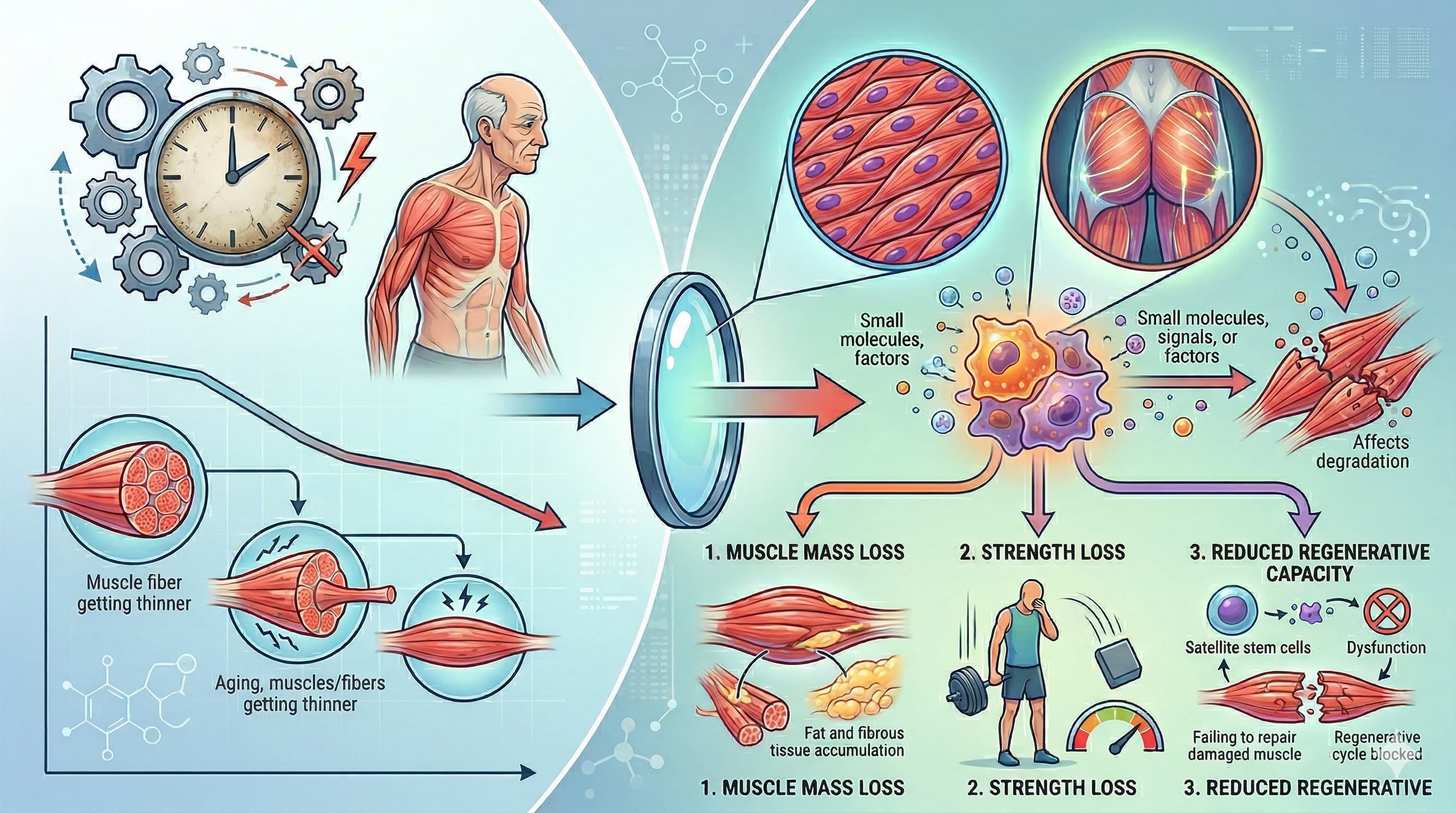
Skeletal muscle aging, clinically recognized as sarcopenia, has traditionally been viewed through the lens of passive “wear and tear.” However, recent evidence definitively shifts this paradigm toward an active, pathology-driving mechanism: cellular senescence. This review synthesizes mounting data demonstrating that the accumulation of senescent cells across various muscle compartments directly orchestrates the progressive loss of muscle mass, strength, and regenerative capacity.

Crucially, senescence in skeletal muscle is not confined to a single cell type. It actively infiltrates muscle stem cells (MuSCs), fibro-adipogenic progenitors (FAPs), immune cells, and even terminally differentiated, post-mitotic myofibers. Once senescent, these cells exhibit profound chromatin alterations, resistance to apoptosis, and the secretion of a toxic senescence-associated secretory phenotype (SASP). The SASP, a cocktail of pro-inflammatory cytokines, chemokines, and extracellular matrix-modifying enzymes, propagates chronic inflammation (“inflammaging”), disrupts niche homeostasis, and induces secondary senescence in neighboring healthy cells.

For example, senescent FAPs lose the ability to secrete WISP1—a vital regenerative signal—while simultaneously driving fibrotic tissue remodeling via transforming growth factor-β (TGF-β) secretion. Concurrently, senescent MuSCs suffer from impaired autophagy and profound regenerative deficits. Even post-mitotic myofibers, previously thought immune to classic replicative senescence, exhibit stress-induced premature senescence driven by p21CIP1 expression, further exacerbating tissue decline.

This mechanistic clarity has accelerated the development of senotherapeutics as a countermeasure. Senolytics (like the BCL-2/BCL-XL inhibitor ABT263 and CAR-T cells targeting uPAR) selectively induce apoptosis in senescent cells, thereby rejuvenating MuSC function in preclinical models. Senomorphics (such as the CCR5 antagonist Maraviroc and natural polyphenols) aim to suppress the SASP without eliminating the cells. While these pharmacological interventions present a formidable frontier against age-related frailty, clinical translation remains gated by target specificity, the complex heterogeneity of the senescent niche, and the potential off-target consequences of broad senescent cell clearance

Source

Related Reading: HIV Drug (Maraviroc) Reverses Muscle Aging by purging “Zombie Cell” Signals

Novelty Assessment

Historically, senescence was primarily associated with replicating cells. The defining novelty consolidated in this review is the robust identification of senescence-like states in post-mitotic, terminally differentiated myofibers. These myofibers do not undergo classic cell-cycle arrest (as they do not divide), but rather exhibit stress-induced premature senescence governed by p21CIP1 (rather than p16INK4a), coupled with DNA damage and chromatin reorganization. Furthermore, the granular mapping of heterogeneous senescent signatures across distinct cell types (MuSCs vs. FAPs vs. Macrophages) using single-nucleus RNA and ATAC sequencing provides unprecedented resolution of the aging muscle niche

Claims & Verification

  • Claim 1: The accumulation of senescent cells and the secretion of the Senescence-Associated Secretory Phenotype (SASP) actively drive the pathology of sarcopenia.
    • Evidence Level: Level C (Human Observational) / Level D (Pre-clinical).
    • Translational Gap: While the presence of senescent cells and SASP markers is well-documented in human cohorts (correlating with frailty and muscle decline), direct causal evidence—ablating these cells to reverse sarcopenia—is entirely restricted to murine models.
    • Verification: Cellular Senescence in Sarcopenia: Possible Mechanisms and Therapeutic Potential (2022)
  • Claim 2: Impaired autophagy in aging Muscle Stem Cells (MuSCs) leads to toxic waste accumulation, initiating cellular senescence and catastrophic regenerative decline.
    • Evidence Level: Level D (Pre-clinical).
    • Translational Gap: FLAG. The mechanistic link between autophagic failure, reactive oxygen species (ROS) accumulation, and MuSC senescence is robustly proven in Atg7 -knockout and geriatric mice. However, translating this functional rescue to human stem cell therapies remains unverified in clinical trials.
    • Verification: Autophagy: a decisive process for stemness (2016)
  • Claim 3: Mitochondrial DNA (mtDNA) release into the cytosol activates the cGAS-STING innate immune pathway in post-mitotic myofibers, triggering premature senescence and SASP independent of cell division.
  • Claim 4: Senescent Fibro-Adipogenic Progenitors (FAPs) lose the ability to secrete the regenerative matricellular signal WISP1, while concurrently upregulating TGF-β to drive intramuscular fibrosis.
  • Claim 5: Senolytics (e.g., ABT263/Navitoclax, uPAR-targeted CAR-T cells) selectively induce apoptosis in senescent cells and rejuvenate skeletal muscle function.
  • Claim 6: Senomorphics (e.g., Maraviroc targeting CCR5) mitigate age-related muscle decline by suppressing the SASP without eliminating the underlying senescent cells.
1 Like

I do think it might be worth a try. I have received mine from India. As soon as I get some baseline blood tests, hand grip strength, etc. I will be starting at 75mg/day for 60 days. If at the end of 60 days I don’t see any results outside of statistical error, I will up the dose to 150 mg/daily. Of course, if someone posts some proof that Mavavoric kills us, I will stop.

The half life is rather long. If I were you I’d start with a pulsed protocol 2x or 3x a week first, and only ramp up to daily if it’s not working. It sounds like something to be careful finding the sweet spot on.