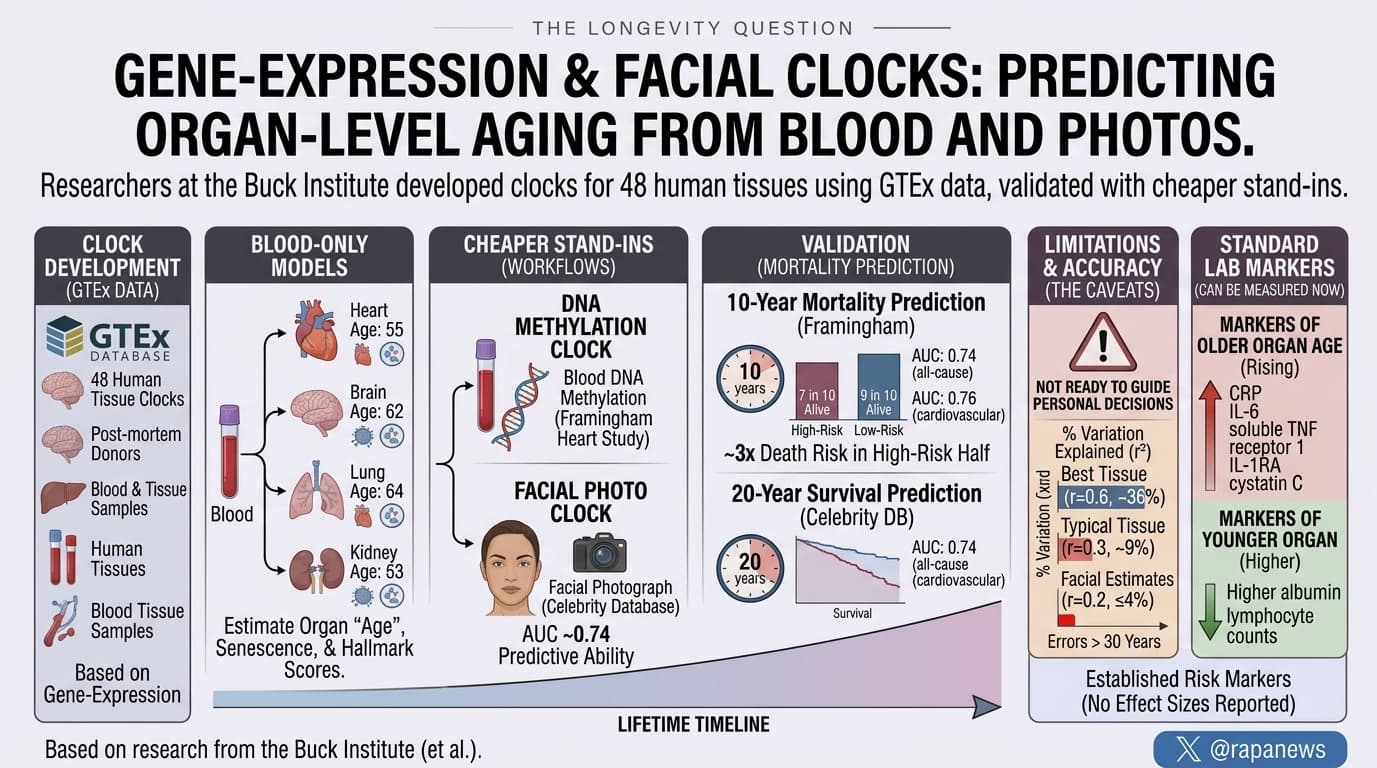
Researchers at the Buck Institute built gene-expression aging clocks for 48 human tissues using GTEx, a post-mortem database where many donors gave both blood and tissue samples. They then trained blood-only models to estimate each organ’s “age,” along with organ-level senescence and hallmark-of-aging scores. They went on to build cheaper stand-ins from blood DNA methylation and from facial photographs. Methylation-derived organ ages predicted ten-year mortality in the Framingham Heart Study (AUC about 0.74; 0.76 for cardiovascular death). Face-derived organ ages predicted twenty-year survival in a celebrity photo database (AUC 0.74). Blood tracked organ age moderately well for some tissues and poorly for others.
Every organ ages on its own schedule. Your arteries may be a decade ahead of your liver, and your brain may lag behind both. Measuring this directly means taking tissue samples, which nobody does to healthy people out of curiosity. A team at the Buck Institute for Research on Aging in California proposes a workaround that runs through the bloodstream and, more ambitiously, through a photograph of your face.
The group, led by David Furman, started with GTEx. This reference library records gene activity in dozens of tissues from roughly a thousand deceased donors. Because many donors gave both blood and tissue, the researchers could ask a direct question. Does gene activity in someone’s blood carry a readable echo of what is happening in their thyroid, colon or heart? They built aging clocks for 48 tissues, then trained blood-only versions to predict each organ clock.
The answer was a qualified yes. A handful of tissues worked well: thyroid, breast, sigmoid colon, tibial artery, skin and oesophagus. For these, blood-based estimates tracked age reasonably closely in people the models had not seen. Blood-based estimates for 34 of 45 workable tissues cleared a modest bar. For others, accuracy collapsed toward nothing on new data. Typical errors were 8 to 13 years.
The team then scored each organ for signatures of cellular senescence and for the broader hallmarks of aging. Senescence is the state in which damaged cells stop dividing and secrete inflammatory signals. The standard senescence gene sets showed no relationship with age at all. The scores only began to rise with age after the researchers split each gene set by whether its genes went up or down with age.
Next came the cheap proxies. The team used 605 participants from a trial run by Edifice Health, a company that employs two of the authors. With them, they trained models that estimate blood-derived organ ages from facial photos taken on an iPad. A parallel model did the same from blood DNA methylation in the Health and Retirement Study. Neither stand-in is precise, but both carried some signal into survival. In the Framingham Heart Study, methylation-based organ ages split people into higher and lower ten-year death risk. In a set of celebrity photos from IMDb and Wikipedia, face-based organ ages predicted twenty-year survival with similar accuracy.
Within one person, organ ages mostly moved together, with an average correlation of about 0.44 between organs, yet some pairs ran in opposite directions. A mediation analysis suggested that genes in one tissue might influence aging genes in another through the blood. Most of these links stayed within a single organ. A drug-matching screen flagged compounds whose known gene targets overlap the clock genes, among them N-acetylglucosamine.
The pitch is appealing: a selfie or a cheap blood test that flags which organ is running fast, followed by a targeted fix. What the data show today is more modest. This is an unreviewed preprint built on post-mortem tissue. Prediction errors run to decades for the facial models, and it has not been tested whether organ age adds anything beyond a person’s actual age. It is an interesting research direction, time will tell if it becomes accurate enough to be valuable.
Actionable Insights
The useful lessons are about how to read the consumer products likely to follow.
First, organ-age readouts from blood or photos are not ready to guide personal decisions. In the best tissues, blood explained about 36 percent of the variation in organ age (correlation 0.6). In a typical usable tissue it explained about 9 percent (correlation 0.3). Facial estimates often explained 4 percent or less (correlation 0.2), with errors that could exceed 30 years. A single reading is mostly noise.
Second, several standard lab markers tracked with older organ ages across many systems, and you can already measure them. Rising CRP, IL-6, soluble TNF receptor 1, IL-1RA and cystatin C went with older organ ages. Higher albumin and lymphocyte counts went with younger ones. These are established risk markers in their own right. The paper reports no effect sizes for them.
Third, the mortality split sounds dramatic. In Framingham’s test set, about 7 in 10 of the high-risk half were alive at ten years, against roughly 9 in 10 of the low-risk half. That is close to three times the death risk. Chronological age probably drives much of that gap.
Fourth, ignore the compound list. N-acetylglucosamine and the others were flagged by overlap between gene lists, not by any test of whether they change organ aging.
Context and Source
- Open Access Paper: Inferring organ aging, hallmark of aging and senescence scores from blood and facial photographs
- Authors: Kevin Schneider, Minja Belic, Matias Fuentealba, Fiona Senchyna, David Furman
- Institution: Buck Institute for Research on Aging, Novato, California
- Country: United States
- Venue: bioRxiv preprint.