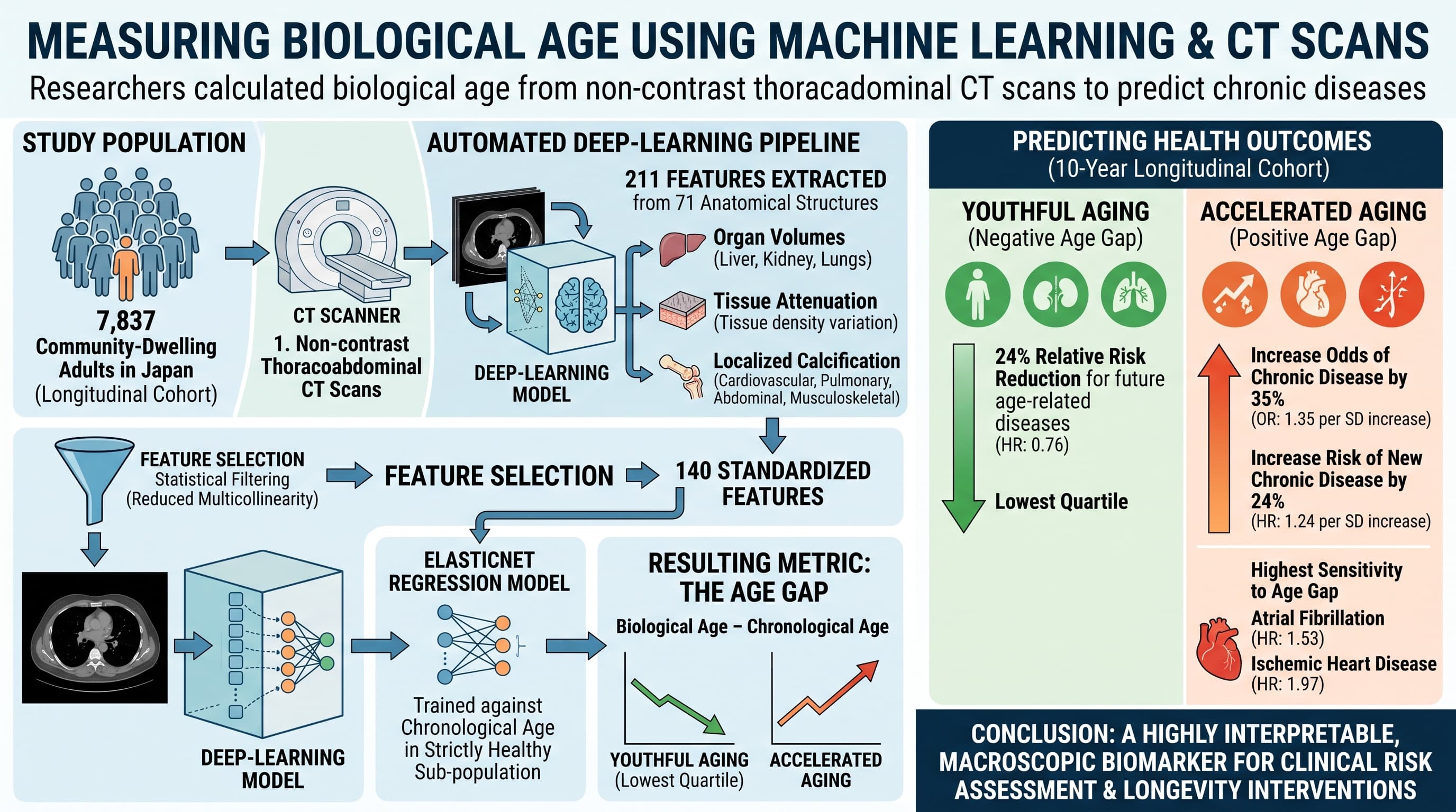
Researchers developed a machine learning model to calculate biological age by analyzing 140 body composition features from standard noncontrast thoracoabdominal computed tomography scans. The resulting metric, termed the “age gap”, successfully identifies accelerated aging and predicts the onset of major chronic diseases across a longitudinal cohort, providing a highly interpretable, macroscopic biomarker for both clinical risk assessment and longevity interventions.
The longevity field has spent the last decade chasing cellular and molecular clocks, primarily focusing on epigenetic methylation patterns. While scientifically profound, these molecular clocks often lack anatomical interpretability and fail to tell clinicians exactly which organ system is driving a patient’s accelerated aging. This study pivots the focus from the microscopic to the macroscopic by utilizing opportunistic computed tomography imaging to quantify systemic aging through gross anatomical structural changes.
Using a large longitudinal health-screening cohort of 7,837 community-dwelling adults in Japan, researchers deployed an automated deep-learning pipeline to extract 211 quantitative body composition features from 71 anatomical structures. These features included organ volumes, tissue attenuation properties, and localized calcification metrics spanning the cardiovascular, pulmonary, abdominal, and musculoskeletal systems. After statistical filtering to reduce multicollinearity, 140 standardized features were retained to train an ElasticNet regression model against chronological age in a strictly healthy sub-population.
By defining the reference trajectory of normal aging in healthy individuals, the researchers established an “age gap” metric, calculated as the age-adjusted residual difference between a subject’s computed tomography-predicted biological age and their true chronological age. A positive age gap indicates accelerated biological aging.
The predictive validity of this age gap was then tested on independent longitudinal datasets. The data shows that the age gap reliably captures disease-specific heterogeneity. Systemic diseases like hypertension and metabolic disorders were strongly linked to multi-organ alterations, specifically visceral adiposity and reduced pancreatic tissue attenuation. Conversely, localized diseases mapped to organ-specific structural decay, such as lower vertebral attenuation in osteoporosis and elevated Agatston scores for cardiovascular conditions.
The operational advantage of this approach is its scalability and reliance on existing medical data. Noncontrast thoracoabdominal scans are routinely performed worldwide. Approximately 93 million CT examinations are performed annually in the United States across roughly 62 million patients.
By running automated segmentation models like TotalSegmentator in the background of routine radiological workflows, healthcare systems could generate opportunistic biological age reports without requiring novel blood draws or expensive molecular assays. This shifts biological age assessment from a niche biohacking commodity into the realm of standard preventative radiology, allowing for targeted lifestyle modifications years before symptomatic chronic disease onset.
Actionable Insights
This study identifies specific anatomical variables that drive the biological aging clock, offering practical targets for longevity interventions. The data points heavily to tissue quality rather than just tissue volume. Specifically, lower tissue attenuation in skeletal muscle and the pancreas indicates lipid infiltration, a major driver of metabolic aging.
To improve this biological age metric, individuals must prioritize the reduction of visceral and ectopic fat. Strategies include strict glycemic control to prevent pancreatic lipid deposition and resistance training to preserve muscle density (preventing myosteatosis).
The magnitude of this intervention is substantial. The study reveals that individuals in the lowest quartile of biological age (the “youthful” group) experienced a 24 percent relative risk reduction for future age-related diseases over a 10-year period compared to the normal reference group (Hazard Ratio: 0.76). Conversely, every 1 standard deviation increase in the biological age gap increased the overall odds of possessing a chronic disease by 35 percent (Odds Ratio: 1.35) and increased the risk of developing a new chronic disease by 24 percent (Hazard Ratio: 1.24). Atrial fibrillation and ischemic heart disease showed the highest sensitivity to this age gap, with hazard ratios of 1.53 and 1.97 per standard deviation increase, respectively.
You Can Try it out Today - The Tools Are Available Online
You can unlock this macroscopic biological aging data using tools already at your disposal. If you have an existing computed tomography or magnetic resonance imaging scan in a standard NIFTI or DICOM zip format, you can bypass standard blood-based epigenetic clocks and calculate your anatomical biological age for free using a two-step open-source workflow.
First, process your scan through TotalSegmentator, an automated deep learning tool that maps out anatomical structures. TotalSegmentator utilizes advanced neural networks to evaluate three-dimensional data, automatically detecting and outlining dozens of distinct internal structures across the body, including major organs, bones, muscles, blood vessels, and various tissue types. The output delivers detailed segmentation masks that precisely isolate the physical boundaries of these regions, alongside quantitative statistics for each localized structure, such as exact volume and tissue intensity. This comprehensive physical profile is frequently utilized by clinicians and researchers for organ volumetry, body composition profiling, surgical planning, and opportunistic disease characterization.
Second, extract the 140 required body composition features generated by TotalSegmentator and feed them into the CT_BIOAGE machine learning model. The researchers behind this model have made their trained algorithm and source code publicly available for this exact purpose. The model translates your structural tissue data into a final biological age gap. A positive age gap identifies accelerated systemic aging driven by macroscopic factors like visceral adiposity, vascular calcification, and ectopic lipid deposition. This two-step process provides organ-specific, actionable data to target your longevity interventions directly at the tissues driving your biological clock.
Context/Source
- Open Access Paper: CT-based biological age as a biomarker of aging and age-related disease assessment
- Institution: Southeast University, The University of Tokyo, Chiba University
- Country: China, Japan
- Journal Name: npj Aging
- Impact Evaluation: The impact score of this journal is approximately 4.9, evaluated against a typical high-end range of 0 to 60+ for top general science, therefore this is a Medium impact journal.
