Why do falls rise with age? Cerebellar Neuron Firing Problems (and Potential Therapeutics)

Aging inevitably strips away motor coordination, balance, and physical strength, directly escalating the risk of debilitating falls. This functional decline is traditionally attributed to the irreversible death of neurons and muscle tissue. However, new research from McGill University in Canada, published in PNAS, dismantles this assumption. The study demonstrates that within the cerebellum—the brain’s primary motor coordination hub—the critical output neurons are not dying; they are simply losing their electrical pacemaker rhythm.

The investigation focused on Purkinje cells, which act as the exclusive output pathway of the cerebellar cortex and normally fire high-frequency, spontaneous action potentials to coordinate movement. The researchers discovered that the total number of these neurons remains completely stable in the anterior cerebellum of aged mice. Instead of cellular death, the underlying pathology is an age-dependent drop in their intrinsic firing frequency.

To establish causality, the team utilized a chemogenetic (DREADD) approach to directly manipulate these firing rates in live animals. When researchers artificially suppressed the Purkinje firing rate in young, healthy mice, the animals instantly became clumsy, perfectly phenocopying the motor decline of old age. More importantly, when the researchers upregulated the firing rate in 18-month-old mice—effectively restoring youthful electrical pacing—the aged mice exhibited immediate and significant improvements in both gross motor balance on a rotarod and fine motor dexterity during string-pulling tasks.

This represents a fundamental paradigm shift for longevity research. It proves that a significant fraction of age-related motor frailty is driven by a reversible electrophysiological deficit, not irreversible neurodegeneration. The neural architecture remains intact, but the electrical signal weakens. Identifying Purkinje cell firing rates as an actionable bottleneck offers a highly specific therapeutic target to rescue motor function, prevent falls, and extend physical healthspan in the elderly.

Source:

More Reading: Pharmacological Interventions for Purkinje Cell Preservation

1 Like
  • Mechanistic Deep Dive:
  • The primary electrical defect is a significant reduction in Purkinje cell intrinsic simple-spike frequency. No alterations to firing regularity were observed in the anterior vermis.
  • Structural degradation was noted, including a reduction in overall dendritic complexity and shrinkage of the cerebellar molecular layer. These morphological changes strongly suggest a concurrent loss of parallel fiber synaptic inputs.
  • Calbindin protein expression, required for cellular calcium buffering, significantly decreases with age. This points to failing calcium homeostasis as a major driver of the electrical dysfunction.
  • Translational Pathway: Purkinje cells operate under extreme metabolic demand to sustain their continuous, high-frequency pacemaking. The functional decline observed here is highly likely downstream of mitochondrial decay and altered voltage-gated ion channel (Nav/Kv) distribution. [Confidence: High]
  • Novelty: * This paper proves that age-related motor coordination decline is partially a reversible, electrophysiological tuning problem. It overrides the dogma that physical frailty in the nervous system is solely due to irreversible neuronal apoptosis.

Part 3: Claims & Verification

1. Claim: Motor coordination and balance progressively decline as a function of normal aging.

2. Claim: Purkinje cell number does not significantly decrease during normal aging, specifically in the anterior cerebellum.

  • External Verification: Aging of the human cerebellum: a stereological study (2003)
  • Evidence Level: Level D (Pre-clinical). [FLAG: High Conflict]
  • Translational Gap: Massive. While this paper asserts no significant cell death in 18-month-old mice, highly rigorous human stereological studies demonstrate a selective 40% loss of Purkinje cells specifically in the anterior lobe of the aging human cerebellum. The murine model utilized in this study completely fails to recapitulate this specific hallmark of human pathology.

3. Claim: The cerebellar molecular layer shrinks and Purkinje cell dendritic complexity decreases with age.

  • External Verification: Aging of the human cerebellum: a stereological study (2003)
  • Evidence Level: Level C (Human Observational / Postmortem).
  • Assessment: Human postmortem data confirms a ~30% volume loss in the cerebellar anterior lobe, primarily driven by cortical volume loss. This aligns with the dendritic retraction and molecular layer shrinkage observed in the paper’s animal models.

4. Claim: Purkinje cell intrinsic simple-spike firing rate spontaneously decreases with age.

5. Claim: Increasing Purkinje cell firing rates rescues motor coordination in aged subjects.

6. Claim: Calbindin protein expression in Purkinje cells declines with age, suggesting impaired calcium buffering.

Pharmacological Interventions for Purkinje Cell Preservation

Cerebellar Purkinje cells (PCs) are among the most metabolically demanding neurons in the mammalian brain, spontaneously firing at 40–100 Hz to maintain motor coordination. Their age-related decline is characterized by a progressive reduction in intrinsic firing rate, dendritic retraction, loss of intracellular calcium buffering capacity, and eventual apoptosis.

No FDA-approved therapeutic is specifically indicated to halt Purkinje cell aging. However, several compounds have demonstrated targeted neuroprotection in pre-clinical models of accelerated cerebellar aging (ataxias) and standard murine aging.

Part 1: Demonstrated Therapeutics (Pre-clinical & Clinical)

  • Mitochondrial-Targeted Antioxidants (MitoQ)
  • Ceftriaxone (Repurposed Beta-Lactam Antibiotic)
  • Hericium erinaceus (Lion’s Mane Extract)
    • Mechanism: Stimulates Nerve Growth Factor (NGF) and Brain-Derived Neurotrophic Factor (BDNF) synthesis, providing broad-spectrum neurotrophic support.
    • Evidence: In normal, frail aging mice, dietary supplementation ameliorated cerebellar volume reduction and specifically decreased the percentage of shrunken, degenerating Purkinje neurons compared to age-matched controls. [Confidence: Medium].
    • Citation: Neuroprotective Metabolites of Hericium erinaceus Promote Neuro-Healthy Aging (2021)
  • 4-Aminopyridine (Dalfampridine) – Functional Mitigation
    • Mechanism: Broad-spectrum voltage-gated potassium channel (Kv) blocker.
    • Evidence: This compound does not prevent cellular apoptosis. However, by blocking potassium efflux, it lowers the threshold for action potentials and artificially forces higher firing rates. This directly mitigates the primary electrophysiological symptom of PC aging, temporarily rescuing motor coordination. [Confidence: High for symptomatic relief; Zero for structural preservation].

Part 2: Theoretically Appropriate Compounds (Mechanistic Targets)

  • Elamipretide (SS-31)
    • Rationale: Purkinje cell functional decline is fundamentally an energy deficit pathology; structural decay of the inner mitochondrial membrane (IMM) precedes the loss of electrical pacing. SS-31 selectively binds to cardiolipin on the IMM, stabilizing its curvature and restoring electron transfer efficiency without acting as a blunt-force reactive oxygen species (ROS) scavenger. Given the reliance of PCs on continuous mitochondrial ATP output, SS-31 represents a premier theoretical candidate to delay the electrophysiological decay of the cerebellum. [Confidence: High theoretical rationale; Direct in vivo PC data lacking].
  • Systemic Sex Steroids (Bioidentical HRT)
    • Rationale: Cerebellar Purkinje cells express high densities of sex steroid receptors and rely on both circulating hormones and locally synthesized neurosteroids for trophic support. Pre-clinical stereological data indicate that age-related PC apoptosis is primarily triggered downstream of the age-dependent crash in systemic steroidogenesis. Maintaining youthful steroid profiles (estradiol/testosterone/progesterone) represents a highly probable prophylactic intervention against structural PC degradation.
    • Citation: Age-related Purkinje cell death is steroid dependent (2011)
  • Intracellular Calcium Chelators / Calpain Inhibitors
    • Rationale: Aging PCs reliably downregulate Calbindin, their primary intracellular calcium buffer. This reduction leaves the cell defenseless against calcium transients generated by normal synaptic activity, ultimately triggering calcium-dependent apoptotic enzymes (calpains). Small-molecule calpain inhibitors or highly targeted calcium chelators address this specific bottleneck of PC aging.

Knowledge Gaps & Translational Limitations

  1. The Delivery Barrier: The blood-brain barrier (BBB) severely limits the cerebellar penetrance of many systemic neuroprotectants, particularly large peptides. Achieving localized therapeutic concentrations without systemic toxicity remains a challenge.
  2. In Vivo Verification: There are currently no non-invasive fluid biomarkers or accessible imaging modalities to measure real-time Purkinje cell firing rates or specific PC apoptosis in living humans. Clinical trials for structural neuroprotection must rely on lagging phenotypic indicators (gross motor coordination tests), complicating the precise measurement of early-stage target engagement.
1 Like

Looking deeper at the possible therapeutics for this neurodegeneration issue, it doesn’t look very good. MitoQ has failed in all the clinical trials its been tried in related to neurodegeneration (Parkinson’s etc.) but may still show results in terms of prevention. Ceftriaxone is IV delivery, which is impractical, and it can’t be used on an ongoing (daily) basis. Lions mane mushrooms “might” provide some benefits, but still unproven. The SS-31 peptide has some potential, but sourcing high quality at reasonable cost is an issue.

Executive Summary: The Translational Reality of MitoQ in Neurodegeneration

Despite possessing a mountain of highly successful pre-clinical data in rodent models of Alzheimer’s, ALS, traumatic brain injury, and Huntington’s disease, MitoQ’s human clinical trial record for neurodegenerative disease is exceptionally sparse and currently stands at a 100% failure rate for disease modification.

The stark reality of mitochondrial-targeted antioxidants is a massive translational gap. While the molecule successfully concentrates in the mitochondria of isolated cells and rodent brains, scaling the dose to achieve therapeutic central nervous system (CNS) penetrance in living humans frequently triggers dose-limiting gastrointestinal toxicity before neuroprotection can be achieved. Furthermore, quenching reactive oxygen species (ROS) appears insufficient to halt the cascading structural collapse of advanced human proteopathies (like Parkinson’s).


Active / Ongoing Clinical Trials

Because MitoQ failed to alter the trajectory of established Parkinson’s, current clinical research has pivoted away from “late-stage disease modification” and toward early-stage vascular dementia, Mild Cognitive Impairment (MCI), and brain hypoperfusion.

3. Mild Cognitive Impairment (MCI) & Cerebrovascular Blood Flow

4. The Mito-Frail Trial: Cognitive and Physical Dysfunction


The Biohacker & Longevity Takeaway

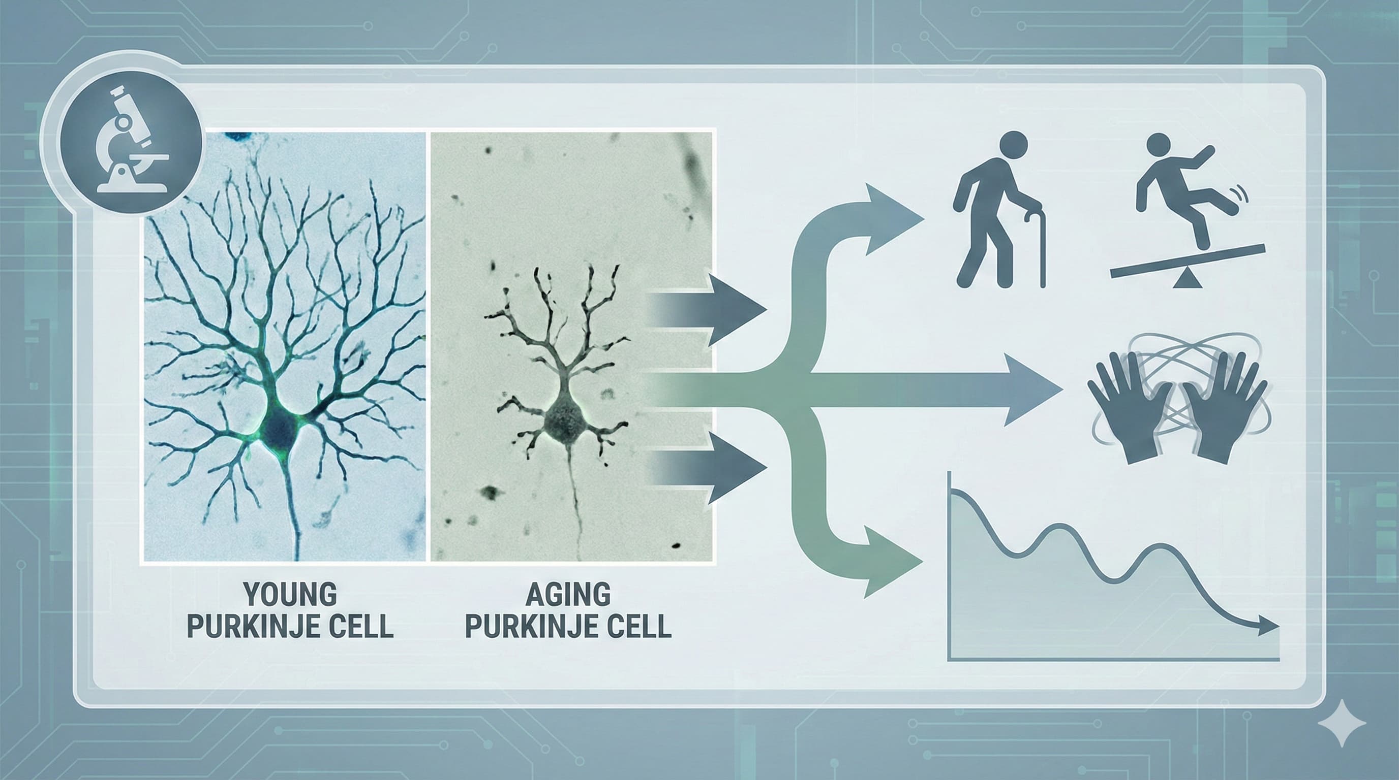
If your goal is structural neuroprotection (e.g., preserving Purkinje cells or dopaminergic neurons), human clinical data does not currently support the use of MitoQ. The failure of the PROTECT trial highlights two critical pharmacological hurdles:

  1. The Blood-Brain Barrier (BBB) Bottleneck: Achieving therapeutic concentrations of MitoQ inside human brain mitochondria requires pushing systemic oral doses to levels that trigger severe GI toxicity (80+ mg). The standard commercial dose of 10–20 mg/day is profoundly unlikely to reach the CNS in concentrations high enough to halt neurodegeneration.
  2. Too Little, Too Late: In neurodegenerative pathologies, by the time clinical symptoms manifest, the structural network collapse is too severe to be reversed simply by capping mitochondrial reactive oxygen species.

Currently, MitoQ’s validated human clinical utility remains strictly confined to peripheral vascular health (improving endothelial flow-mediated dilation in older adults). It should not be prescribed or relied upon as a primary neuroprotective agent.


The Ceftriaxone Translational Protocol:

  • The Clinical Precedent: Ceftriaxone is an FDA-approved, third-generation cephalosporin antibiotic. Its neuroprotective mechanism is completely independent of its antimicrobial properties; it transcriptionally upregulates the GLT-1 (EAAT2) glutamate transporter on astrocytes, enhancing the clearance of excitatory glutamate from the synaptic cleft and preventing calcium-induced excitotoxicity in neurons.

Pharmacokinetics (PK/PD):

  • Bioavailability: Zero to <1% orally. The drug is completely degraded in the GI tract. It strictly requires intravenous (IV) or intramuscular (IM) administration.

  • Phase I / Clinical Safety: Extremely well characterized as a short-term antibiotic (Rocephin). However, chronic daily administration is highly problematic. The primary long-term risk is biliary pseudolithiasis (gallbladder sludging), caused by the precipitation of ceftriaxone-calcium salts.

  • NOAEL / LD50: Acute toxicity is exceptionally low. The intravenous LD50​ in mice is ~2,000 mg/kg. Long-term tox studies in non-human primates established a NOAEL of approximately 100 mg/kg/day due to gastrointestinal and biliary complications.

The Hericium erinaceus (Lion’s Mane)Translational Protocol:

  • The Clinical Mechanism: Hericium erinaceus (Lion’s Mane) acts as a neurotrophic stimulator. Its primary bioactive compounds—specifically erinacines—are small-molecule cyathane diterpenoids capable of crossing the blood-brain barrier (BBB). Once in the central nervous system, they stimulate the synthesis of Nerve Growth Factor (NGF) and Brain-Derived Neurotrophic Factor (BDNF), protecting neurons against endoplasmic reticulum stress, excitotoxicity, and apoptosis.
  • Human Equivalent Dose (HED): Pre-clinical models evaluating age-related cognitive decline (e.g., SAMP8 mice) and neurodegeneration utilize Erinacine A-enriched mycelium doses ranging from 108 mg/kg to 300 mg/kg per day.
    • Human Actionable Target: Using a moderate efficacy dose of 215 mg/kg: Mouse Dose 215 mg/kg × (Mouse Km​ 3 / Human Km​ 37) = 17.43 mg/kg. For a 70 kg human, this translates to ~1,220 mg/day (1.2 grams) of standardized mycelium extract.

Pharmacokinetics (PK/PD) of Erinacine A:

  • Bioavailability: ~24.39% orally. While modest, it is highly lipophilic and sufficient to achieve CNS penetrance.
  • Half-life: Approximately 7.3 hours in mammalian models. Following oral administration, Erinacine A is detectable in cerebrospinal fluid (CSF) within 15 minutes and reaches peak concentrations in brain tissue between 1 and 8 hours.
  • Clearance: Rapid hepatic processing. Over 75% of the compound is metabolized into secondary metabolites (like Erinacine B) within 60 minutes via the liver.

Feasibility & ROI:

  • Sourcing: Widely available over-the-counter (OTC) globally.
  • Cost vs. Effect: ~$20–$50/month. The ROI for generalized neuroprotection and cognitive maintenance is high, provided the user sources the correct formulation (mycelium, not fruiting body). The ROI for reversing acute cerebellar ataxia or severe motor decline is zero.

The Strategic FAQ

1. Does standard Lion’s Mane powder from the grocery store protect Purkinje cells? Answer: Highly unlikely. The bioactive compounds proven to cross the BBB and stimulate NGF are erinacines, which are almost entirely restricted to the mushroom’s mycelium (the root-like structure). Standard culinary powders and cheap supplements use the fruiting body (the visible mushroom cap), which primarily contains hericenones—compounds that largely fail to stimulate clinically significant neurogenesis.

2. If Erinacine A promotes NGF, does it actually rescue Purkinje cell firing rates as discussed in the McGill paper? Answer: Unknown. Pre-clinical data proves Lion’s Mane prevents structural Purkinje cell death (apoptosis) and stimulates neurite outgrowth in the presence of toxins. However, there is absolutely no ex vivo electrophysiological data proving it restores the intrinsic, high-frequency electrical pacemaking of aged Purkinje cells. It protects the hardware, but we do not know if it tunes the electrical signal.

3. What is the primary physiological bottleneck for this supplement? Answer: First-pass liver metabolism. While Erinacine A has an absolute oral bioavailability of ~24%, LC-MS/MS data shows that roughly 75% of the circulating compound is aggressively metabolized into secondary metabolites (like Erinacine B) within the first 60 minutes of ingestion.

4. Can stimulating Nerve Growth Factor (NGF) cause neuropathic pain? Answer: Theoretically, yes; clinically, no. Elevated NGF in peripheral tissues is highly correlated with hyperalgesia (increased pain sensitivity) and is a target for anti-pain biologics. However, systemic oral dosing of Lion’s Mane has never been reliably linked to inducing nerve pain in human cohorts, likely because its effects are modulatory rather than artificially flooding the system.

5. Is there definitive human clinical data for severe neurodegeneration? Answer: No. Small-scale, double-blind human trials show mild improvements in spatial memory and cognitive function in older adults with Mild Cognitive Impairment (MCI). There are no massive Phase III trials proving it alters the disease trajectory of hard neurological pathologies like Spinocerebellar Ataxia, ALS, or advanced Alzheimer’s.

6. Does Lion’s Mane carry bleeding risks? Answer: Mildly. The mushroom possesses mild anti-platelet aggregation properties. While entirely safe for a healthy biohacker, it introduces an additive bleeding risk if combined with prescription anticoagulants (e.g., warfarin, apixaban, clopidogrel).

7. Is this a viable replacement for targeted pharmacological therapies like 4-Aminopyridine for motor decline? Answer: Absolutely not. 4-AP is a direct voltage-gated potassium channel blocker that immediately lowers the action potential threshold, forcing neuronal firing within hours. Lion’s Mane is a slow-acting neurotrophic agent that requires weeks to alter gene transcription and NGF levels. It offers zero immediate symptomatic relief for motor coordination.

8. Why do some clinical trials and user anecdotes show zero effect? Answer: Lack of standardization in the supplement industry. If a product is not explicitly standardized to a guaranteed percentage of Erinacine A (e.g., 5 mg/g) and utilizes unfermented fruiting bodies rather than liquid-cultured mycelium, the user is consuming inert dietary fiber.

1 Like

The 4-Aminopyridine (Dalfampridine) Translational Protocol:

  • The Clinical Mechanism: 4-Aminopyridine (4-AP / Dalfampridine) is a broad-spectrum, voltage-gated potassium channel (Kv) blocker. By blocking potassium efflux, it delays the repolarization phase of the action potential. In demyelinated or aging neurons, this prolongs the action potential duration, lowers the firing threshold, and artificially restores the high-frequency pacing of Cerebellar Purkinje cells. It is purely an electrophysiological “hardware patch,” not a structural repair mechanism.
  • Human Equivalent Dose (HED): Pre-clinical murine models that successfully restored Purkinje cell firing precision in ataxia (e.g., SCA6 and tottering mice) frequently utilized an intraperitoneal dose of ~3.0 to 4.0 mg/kg.
    • Human Actionable Target: Using a standard 3.5 mg/kg mouse dose: Mouse Dose 3.5 mg/kg × (Mouse Km​ 3 / Human Km​ 37) = 0.283 mg/kg. For a 70 kg human, this translates to ~19.8 mg/day. This perfectly mathematically aligns with the strict FDA-approved maximum clinical dosage for humans of 10 mg extended-release twice daily (20 mg/day max).

Pharmacokinetics (PK/PD):

  • Bioavailability: ~96% following oral administration. It is highly lipid-soluble and readily crosses the blood-brain barrier (BBB).
  • Half-life: ~5.2 to 6.5 hours for the extended-release (ER) matrix tablets.
  • Clearance: Uniquely, 4-AP bypasses the liver. ~95.9% of the drug is excreted entirely unchanged in the urine via active renal secretion.

Safety & Toxicity:

  • Phase I / Clinical Safety: 4-AP has a notoriously narrow therapeutic index. The primary, dose-limiting toxicity is tonic-clonic seizures. Clinical data indicates that peak plasma concentrations (Cmax​) exceeding 100 ng/mL massively increase seizure risk. Doses exceeding 25 mg/day in humans are highly dangerous.
  • NOAEL / LD50: 4-AP is extremely toxic in acute overdoses (historically used as an avicide/bird poison). The acute oral LD50​ in rodents is exceptionally low, ranging from 20 to 29 mg/kg. Subchronic NOAEL in rats is strictly limited to 1–5 mg/kg/day.
  • Metabolic Signals: 4-AP is not a substrate, inhibitor, or inducer of Cytochrome P450 (CYP450) enzymes. The critical safety signal is renal function; it is strictly contraindicated in patients with moderate or severe renal impairment (CrCl ≤ 50 mL/min) because delayed clearance inevitably causes drug accumulation and subsequent seizures.

Feasibility & ROI:

  • Sourcing: Available by prescription only (Rx) as generic dalfampridine or brand-name Ampyra. Immediate-release compounded versions exist but carry a much higher seizure risk due to rapid plasma spiking.
  • Cost vs. Effect: Generic dalfampridine costs $50–$200/month. For a patient with diagnosed Multiple Sclerosis or clinical Cerebellar Ataxia, the ROI for improved ambulation is high. For a healthy longevity biohacker looking to marginally improve athletic coordination or “prevent” aging, the ROI is catastrophically negative. The functional gains in a healthy nervous system are negligible, while the risk of inducing a life-threatening seizure is severe.

Part 5: The Strategic FAQ

1. Does 4-Aminopyridine repair the physical retraction of Purkinje cell dendrites seen in aging? Answer: No. 4-AP provides zero structural repair. It does not stop apoptosis, rebuild dendritic spines, or restore lost parallel-fiber synaptic inputs. It simply forces the surviving, structurally degraded neurons to fire their electrical signals more aggressively.

2. If this drug forces Purkinje cells to fire faster, does it accelerate their metabolic burnout and death? **Answer:**This is a major subject of debate. Purkinje cells have immense ATP demands. Forcing an aged, metabolically compromised cell to fire at youthful 40–100 Hz frequencies increases its mitochondrial oxygen demand. While some murine data suggests the increased electrical activity upregulates BDNF (providing secondary neuroprotection), there is a highly plausible risk that chronic 4-AP accelerates excitotoxic burnout in cells already suffering from calcium-buffering deficits.

3. What is the absolute threshold for severe toxicity or seizures in humans? Answer: Toxicity is strictly concentration-dependent, not cumulative over months. Plasma concentrations exceeding 100 ng/mL cross the seizure threshold. In human trials, doses of 30 to 35 mg twice daily (60–70 mg/day total) consistently induced severe neurological adverse events, including encephalopathy and status epilepticus.

4. Can I use the immediate-release (compounded) version instead of the expensive extended-release formulation? Answer: It is highly discouraged. Immediate-release 4-AP causes rapid, dangerous spikes in blood plasma levels, frequently pushing the user temporarily over the 100 ng/mL seizure threshold before rapidly clearing. The FDA-approved extended-release (ER) matrix keeps the plasma concentration in a narrow, flat therapeutic window.

5. How does this drug bypass the blood-brain barrier (BBB) so efficiently compared to other longevity peptides? Answer: 4-Aminopyridine is a very small (molecular weight 94.1 g/mol), highly lipophilic, uncharged mono-amine. Unlike large peptides or complex biologics, it diffuses effortlessly across the lipid bilayers of the BBB without requiring specialized active transport channels.

6. Does 4-AP upregulate BDNF or NGF like Lion’s Mane does? Answer: Only indirectly. 4-AP is not a neurotrophin. However, prolonged depolarization and increased neuronal firing can trigger activity-dependent calcium influx, which in turn acts as a transcription signal to upregulate Brain-Derived Neurotrophic Factor (BDNF) in localized circuits. It is a secondary consequence of the electrical stimulation, not a primary mechanism.

7. Is there any evidence that 4-AP extends lifespan in animal models? Answer: None. All clinical and pre-clinical trials evaluate 4-AP strictly for healthspan metrics (motor coordination, walking speed, nystagmus reduction). It has never been demonstrated to extend median or maximum lifespan in any model organism.

8. Are there any dietary restrictions or microbiome concerns with 4-AP? Answer: No. Because 4-AP is almost entirely absorbed in the upper GI tract and excreted unchanged in the urine, it does not act as an antimicrobial, nor does it rely on the gut microbiome for activation or clearance. It can be taken with or without food.

9. Interaction Check: How does 4-AP interact with the standard longevity stack (Rapamycin, Metformin, Acarbose, 17-alpha estradiol, PDE5i)? Answer: Pharmacokinetically, it is entirely safe. Because 4-AP is not metabolized by the liver, it does not compete with Rapamycin or PDE5 inhibitors at the CYP3A4 enzyme, nor does it interact with the carbohydrate-blocking mechanisms of Acarbose. However, pharmacodynamically, there is a deep contradiction with Metformin and Rapamycin. Metformin (via Complex I inhibition) and Rapamycin (via mTOR inhibition) act to mimic an energy-depleted state, slowing cellular processes to induce autophagy. 4-AP does the exact opposite in the brain: it ramps up electrical firing, forcing the cell into a state of maximum ATP consumption and high metabolic demand.

10. If my kidneys are slightly compromised by age, how does that alter the risk profile? Answer: It makes the drug extraordinarily dangerous. Because ~96% of the drug is cleared by the kidneys, even mild age-related renal decline (e.g., an eGFR dropping from 90 to 50 mL/min) means the standard 10 mg dose will stay in the blood twice as long. This causes the drug to stack upon itself with each subsequent dose, inevitably breaching the seizure threshold within days. A strict creatinine clearance test is mandatory before initiation.

The Elamipretide (SS-31) Translational Protocol:

  • The Clinical Mechanism: Elamipretide (SS-31) is a water-soluble, synthetic tetrapeptide (D-Arg-Dmt-Lys-Phe-NH2). Unlike traditional antioxidants that indiscriminately scavenge reactive oxygen species (ROS), SS-31 selectively localizes to the inner mitochondrial membrane (IMM) independent of the membrane potential. It explicitly binds to cardiolipin, a structural lipid essential for organizing the Electron Transport Chain (ETC) into efficient “supercomplexes.” By structurally stabilizing cardiolipin, SS-31 restores ATP synthesis, prevents electron leakage (which causes ROS), and inhibits the opening of the mitochondrial permeability transition pore (mPTP). For metabolically demanding Purkinje cells, this restores the energy budget required to sustain 40–100 Hz intrinsic firing rates without triggering excitotoxic calcium overload or apoptosis.

  • Human Equivalent Dose (HED): Pre-clinical murine models of aging, neuroinflammation, and anesthesia-induced cognitive decline reliably utilize an intraperitoneal (IP) or subcutaneous (SC) dose of 5.0 mg/kg/day to rescue mitochondrial functional deficits.

    • Human Actionable Target: Mouse Dose 5.0 mg/kg × (Mouse Km 3 / Human Km 37) = 0.405 mg/kg. For a 70 kg human, this translates to ~28.35 mg/day.
  • Pharmacokinetics (PK/PD):

    • Bioavailability: Oral bioavailability is effectively zero due to rapid peptide degradation in the GI tract. Systemic administration strictly requires daily subcutaneous (SC) injections or continuous intravenous (IV) infusion. It is highly permeable to the blood-brain barrier (BBB) despite being a peptide.
    • Half-life: Approximately 3 to 4 hours in mammals.
    • Clearance: Uniquely, there is zero hepatic metabolism. Elamipretide and its circulating metabolites are ~100% excreted entirely unchanged via the kidneys within 48 hours.
  • Safety & Toxicity:

    • Phase I / Clinical Safety: Extensive clinical safety data (including Phase III trials for primary mitochondrial myopathy and Barth syndrome) demonstrates the drug is generally well-tolerated. The primary dose-limiting adverse events are strictly localized: severe injection site reactions (erythema, pruritus, induration) and rare systemic hypersensitivity.
    • NOAEL / LD50: An exact acute LD50 is not established in the literature due to its exceptionally low acute toxicity profile. Subchronic pre-clinical toxicity studies indicate a massive safety margin, with NOAEL exceeding standard therapeutic dosing by orders of magnitude.
    • Metabolic Signals: SS-31 is not a substrate, inhibitor, or inducer of Cytochrome P450 (CYP450) enzymes. The primary safety signal dictates that patients with moderate to severe renal impairment must avoid or heavily titrate the drug, as delayed renal clearance will lead to systemic drug accumulation.

Biomarker Verification:

  • There are no standard clinical blood panels to directly measure cardiolipin stabilization. Peripheral target engagement can be approximated by measuring reductions in systemic oxidative stress markers (e.g., plasma malondialdehyde [MDA] or F2-isoprostanes). Functional verification relies entirely on phenotypic proxies, such as maximum oxygen uptake (peak VO2) tests or continuous kinematic tracking of fine motor coordination.

Feasibility & ROI:

  • Sourcing: FDA approval is currently restricted to ultra-rare diseases (e.g., Barth syndrome). For off-label use, SS-31 is exclusively sourced as a lyophilized research chemical on the “grey market.”
  • Cost vs. Effect: Grey market vials typically cost $40–$60 per 50 mg. Maintaining a daily ~28.35 mg human dose exceeds $500/month. The ROI for reversing generalized age-related mitochondrial dysfunction is theoretically exceptional, but the financial cost, lack of quality control in grey-market peptides (endotoxin risk), and the burden of daily SC injections present severe logistical barriers.

The Elamipretide (SS-31) Strategic FAQ

1. Why use SS-31 when cheaper mitochondrial targeted antioxidants like MitoQ exist? Answer: MitoQ relies on a massive positive charge (TPP+) to drive itself into the mitochondria using the membrane potential, where it acts as a blunt ROS scavenger. At high doses, MitoQ can actually depolarize the membrane and impair ATP production. SS-31 does not rely on the membrane potential to enter; it physically binds to cardiolipin to repair the architecture of the electron transport chain. SS-31 prevents ROS from being generated in the first place, rather than just cleaning up the damage afterward.

2. Does SS-31 readily cross the blood-brain barrier to reach cerebellar Purkinje cells? Answer: Yes. Despite being a peptide (which typically struggle with CNS penetrance), SS-31 is highly water-soluble but possesses alternating aromatic residues and basic amino acids that allow it to freely permeate the blood-brain barrier and rapidly concentrate within neuronal mitochondria.

3. Will this peptide rebuild lost Purkinje cell dendritic spines or reverse brain atrophy? Answer: No. SS-31 is a functional “hardware protector,” not a neurogenic stimulant. It rescues the energy production of surviving, metabolically compromised neurons, allowing them to restore their intrinsic firing rates. It does not trigger the creation of new neurons or rebuild lost structural connections.

4. How long does the neuroprotective or metabolic effect last after the injection? Answer: Because the half-life is strictly 3 to 4 hours and the peptide clears the system rapidly via the kidneys, the stabilization of cardiolipin is transient. Maintaining the structural integrity of the inner mitochondrial membrane requires continuous, daily dosing. Once administration ceases, the age-related cardiolipin peroxidation will resume.

5. Can I combine SS-31 with intranasal administration to avoid subcutaneous injections? Answer: Intranasal formulations of SS-31 have been explored in pre-clinical models of stroke and traumatic brain injury with moderate success. However, precise dosing and systemic absorption via the olfactory bulb are highly volatile compared to subcutaneous injections, making it an unreliable route for continuous, systemic longevity protocols.

6. Are there any risks of developing auto-antibodies to SS-31? Answer: It is highly unlikely. SS-31 is a tetrapeptide (only four amino acids long). Molecules this small generally evade detection by the adaptive immune system, minimizing the risk of neutralizing antibodies. The observed hypersensitivity reactions in clinical trials are typically localized inflammatory responses to the injection vehicle or the sheer volume of the SC administration, not systemic anaphylaxis.

7. If I have borderline kidney function, can I still use this? Answer: It is a significant risk. SS-31 undergoes zero hepatic metabolism; it is cleared ~100% by the kidneys. If the glomerular filtration rate (eGFR) is compromised, the peptide will accumulate in the blood. While its acute toxicity is low, chronic accumulation could alter systemic cellular energetics unpredictably.

8. Interaction Check: How does SS-31 interact with the standard longevity stack (Rapamycin, Metformin, Acarbose, 17-alpha estradiol, PDE5i)? Answer: Pharmacokinetically, SS-31 is entirely isolated. It does not touch the liver, meaning there is zero CYP450 competition with Rapamycin, PDE5 inhibitors, or 17-alpha estradiol. However, pharmacodynamically, there is a severe mechanistic contradiction with Metformin. Metformin extends lifespan in pre-clinical models by mildly inhibiting mitochondrial Complex I, intentionally creating an energy deficit (low ATP, high AMP) to trigger the AMPK pathway and induce autophagy. SS-31 does the exact opposite: it hyper-optimizes the electron transport chain to maximize ATP production. Co-administering them means SS-31 will actively fight Metformin’s primary mechanism of action.

9. What is the greatest translational risk of using off-label SS-31? Answer: Sourcing contamination. Because the drug is not commercially available at pharmacies, longevity practitioners rely on lyophilized peptide synthesis from unregulated overseas laboratories. These synthesis processes can frequently leave behind trifluoroacetic acid (TFA) salts, heavy metals, or bacterial endotoxins (LPS). Injecting LPS directly mimics a severe systemic bacterial infection, triggering massive neuroinflammation that entirely negates the mitochondrial protective effects of the peptide.

1 Like

Looking forward to the analysis on this approach.

The most interesting to me is dalfampridine: Confidence: High for symptomatic relief. This drug has actual evidence in humans with MS.

“The researchers discovered that the total number of these neurons remains completely stable in the anterior cerebellum of aged mice.” If this is also correct in humans, dalfampridine might be beneficial. If it relieves the symptoms, it would be great.

At 85, I am acutely aware that I do not have the balance skills that I once had. I do not climb tall ladders, etc. Fortunately, I have never fallen.

I think the study is amazing. It offers some hope that balance can be restored. The drug/supplement least promising is MitoQ. This is an expensive, overhyped supplement that does nothing, IMO. I have wasted my own money on this supplement.

Dalfampridine:
(Claude) "More importantly, the safety profile is a real concern in elderly patients—the seizure risk (dose-dependent), the narrow therapeutic window, renal clearance considerations (creatinine clearance matters a lot since it’s renally excreted), and drug interactions make this non-trivial to use off-label in older adults. " I am going to do a little more research before trying it.

The therapeutic window for dalfampridine is quite narrow, which is a major reason it requires careful dosing.
Approved dosing
The FDA-approved dose is 10mg extended-release twice daily (every 12 hours). This was specifically chosen because it sits at the edge of efficacy and the seizure threshold.
The numbers
Therapeutic plasma concentrations are generally considered to be in the range of roughly 20-100 ng/mL. Seizure risk increases meaningfully above approximately 100 ng/mL, so the margin between “working” and “dangerous” is not comfortable. In the immediate-release form used in research settings, doses above 0.3 mg/kg start producing significant adverse event risk.
“Looking back at the clinical trial data, the seizures occurred predominantly in patients who had identifiable risk factors — prior seizure history, or situations where plasma levels climbed higher than intended. The 0.4% figure from the controlled trials was in a relatively screened population where people with prior seizure history were largely excluded. In the post-marketing data, a pattern emerged where many seizure events were associated with renal impairment driving levels up, accidental double-dosing, or use of the immediate-”

“In patients with normal renal function, no seizure history, and no concomitant medications that lower seizure threshold, the risk at the approved 10 mg ER dose is quite low. The extended-release formulation was partly developed precisely to avoid the peak plasma spikes that the older immediate-release 4-AP produced in research and compounding pharmacy contexts, which carried considerably higher seizure risk.”

3 Likes

The Systemic Sex Steroids (Bioidentical HRT) Translational Protocol:

  • The Clinical Mechanism: Purkinje cells are extraordinary in their endocrine sensitivity; they express high densities of estrogen receptors (ER-alpha and ER-beta), androgen receptors (AR), and progesterone receptors (PR). They also possess the enzymatic machinery to synthesize neurosteroids locally (e.g., aromatase). The systemic crash of gonadal steroids during menopause or andropause removes critical neurotrophic signaling. Estrogen and testosterone directly stabilize Purkinje cell dendritic spines, enhance mitochondrial calcium buffering, and suppress pro-apoptotic proteins (like Bax). Maintaining youthful systemic steroid profiles acts as a structural prophylactic barrier against age-related excitotoxicity and dendritic retraction.

  • Human Equivalent Dose (HED): Pre-clinical neuroprotection models evaluating ovariectomized (OVX) mice reliably demonstrate that a dose of ~0.1 mg/kg of 17-beta-estradiol (E2) prevents cerebellar Purkinje cell loss and preserves motor coordination.

    • Human Actionable Target: Mouse Dose 0.1 mg/kg x (Mouse Km 3 / Human Km 37) = 0.008 mg/kg. For a 70 kg human, this translates to ~0.56 mg/day. This aligns perfectly with standard human transdermal estradiol protocols, which typically utilize 0.5 to 1.0 mg/day patches or gels.
  • Pharmacokinetics (PK/PD):

    • Bioavailability: Transdermal administration (patches, creams) provides excellent bioavailability, bypassing intestinal degradation and the hepatic first-pass effect. Oral administration is highly discouraged for neuroprotection due to massive hepatic metabolism and subsequent systemic coagulopathy risks.
    • Half-life: Transdermal matrices provide continuous steady-state release over 24 to 72 hours.
    • Clearance: Steroids are primarily metabolized in the liver via Cytochrome P450 enzymes (specifically CYP3A4 and CYP1A2) via glucuronidation and sulfation, then excreted in urine.
  • Safety & Toxicity:

    • Phase I / Clinical Safety: Bioidentical Hormone Replacement Therapy (BHRT) is FDA-approved and extensively characterized. Transdermal estradiol carries a significantly lower risk of venous thromboembolism (VTE) compared to oral conjugated equine estrogens.
    • NOAEL / LD50: Acute toxicity is virtually nonexistent (rodent LD50 > 1000 mg/kg). The primary safety limitations are chronic: dose-dependent increases in hormone-sensitive tissue proliferation (endometrial and breast tissue).
    • Metabolic Signals: Unopposed estrogen in intact uteruses carries high oncogenic risk; it must be balanced with bioidentical progesterone. Hepatic function should be monitored due to CYP450 clearance pathways.

Biomarker Verification:

  • Target engagement is highly quantifiable. Verification requires Liquid Chromatography-Tandem Mass Spectrometry (LC-MS/MS) blood panels measuring Free and Total Testosterone, 17-beta-Estradiol (E2), Progesterone, Luteinizing Hormone (LH), Follicle-Stimulating Hormone (FSH), and Sex Hormone-Binding Globulin (SHBG). The goal is titration to the 75th percentile of youthful (age 25) reference ranges.

Feasibility & ROI:

  • Sourcing: Readily available via prescription (Rx) worldwide.
  • Cost vs. Effect: Transdermal generic estradiol and micronized progesterone cost $30 to $80/month. The ROI for generalized neuroprotection, bone density preservation, and cardiovascular health is exceptionally high, representing one of the few clinically validated, disease-modifying interventions available to aging populations.

Part 5: The Strategic FAQ

1. Do systemic steroids actually cross the blood-brain barrier to reach the cerebellum? Answer: Yes. Sex steroids are small, highly lipophilic cholesterol derivatives. They freely diffuse across the blood-brain barrier and the plasma membranes of Purkinje cells to bind with both cytosolic and nuclear receptors.

2. If Purkinje cells synthesize their own neurosteroids, why does systemic hormone decline matter? Answer: Local neurosteroidogenesis relies on an abundant supply of systemic precursor molecules (like circulating cholesterol, pregnenolone, and DHEA). When the gonadal axis fails, systemic precursor availability drops, starving the brain of the raw materials needed to maintain local paracrine hormone production.

3. Does restoring youthful hormones reverse the dendritic retraction seen in aged Purkinje cells? Answer: No. Hormones are structural prophylactics, not resurrection agents. While they can stimulate the formation of new dendritic spines on surviving branches, they cannot resurrect dead Purkinje cells or reconnect parallel fibers that have fully degenerated.

4. Can we simply use Progesterone instead of Estrogen to avoid cancer risks? Answer: They serve different neurobiological functions. Progesterone is highly effective at promoting myelin repair and reducing neuroinflammation (via allopregnanolone conversion). However, 17-beta-estradiol is the primary driver of synaptic plasticity, mitochondrial efficiency, and dendritic spine stabilization in Purkinje cells. Both are required for complete structural support.

5. How does the age-related hormonal crash differentiate Purkinje cell aging in males versus females? **Answer:**Females experience a sudden, catastrophic loss of neurotrophic support during menopause, leading to accelerated windows of structural vulnerability. Males experience andropause—a slow, linear decline in testosterone (which aromatizes into estrogen in the brain). This suggests female neuroprotection protocols must be aggressive and front-loaded at the onset of menopause.

6. Does high Sex Hormone-Binding Globulin (SHBG) confound this therapy? Answer: Absolutely. As organisms age, SHBG levels typically rise. SHBG tightly binds circulating estradiol and testosterone, rendering them biologically inactive. A subject could have “normal” total hormone levels but be effectively starved of neurotrophic support if free hormone levels are suppressed by high SHBG.

7. Will transdermal HRT rescue the specific firing-rate deficit identified in the McGill study? Answer: Unlikely to be a complete rescue. While preventing the loss of intracellular calcium-buffering proteins (like Calbindin) will certainly help the cell manage electrical loads, tuning the actual high-frequency pacing relies on specific voltage-gated ion channels. HRT maintains the hardware; it does not directly act as an ion-channel pacemaker.

8. Why is oral estrogen universally condemned for longevity protocols? Answer: Oral administration subjects the hormone to the “first-pass effect” in the liver. This massively upregulates hepatic production of clotting factors and inflammatory markers (like CRP), dramatically increasing the risk of deep vein thrombosis, stroke, and cardiovascular events. Transdermal administration entirely avoids this hepatic spike.

9. Can we utilize SERMs (Selective Estrogen Receptor Modulators) like Raloxifene for brain protection? **Answer:**No. While SERMs protect bone tissue and antagonize breast tissue receptors, they often act as antagonists in the central nervous system. Using SERMs can actively block endogenous estrogen from reaching Purkinje cells, accelerating neurodegeneration and cognitive decline.

10. Interaction Check: How does systemic BHRT interact with the standard longevity stack (Rapamycin, Metformin, Acarbose, 17-alpha estradiol, PDE5i)? Answer: * 17-alpha estradiol (17a-E2): This is a direct conflict. 17a-E2 is a non-feminizing stereoisomer highly popular in longevity mouse protocols. However, it binds to the same estrogen receptors (albeit with different affinities) as 17-beta-estradiol. Co-administering them creates receptor competition, potentially blunting the specific neuroprotective transcription signaled by the beta-isomer.

  • Metformin/Acarbose/SGLT2i: No negative interaction. Systemic estrogen generally improves insulin sensitivity, creating a synergistic effect with these metabolic regulators.
  • Rapamycin: Rapamycin inhibits mTOR, a pathway critical for cellular growth and protein synthesis. Because estrogen’s neuroprotective mechanism partially relies on stimulating structural growth (dendritic spines) via BDNF, heavy continuous mTOR inhibition could theoretically blunt the structural benefits of the hormone therapy. Pulse-dosing rapamycin minimizes this conflict.
2 Likes

The Testosterone (male) Translational Protocol:

  • The Clinical Mechanism: Cerebellar Purkinje cells (PCs) are densely packed with androgen receptors (AR) and the aromatase enzyme. Testosterone acts as a dual-mechanism neuroprotectant: it binds directly to ARs to upregulate survival proteins like Brain-Derived Neurotrophic Factor (BDNF) and is locally aromatized into 17-beta-estradiol to stabilize dendritic spines and enhance mitochondrial calcium buffering. The age-related decline in systemic testosterone (andropause) starves the brain of these requisite precursor molecules, accelerating the structural degradation of the cerebellar network. Maintaining a youthful testosterone profile provides a prophylactic, structural barrier against age-related excitotoxicity and cellular apoptosis Testosterone and brain aging (2025).

  • Human Equivalent Dose (HED): Pre-clinical murine models demonstrating neuroprotection and preservation of cerebellar volume frequently utilize systemic testosterone doses of approximately 1.5 mg/kg/day.

    • Human Actionable Target: Mouse Dose 1.5 mg/kg × (Mouse $K_m$ 3 / Human $K_m$ 37) = 0.121 mg/kg. For a 70 kg human, this translates to a systemic delivery of ~8.5 mg/day. This mathematical target aligns perfectly with the endogenous production of a healthy 25-year-old male (7–10 mg/day) and represents standard clinical Testosterone Replacement Therapy (TRT) targeting ~100 mg/week of injectable Testosterone Cypionate (factoring in the ester weight) or 50–100 mg/day of 1% transdermal gel (assuming ~10% systemic absorption).
  • Pharmacokinetics (PK/PD):

    • Bioavailability: Oral bioavailability of unmodified testosterone is near zero due to massive hepatic first-pass metabolism. Systemic delivery strictly requires transdermal application (gels/creams) or subcutaneous/intramuscular injection of esterified analogs (e.g., Cypionate, Enanthate) Pharmacokinetics of testosterone.
    • Half-life: Highly dependent on the delivery mechanism. Transdermal gels provide a 24-hour steady state. Testosterone Cypionate has an elimination half-life of ~8 days.
    • Clearance: Metabolized in the liver primarily via 5-alpha reduction (to DHT) and aromatization (to estradiol), followed by glucuronidation/sulfation and excretion in the urine. It does not heavily rely on the CYP3A4 pathway for primary clearance.
  • Safety & Toxicity:

    • Phase I / Clinical Safety: FDA-approved and heavily characterized. The primary dose-limiting, chronic adverse events are secondary erythrocytosis (elevated hematocrit/viscosity) and the acceleration of pre-existing hormone-sensitive prostate pathologies Long vs short acting testosterone treatments: A look at the risks (2023).
    • NOAEL / LD50: Acute toxicity is virtually nonexistent. The acute oral LD_50 of testosterone esters in rodents exceeds 1,000 mg/kg. Toxicity is almost exclusively chronic and dose-dependent.
    • Metabolic Signals: Testosterone does not significantly inhibit or induce major Cytoddddchrome P450 enzymes. The critical safety signals are hepatic strain (strictly associated with obsolete oral 17-alpha-alkylated derivatives, which are heavily condemned) and suppression of the hypothalamic-pituitary-gonadal (HPG) axis, leading to testicular atrophy.

Biomarker Verification:

  • Target engagement is definitively measured via liquid chromatography-tandem mass spectrometry (LC-MS/MS) blood panels. Required markers: Total Testosterone, Free Testosterone (via equilibrium dialysis), 17-beta-Estradiol (Ultrasensitive), Sex Hormone-Binding Globulin (SHBG), Dihydrotestosterone (DHT), and Complete Blood Count (CBC) to monitor hematocrit. The objective target is the 75th percentile of the youthful (age 20-25) reference range for Free Testosterone.

Feasibility & ROI:

  • Sourcing: Widely available by prescription (Rx) globally.
  • Cost vs. Effect: Generic Testosterone Cypionate or compounded transdermal creams cost $20 to $50/month. The ROI for generalized neuroprotection, sarcopenia prevention, and metabolic optimization is exceptionally high, establishing it as a foundational pillar of male longevity protocols.

Part 5: The Strategic FAQ

1. Does testosterone protect Purkinje cells directly, or only after converting to estrogen?

Answer: Both. Testosterone directly binds to androgen receptors on Purkinje cells to stimulate BDNF production. Concurrently, it serves as the necessary systemic substrate for local cerebellar aromatase to produce 17-beta-estradiol, which prevents dendritic spine retraction. Both pathways are structurally required.

2. Will standard TRT rescue the intrinsic firing-rate deficit identified in the McGill study?

Answer: Unlikely. Exogenous testosterone acts as a structural prophylactic (maintaining the physical hardware of the cell). It is not an ion-channel modulator. While it will preserve the cell’s physical architecture and calcium-buffering proteins against excitotoxicity, it will not directly force a degraded cell to suddenly resume firing at youthful 40–100 Hz frequencies.

3. Does TRT increase the risk of prostate cancer?

Answer: Current clinical consensus states that TRT does not cause de novo prostate cancer. However, it will act as an aggressive growth factor for pre-existing, subclinical, hormone-receptor-positive prostate carcinomas. Aggressive PSA screening is mandatory prior to and during therapy.

4. Why is oral testosterone universally condemned in longevity?

Answer: Unmodified testosterone is destroyed by the liver. To make it orally bioavailable, early pharmaceuticals added a 17-alpha-alkylated group. This molecular alteration forces the liver to process the drug, causing massive hepatic toxicity, crashing HDL cholesterol, and spiking systemic inflammation.

5. Do exogenous injections cause massive hormonal spikes that harm the brain?

Answer: Only if administered poorly. The obsolete protocol of injecting 200 mg every two weeks creates massive, supraphysiological peaks followed by hypogonadal troughs, triggering mood instability, aromatase overdrive, and neuroinflammation. Modern protocols utilize daily transdermal gels or highly frequent micro-injections (e.g., daily or 2x/week subcutaneous) to mimic natural, stable circadian releases.

6. How does Sex Hormone-Binding Globulin (SHBG) confound this therapy?

Answer: SHBG binds tightly to circulating testosterone, rendering it biologically inactive. An aging male might present with “normal” Total Testosterone, but if his SHBG is highly elevated, his Free Testosterone is crushed, effectively starving his central nervous system of neurotrophic support. Therapy must be dosed to optimize the Free metric.

7. Does maintaining youthful testosterone extend maximum lifespan?

Answer: No. Paradoxically, castration actually extends maximum lifespan in several mammalian models (by lowering basal metabolic rate and suppressing systemic mTOR signaling). Exogenous testosterone optimizes healthspan—preventing frailty, dementia, and sarcopenia—but it does not increase the absolute maximum chronological lifespan of the organism.

8. What happens to endogenous production once TRT is initiated?

Answer: It completely shuts down. Exogenous testosterone introduces a negative feedback loop at the hypothalamus, dropping Luteinizing Hormone (LH) and Follicle-Stimulating Hormone (FSH) to near zero. Co-administration of Human Chorionic Gonadotropin (HCG) is frequently utilized to maintain endogenous testicular volume and upstream neurosteroid pathways.

9. Can women utilize testosterone for Purkinje cell protection?

Answer: Yes. Female Purkinje cells also express androgen receptors. However, the female HED must be titrated to roughly 1/10th of the male dose to prevent severe virilization (clitoromegaly, hirsutism, vocal cord thickening).

10. Interaction Check: How does systemic TRT interact with the standard longevity stack (Rapamycin, Metformin, Acarbose, 17-alpha estradiol, PDE5i)?

Answer:

  • Rapamycin: Direct mechanistic tension. Rapamycin extends lifespan by inhibiting mTOR (suppressing growth). Testosterone forcefully activates mTOR to drive muscle protein synthesis and neural hypertrophy. Stacking them requires precise, pulse-dosed rapamycin to avoid neutralizing the benefits of the hormone.
  • Metformin/Acarbose/SGLT2i: Synergistic. TRT vastly improves lean tissue mass, serving as a glucose sink and improving systemic insulin sensitivity.
  • 17-alpha estradiol (17a-E2): Mild conflict. TRT aromatizes into 17-beta estradiol. Flooding the system with 17a-E2 creates receptor competition at the estrogen receptors on Purkinje cells, potentially blunting the specific transcription signaled by the beta-isomer.
  • PDE5 inhibitors: Highly synergistic. PDE5 inhibitors improve endothelial nitric oxide signaling, compounding the cardiovascular and neuro-vascular benefits of healthy testosterone levels.
2 Likes

A new study, reminding us that not all mouse studies translate well to humans:

MitoQ Reverses Muscle Aging in Mice, But Human Translation Stalls in Healthy Seniors

Part 1: The Executive Summary

Mitochondrial dysfunction and the resulting reactive oxygen species (mitoROS) are well-established hallmarks of aging, directly contributing to skeletal muscle deterioration and frailty. This study investigates whether chronic supplementation with the mitochondria-targeted antioxidant MitoQ can rescue age-related physical dysfunction. MitoQ utilizes a lipophilic cation combined with ubiquinol to penetrate the inner mitochondrial membrane, positioning it directly at the site of mitoROS generation.

The researchers utilized a dual-phase translational approach, beginning with aged mice and progressing to a human pilot cohort. In 27-month-old male mice, four weeks of MitoQ administration successfully reversed age-associated deficits in grip strength, endurance, and motor coordination. These functional improvements occurred alongside a reduction in mitochondria-specific superoxide production and lower levels of pro-inflammatory cytokines (IL-6, TNF-a) in skeletal muscle tissue.

However, the transition from murine models to human subjects yielded highly attenuated results. In a 6-week randomized, double-blind, placebo-controlled crossover trial involving healthy older adults (aged 60–79), MitoQ failed to produce significant improvements across a comprehensive battery of physical function tests, including strength, dexterity, and mobility. A post-hoc, exploratory subgroup analysis of the oldest participants (≥70 years) suggested marginal improvements in peak leg extension power and grip strength. The disparity between the robust animal data and the null human data strongly suggests that MitoQ’s efficacy may be strictly limited to individuals with pre-existing, severe mitochondrial dysfunction or clinical frailty, rather than generally healthy seniors. [Confidence: High]

Context:

2 Likes

Asking Chatgpt - this seems tuned to my specific activities, etc.

How do you Increase Purkinje cell firing rates?


Purkinje cells are the main output neurons of the cerebellar cortex. They’re unusual because they:

  • Fire spontaneously (intrinsic pacemaker activity ~30–100 Hz baseline)
  • Are GABAergic (inhibitory) onto deep cerebellar nuclei
  • Strongly shape coordination, timing, motor learning, and even cognitive timing

Given your interest in performance physiology and neuroplasticity, I’ll frame this in practical, mechanistic tiers.


:one: Immediate / Physiologic Ways (Non-pharmacologic)

A. Increase Excitatory Input

Purkinje cells fire more when they receive stronger input from:

  • Parallel fibers (granule cells → mossy fiber pathway)
  • Climbing fibers (from inferior olive)

What activates these?

:heavy_check_mark: Complex motor learning

  • Precision balance work
  • Slacklining (very cerebellar-heavy)
  • Advanced kayaking roll/bracing drills
  • Eyes-closed balance tasks
  • Variable terrain hiking / skiing

Motor error + correction → climbing fiber activation → increased firing + plasticity.

This is probably the most robust physiologic method.


B. Increase Arousal / Neuromodulators

Purkinje firing rate increases with:

  • Norepinephrine
  • Acetylcholine
  • Moderate dopamine tone

You naturally increase these via:

  • HIIT (your 4×4 intervals)
  • Cold exposure
  • Novel skill training
  • Social performance stress (moderate)

Overtraining suppresses cerebellar excitability, so balance matters.


C. Reduce Neuroinflammation

You’ve asked before about brain inflammation.

Purkinje cells are particularly vulnerable to:

  • Oxidative stress
  • Microglial activation
  • Cytokines (IL-1β, TNF-α)

Reducing inflammation can restore baseline firing rate:

  • Sleep regularity
  • Zone 2 aerobic volume
  • Omega-3s
  • Creatine (mitochondrial buffering)
  • Possibly low-dose rapamycin cycles (emerging data — complex effect)

:two: Cellular / Biophysical Mechanisms

Purkinje firing rate depends on:

  • Naᵥ channel density
  • P/Q-type calcium channels
  • SK potassium channel conductance
  • Intrinsic pacemaker currents (HCN channels)

To increase firing rate, you generally:

  • Increase Na⁺ conductance
  • Reduce SK channel activity
  • Enhance excitatory glutamatergic input

But this is pharmacologic territory.


:three: Pharmacologic / Experimental (Not casual use)

These increase Purkinje excitability in research models:

  • 4-aminopyridine (blocks potassium channels)
  • SK channel blockers
  • AMPA receptor potentiators
  • β-adrenergic agonists

However:

  • Too much firing → ataxia
  • Too little firing → ataxia
  • Purkinje cells operate in a narrow optimal window

Cerebellar timing precision > raw firing rate.


:four: Long-Term Structural Enhancement

Purkinje firing robustness depends heavily on:

:brain: Mitochondrial density

They are extremely energy-hungry neurons.

Things that support this:

  • Endurance training
  • BDNF elevation (exercise, learning)
  • Intermittent hypoxia (carefully dosed)

You’ve previously asked about BDNF + inflammation — that combo is highly relevant here.


:five: The Important Caveat

Increasing Purkinje firing does NOT necessarily improve performance.

Why?

Purkinje cells inhibit deep cerebellar nuclei.

If you increase Purkinje firing:
→ You increase inhibition
→ Which may reduce motor output precision if not balanced.

Optimal cerebellar function = proper timing modulation, not maximal firing.

1 Like