The Scale Lies: Hidden Belly Fat Grows Fivefold With Age While Body Weight Barely Budges

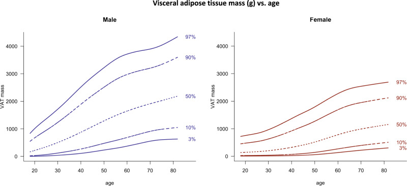
Austrian researchers scanned 10,894 randomly selected adults aged 18 to 81 with a GE Lunar Prodigy DXA scanner. They published percentile charts for fat mass, lean mass, fat distribution and visceral adipose tissue (VAT), the fat packed around the abdominal organs. Men carry roughly twice the visceral fat of women at most ages. VAT rises about fivefold in both sexes between the twenties and the seventies and does not plateau in old age. Appendicular lean mass, a proxy for limb muscle, falls in men after 50 but stays flat in women. The paper also gives lean mass reference values adjusted for how much fat a person carries. This is a normative reference study. It tests no intervention and measures no health outcomes.

Step on a bathroom scale at 25 and again at 75, and an average Austrian man will see about 7 kg of difference. A DXA scan tells a different story. Over the same span, the fat wrapped around his liver, intestines and other abdominal organs rises from about 425 g to just over 2 kg. That is close to a fivefold increase hidden inside a 9 percent change in body weight.

This is the headline from the LEAD cohort, a population study from Vienna that recruited adults at random from the Austrian residents’ register. Between 2011 and 2019, 10,894 people aged 18 to 81 had whole-body DXA scans on the same machine. The researchers then built percentile curves, similar to growth charts for children, showing where any adult sits relative to peers of the same age and sex.

The visceral fat curves matter most. Visceral fat is metabolically active tissue. Earlier research has linked it with insulin resistance, type 2 diabetes, high blood pressure, heart disease and death from all causes, more strongly than total body fat or BMI. In LEAD, visceral fat climbed across every age group in both sexes, with no plateau after 70. A smaller multicentre study had reported a decline in the oldest group, and the Austrian data contradict it.

The timing differs by sex. Men gain visceral fat fastest between their thirties and forties. Women gain it fastest between their fifties and sixties, which lines up with the menopausal transition. The researchers did not record menopausal status, so that link is an inference, not a finding. At every age, men carried about twice as much visceral fat as women, while women carried more total fat, most of it on the hips and thighs.

Muscle tells a quieter story. Limb lean mass in men held steady until about 50 and then fell about 7 percent by the oldest group. In women it barely moved across the adult lifespan. The paper also makes a useful methodological point: people with more fat also carry more lean tissue, because a heavier body needs more muscle to move it. To address this, the team published lean mass percentiles stratified by fat level. A heavy person with “average” muscle may actually be under-muscled for their size.

There is a trap in how charts like these get used. A reference range describes what is typical, not what is healthy. The median 70-year-old Austrian man carries around 2 kg of visceral fat, and in a population where cardiometabolic disease is common, the average is not a target. The numbers are also specific to one scanner model and software. A result from a GE iDXA or a Hologic machine cannot be dropped onto these curves.

The study’s value is practical. Anyone getting a DXA scan on a Lunar Prodigy now has a large, well-sampled European yardstick for visceral fat and muscle. The study cannot say whether changing your position on the chart changes your future. That question needs longitudinal and interventional data this paper does not have.

Actionable Insights

This paper tests no treatment, so every practical message below is an inference from population patterns, not proof that acting on it helps.

  1. Body weight is a poor tracker of the fat that matters. In men, weight rose 9 percent between the youngest and oldest groups, but visceral fat rose 380 percent. In women, weight rose 15 percent and visceral fat rose about 390 percent. As a standardized effect size, the age difference in visceral fat is huge: roughly 2.4 standard deviations in men and 1.9 in women. For comparison, 0.8 is usually called a “large” effect.
  2. If you get a DXA scan, visceral fat is probably the most informative single number. Compare it only against references from the same scanner model.
  3. Men should start watching early. The steepest rise happens between the thirties and forties, about 480 g per decade.
  4. Women should watch the fifties and sixties, when the rise accelerates to about 300 g per decade.
  5. Men lose about 7 percent of limb lean mass after 50, a moderate effect of about 0.66 standard deviations. Resistance training is the obvious countermeasure, though this study does not test it.
  6. Spread is enormous. At 60, men range from roughly 0.8 kg to 3 kg of visceral fat between the 10th and 90th percentiles. Age is not destiny.

Context and Source

  • Full title: Reference values of body composition parameters and visceral adipose tissue (VAT) by DXA in adults aged 18–81 years: results from the LEAD cohort
  • Institution: Ludwig Boltzmann Institute for COPD and Respiratory Epidemiology (Vienna), with Otto Wagner Hospital, Sigmund Freud University, NUTRIM at Maastricht University Medical Center, ETH Zurich and CIRO
  • Country: Austria (cohort and lead institution), with Dutch and Swiss collaborators
  • Journal: European Journal of Clinical Nutrition, 2020, volume 74, pages 1181 to 1191
  • Impact evaluation: The journal’s 2025 Journal Impact Factor is 3.2, with a 5-year JIF of 4.1. The impact score of this journal is 3.2, evaluated against a typical high-end range of 0 to 15 for nutrition and dietetics journals (0 to 60+ for top general science), therefore this is a Medium impact journal.

Related reading

What your visceral fat says about your body composition and metabolic health:

50–100 g → Exceptionally low

Very lean and low body fat overall. Peak metabolic health. Top 1-3% for men and women aged 20-30.

101–200 g → Very low

A lean profile. Great metabolic health and insulin sensitivity. Top 10% for men around 30 and women around 50.

201–300 g → Low

Still lean and insulin sensitive. Top 10% for men around 40, average for women aged 18-29.

301–400 g → Low to moderate

Still below the 425g average for men aged 18-29. For a woman in her 20s, higher than average.

401–500 g → Moderate

Average amount of visceral fat for men. At age 50, top 10% for men, but highest 10% for women. Can still have normal metabolic health and insulin sensitivity.

501–700 g → Increasing accumulation

Suboptimal insulin sensitivity and metabolic health. For men in their 60s, top 10%. For women, middle of her age group.

701–1,000 g → Poor metabolic health territory

Early signs of insulin resistance and poor glucose control. More common in people over 70. In young people, this occurs mostly in people with obesity.

Over 1,000 g → High visceral fat burden

This corresponds to obesity, insulin resistance, and metabolic syndrome. More common in older than younger people.

Men tend to carry more visceral fat than women, and visceral fat generally increases with age in both sexes.

Consensus position

A 2019 position statement in Lancet Diabetes and Endocrinology from the International Atherosclerosis Society and the International Chair on Cardiometabolic Risk treats visceral and ectopic fat as core drivers of atherosclerosis and cardiometabolic disease. It still stops short of endorsing a universal VAT cutoff. The Dallas group’s framing is that VAT is a pathological depot that accumulates when subcutaneous storage is overwhelmed, is more prone to lipolysis, and secretes more inflammatory cytokines. thelancetjacc

A refinement worth tracking is that liver fat may carry as much signal as VAT, or more. In combined Dallas Heart Study and UK Biobank data, people with high VAT had more atherosclerosis whether their liver fat was high or low, while low VAT with high liver fat did not. The two depots are partly separable, and DXA sees only one of them

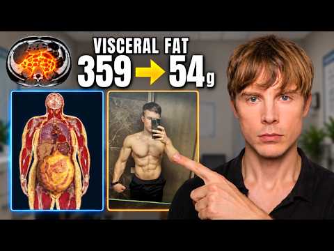
How I Lost 85% of My Visceral Fat (Not Basic Weight Loss)

I. Executive Summary

This presentation delivers a clinical and mechanistic blueprint for reducing visceral adipose tissue (VAT) from standard baseline ranges to sub-100 gram thresholds. Visceral adiposity represents a pathogenic depot surrounding internal organs that strongly correlates with cardiometabolic disease, independent of subcutaneous fat volume. Moving from lean status to minimal visceral adiposity relies on distinct physiological levers rather than gross caloric restriction. Caloric deficits demonstrate diminishing returns for visceral mobilization beyond 500 to 700 kcal per day, whereas targeted exercise regimens maintain a dose-dependent relationship with intra-abdominal lipid mobilization.

The primary physical intervention combines Zone 2 low-intensity steady-state cardiovascular conditioning (60% to 70% maximum heart rate) and High-Intensity Interval Training (HIIT) with moderate resistance training. Aerobic modalities consistently outperform isolated resistance regimens in depleting VAT by elevating systemic lipid oxidation and metabolic turnover. Nutritional strategies prioritize high total polyphenol density (green tea catechins like EGCG, coffee chlorogenic acids, anthocyanin-rich fruits, walnuts, and duckweed) along with elevated soluble and insoluble fiber (50+ grams per day). High-carbohydrate intakes (300+ grams per day) do not impede visceral lipid clearance provided energy balance and whole-food micronutrient composition are preserved.

At the cellular signaling level, manipulating amino acid availability—specifically optimizing the dietary glycine-to-methionine ratio—serves as an endocrine and hepatic modulator. Restricting methionine intake to approximately 1.4 to 1.6 grams of total protein per kilogram of body weight, combined with supplemental glycine (10 grams per day), mimics caloric restriction phenotypes, promotes hepatic lipid clearance, and enhances phase II conjugation pathways without compromising lean mass. Quantitative progress requires imaging modalities such as Dual-Energy X-ray Absorptiometry (DEXA) or Magnetic Resonance Imaging (MRI), with waist circumference serving as an accessible proxy metric.

II. Insight Bullets

  • Visceral adipose tissue wraps internal organs and exhibits markedly higher inflammatory and cardiometabolic pathogenicity than subcutaneous adipose tissue.
  • Transitioning from lean body composition to minimal visceral adiposity requires targeted metabolic interventions rather than severe caloric restriction.
  • Caloric deficits show a plateau effect for visceral fat depletion around 500 to 710 kcal per day (3,500 to 5,000 kcal per week).
  • Exercise expenditure maintains a linear, dose-dependent relationship with visceral fat mobilization without the early plateau seen in dietary caloric deficits (Ismail et al., 2012).
  • Aerobic exercise and High-Intensity Interval Training (HIIT) are superior to isolated resistance training for direct intra-abdominal fat clearance (Ismail et al., 2012).
  • Combined exercise programming utilizing Zone 2 endurance base training with 4x4 interval protocols accelerates metabolic flexibility and mitochondrial lipid utilization.
  • Green tea catechins, particularly epigallocatechin gallate (EGCG), augment exercise-induced abdominal fat reduction through AMP-activated protein kinase (AMPK) activation (Maki et al., 2009).
  • Daily intake of 1 to 3 cups of green tea provides bioactive polyphenol levels capable of accelerating total fat oxidation during exercise.
  • Coffee consumption delivers thermogenic caffeine alongside chlorogenic acid polyphenols that assist visceral fat oxidation.
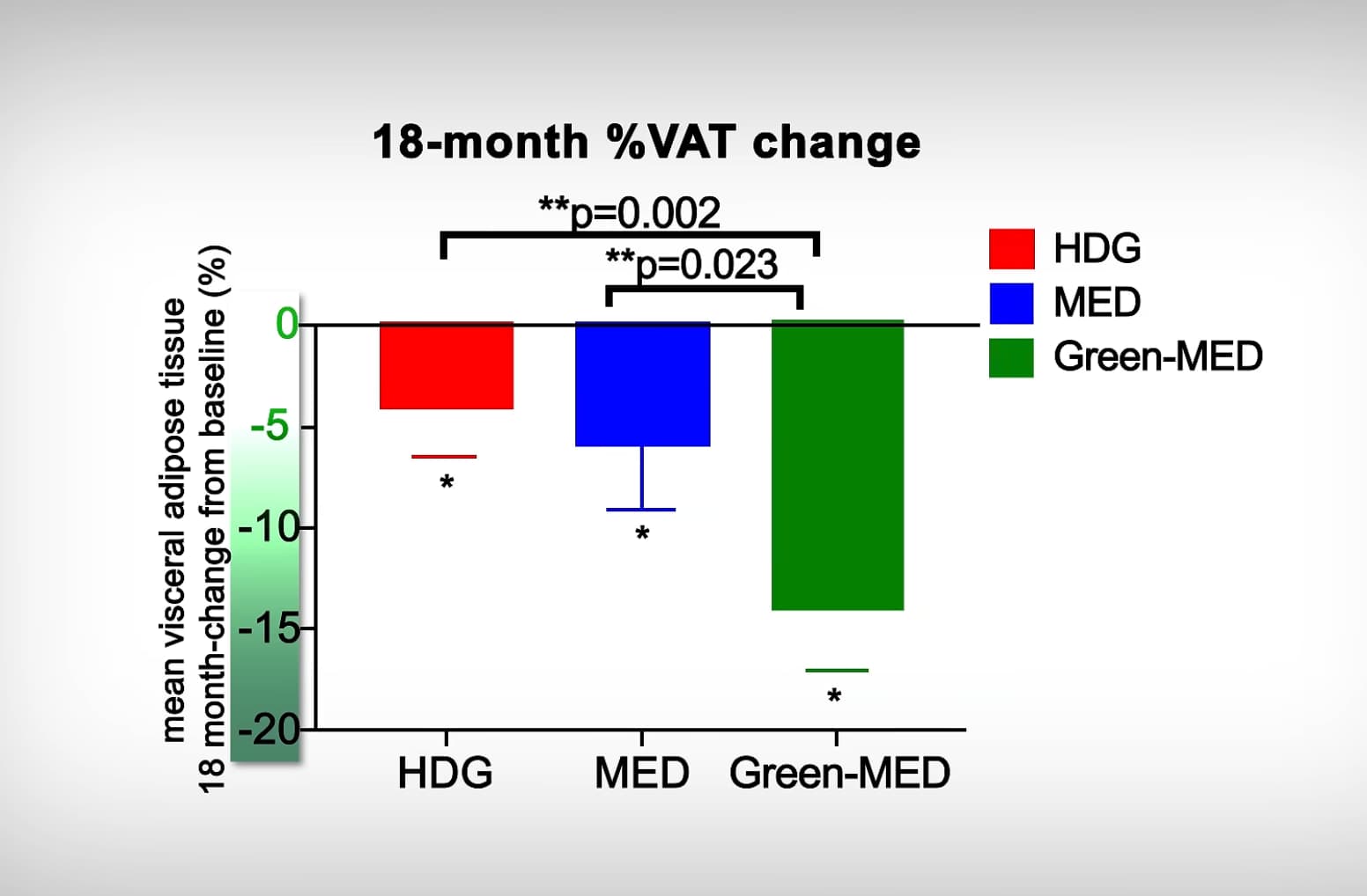
  • The 18-month DIRECT-PLUS randomized controlled trial demonstrated that a high-polyphenol Mediterranean diet drives double the visceral fat loss of standard Mediterranean diets (Zelicha et al., 2022).
  • Incorporating 100 grams of Wolffia globosa duckweed shakes was utilized in clinical trials to achieve high daily polyphenol targets (Zelicha et al., 2022).
  • Elevating dietary fiber intake to 50 grams or more daily supports gut microbial diversity, short-chain fatty acid generation, and visceral fat reduction.
  • High carbohydrate intake (300+ grams per day) does not impede visceral fat loss when derived from unprocessed whole-food matrices.
  • Isocaloric trials demonstrate comparable visceral fat reduction between low-carbohydrate and high-carbohydrate diets when calories and protein remain controlled.
  • Low circulating plasma glycine correlates with insulin resistance, non-alcoholic fatty liver conditions, and elevated visceral adiposity (Alves et al., 2019).
  • Dietary methionine restriction elevates whole-body fat oxidation and clears intrahepatic lipids independent of weight loss in metabolic syndrome cohorts (Plaisance et al., 2011).
  • Supplemental glycine functions as a biochemical mimetic of methionine restriction by enhancing metabolic conjugation and one-carbon cycle flux.
  • Moderating daily protein intake to 1.4–1.6 g/kg avoids excessive methionine consumption while fully sustaining skeletal muscle protein synthesis.
  • Supplementing 10 grams of glycine daily helps optimize the glycine-to-methionine balance in athletic individuals consuming animal proteins.
  • Magnetic Resonance Imaging (MRI) serves as the gold-standard diagnostic tool for visceral adipose measurement, followed closely by DEXA imaging.
  • DEXA scan measurements generally match MRI visceral fat estimates within an acceptable margin of 50 to 100 grams.
  • Age-adjusted target values place optimal visceral fat in the bottom 10th percentile, corresponding to roughly 100 grams for young adult males.
  • Waist circumference provides a cost-effective surrogate metric for visceral adiposity, targeting sub-90 cm for men and sub-75 cm for women.
  • Waist circumference measured at the umbilicus linearly tracks cardiovascular morbidity and all-cause mortality risk.
  • Outward leanness (visible abdominal musculature) can conceal metabolically active visceral fat depots unless quantified via objective imaging.