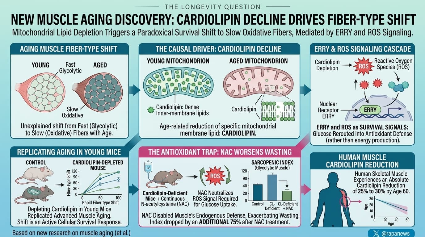
Researchers at the University of Copenhagen report that cardiolipin declines in aging mouse muscle, and in a small set of human biopsies. Cardiolipin is a lipid found almost exclusively in the inner mitochondrial membrane, and the gene for the enzyme that makes it, CRLS1, falls with age as well. Deleting Crls1 in mouse skeletal muscle reproduced several features of muscle aging, most notably a shift from fast glycolytic fibers toward slower oxidative fibers. That shift is relayed to the nucleus by the receptor ERR-gamma, and it diverts glucose into antioxidant pathways. High-dose N-acetylcysteine (NAC) worsened muscle loss rather than helping. Partially restoring cardiolipin reversed weight loss and prevented premature death.
Muscle ages in a way that has long looked contradictory. The mitochondria that power our cells work less well as we get older, yet aging muscle drifts toward slow-twitch fibers, the type most packed with mitochondria. If the power plants are failing, why would muscle lean harder on the fibers that need them most?
A team led by Fabian Finger and Zachary Gerhart-Hines at the University of Copenhagen thinks the answer lies in cardiolipin. It is an unusual four-tailed fat found almost only in the inner membrane of mitochondria. Cardiolipin shapes the folded cristae where energy is made and helps the respiratory machinery lock together into efficient units. In mice, cardiolipin in leg muscle fell by roughly a quarter between young adulthood and two years of age. Expression of Crls1, the gene for the enzyme that makes it, fell too. A handful of human biopsies pointed the same way.
To test whether the loss matters, the researchers deleted Crls1 only in skeletal muscle. The mice failed to build muscle and ended up about 40 percent lighter than their littermates. Their mitochondria had collapsed cristae and produced less energy per unit. Most strikingly, their fast, sugar-burning muscles began to look like slow, fat-burning ones. They doubled their mitochondrial content and shed the most glycolytic fiber type.
The relay turned out to be a nuclear receptor called estrogen-related receptor gamma, or ERR-gamma, which despite its name does not respond to estrogen. When cardiolipin fell, ERR-gamma rose. In cultured muscle cells, blocking it prevented the fiber switch and cancelled around 80 percent of the gene changes triggered by cardiolipin loss. ERR-gamma was also elevated in the fast muscle of old mice.
Why would muscle make this switch? The team’s answer is defense. Damaged mitochondria leak reactive oxygen species. The remodeled muscle pulled in two to four times more glucose, then diverted much of it into side pathways that build antioxidant capacity instead of burning it for fuel. The knockout mice even cleared blood sugar better than normal mice.
That idea produced the study’s most provocative result. Giving the mice the antioxidant N-acetylcysteine in their drinking water made their muscle wasting worse, not better. The authors argue that mopping up oxidants removed the very signal the muscle was using to protect itself.
There was also a hint of reversibility. When the researchers stopped triggering the deletion in adult mice, muscle stem cells gradually supplied fibers with working copies of the gene. Cardiolipin climbed from 27 to 67 percent of normal, and weight loss partly reversed. Every recovering mouse survived, while more than half of those left deficient died.
The caveats are large. The genetic model removes about 85 percent of muscle cardiolipin, three to four times what aging does. Its mice die young from failing breathing muscles, not from old age. The human data come from nine men. Whether topping up cardiolipin in normally aging muscle would help anyone is untested. What the work does offer is a plausible reason why old muscle shifts the way it does.
Actionable Insights
This is a mouse mechanism paper, so the practical lessons are indirect.
- Be cautious with megadose antioxidants. The mice received NAC at roughly 2,500 to 3,000 mg per kg per day. That is a human-equivalent dose of about 14 to 18 grams daily, around ten times common supplement doses. It deepened muscle loss by about a third in the slow calf muscle and by about half in the fast one. This fits human trials showing that antioxidant pills can blunt training gains. Ordinary doses were not tested.
- Cardiolipin decline with age appears real but modest. In humans, the synthase gene was about 40 percent lower in older muscle. That is a large effect on paper (Cohen’s d around 2.5, where 0.8 already counts as large), but it comes from only nine people, so the true effect could be much smaller.
- The rescue numbers look dramatic, but they come from a severe genetic disease model. Restoring cardiolipin from 27 to 67 percent of normal took survival from about 44 to 100 percent. Human skeletal muscle experiences an absolute cardiolipin reduction of 25% to 30% by age 60.
- Do not buy cardiolipin supplements or grey-market ERR agonists based on this paper. The one cardiolipin-binding drug, elamipretide (SS-31), received FDA accelerated approval in September 2025 for improving muscle strength in Barth syndrome. Early review of the evidence suggests that SS-31 may be helpful to maintain muscle cardiolipin, but does not replace cardiolipin, so would not reverse the problem identified in this paper.
- Exercise remains the best-supported way to build healthy muscle mitochondria.
Context/Source
- Open Access Paper: Mitochondrial membrane lipid cardiolipin controls fiber-type adaptations in aging muscle via estrogen-related receptor gamma
- Institution: Novo Nordisk Foundation Center for Basic Metabolic Research, University of Copenhagen (lead). Collaborators include the University of Southern Denmark, University of Utah, MRC Mitochondrial Biology Unit Cambridge, Princeton, Amsterdam UMC, Karolinska Institute and the German Institute of Human Nutrition.
- Country: Denmark
- Journal: Nature Aging (Springer Nature).
- Journal Impact Factor: The impact score of this journal is 25.0, evaluated against a typical high-end range of 0-60+ for top general science, therefore this is a High impact journal.