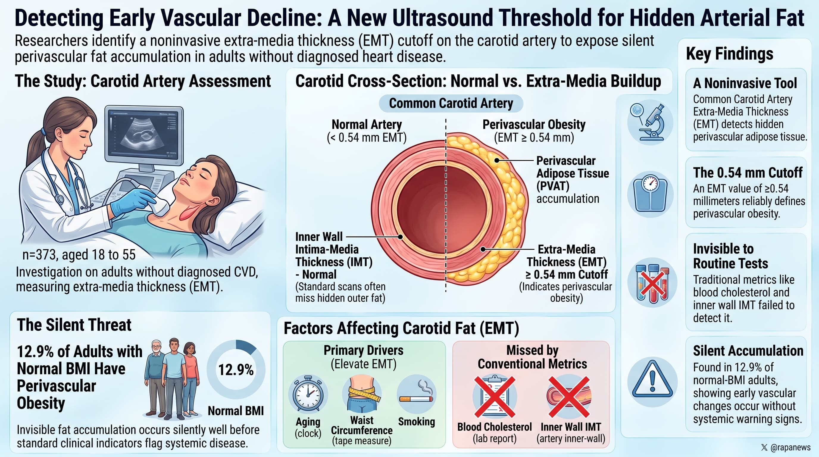
Researchers investigating early cardiovascular decline have identified a noninvasive ultrasound threshold for common carotid artery extra-media thickness, exposing hidden perivascular adipose tissue accumulation in adults without diagnosed cardiovascular disease. By evaluating 373 participants aged 18 to 55, investigators established that an extra-media thickness equal to or exceeding 0.54 millimeters marks perivascular obesity. This outside-in arterial expansion was driven by advancing age, waist circumference, and smoking, while conventional metrics like blood cholesterol and inner wall intima-media thickness failed to detect it. Crucially, 12.9 percent of individuals with normal body mass index exhibited perivascular obesity, demonstrating that localized vascular fat accumulation can occur silently well before standard clinical indicators flag systemic disease.
Most people have a rough idea of their waist size. Far fewer have thought about the fat that wraps around their arteries. This perivascular adipose tissue sits directly against the outer wall of blood vessels. Unlike the fat under the skin, it communicates chemically with the vessel it surrounds. In lean, healthy people it appears to help arteries relax. When it expands and becomes inflamed, that relationship can turn harmful, and a growing body of research links dysfunctional perivascular fat to stiffer, more inflamed arteries and to atherosclerosis.
Measuring this fat is the hard part. CT and MRI can do it, but they are expensive, not always available, and CT involves radiation. A team at Sechenov University in Moscow wanted to know whether a routine neck ultrasound could stand in for those scans. Their measurement, carotid extra-media thickness, captures the layer outside the muscular middle of the carotid artery wall: the artery’s tough outer coat plus the fat around it.
The researchers scanned 373 adults aged 18 to 55 with no known heart or vascular disease. Most were young, with a median age of 23. Thicker EMT went along with older age, higher blood pressure, larger waists and BMI, higher blood sugar and smoking. When these factors were analyzed together, age, obesity and smoking remained independently linked to EMT.
To set a cutoff, the team selected 185 participants with none of the usual risk factors and found the value exceeded by only the top 10 percent of them: 0.54 mm. Anyone at or above that line was labeled as having “perivascular obesity.” By that definition, roughly a quarter of the whole sample qualified. That included nearly two-thirds of people aged 41 to 55 and about one in eight people of normal weight.
That last figure carries the authors’ main message: a hidden fat problem that the bathroom scale cannot see. The idea is appealing, but the evidence is weaker than it looks. A cutoff set at the 90th percentile of healthy people will flag about one in ten healthy people by design. Finding 13 percent in the normal-weight group is close to what the method would produce on its own.
The age finding has a similar problem. The reference group was dominated by people in their twenties, and EMT rises steeply with age, so a single cutoff drawn from that group will flag most middle-aged people almost automatically. The outer arterial coat also thickens and stiffens with age whether or not fat accumulates, so EMT may be partly a measure of vascular aging rather than fat.
Several numbers in the paper do not agree with each other. The study did not check EMT against CT or MRI fat measurements in the same people, reported no data on scan reproducibility, and included no follow-up to show that a high EMT predicts heart attacks, strokes or anything else.
The underlying question is still worth pursuing. A cheap, radiation-free marker of vessel-hugging fat could help identify risk that BMI misses. This study offers a starting number, not a validated test.
Actionable Insights
This was a one-time snapshot with no treatment arm, and it reports associations only, and their size is worth knowing.
- Waist size was the only body measurement that stayed independently tied to crossing the 0.54 mm line. Each one-standard-deviation increase in waist (roughly 10 to 14 cm, estimated from the reported ranges) came with about 2.8 times the odds. For comparison, being one standard deviation older (roughly 8 to 10 years) came with about 5 times the odds.
- Obesity added about 0.22 mm to EMT on average. At the reported age slope of 0.006 mm per year, that equals about 36 years of age-related thickening. This figure is hard to reconcile with other data in the paper.
- Smoking added about 0.05 mm, roughly 9 years’ worth, and the result was borderline.
- Body-size measures correlated with EMT at about 0.4, so they account for only around 16 percent of the variation between people.
Practical takeaway: track your waist, not just your weight, and do not smoke. Do not pay for an EMT scan expecting a meaningful answer, because the 0.54 mm threshold has not been validated against any health outcome.
Context and Source
- Open Access Paper: Common Carotid Artery Extra-Media Thickness as a Marker of Perivascular Obesity and Cardiometabolic Risk in Individuals Without Cardiovascular Disease
- Institution: Sechenov First Moscow State Medical University
- Country: Russia
- Journal: Journal of Clinical Medicine Research (Elmer Press, Canada), 2026
- Impact evaluation: The impact score of this journal is 2.2, evaluated against a typical high-end range of 0-90+ for top general and internal medicine journals, therefore this is a Low impact journal.