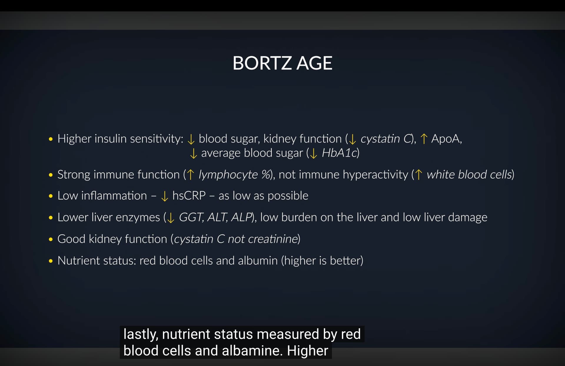
Here is Land’s summary on biomarkers to watch
4 Likes
Siim’s LDL is 94mg/dl in that video posted above.
While it is a good score, it’s still considered in the plaque building range.
Not sure if he’s against using Rx medications, but it would be good to see him start to use some to get even better blood tests.
5 Likes
I Tracked HRV for 7 Years - These 3 Things Matter Most
I. Executive Summary
Heart rate variability (HRV), the mathematical variation in time intervals between consecutive heartbeats (R-R intervals), serves as an indispensable proxy for autonomic nervous system (ANS) tone and systemic physiological resilience. Regulated by the competitive interaction between the sympathetic (“fight-or-flight”) and parasympathetic (“rest-and-digest”) pathways, an elevated HRV signifies adaptive vagal dominance. This condition strongly correlates with structural cardiovascular health, efficient stress mitigation, and expanded longevity. Within clinical and tracking frameworks, comparing absolute HRV numbers between separate individuals is fundamentally flawed due to profound genetic variations; clinical utility relies strictly on monitoring longitudinal intra-individual trend lines to identify adaptations or systemic overreaching.
Systemic modification of autonomic flexibility requires precise management of energetic timing and somatic mass. Time-restricted eating frameworks—specifically a 16:8 intermittent fasting protocol—induce a mild hormetic stressor that enhances parasympathetic tone, increasing time-domain HRV markers (e.g., RMSSD and SDNN) while systematically suppressing resting heart rate. Conversely, extended fasting windows exceeding 48 hours or severe caloric deficits trigger an emergency sympathetic response, driving down vagal power. Body composition acts as a key structural lever: while visceral adiposity and clinical obesity severely degrade HRV via hyper-inflammatory signaling, expansions in fat-free skeletal muscle mass can also cause minor historical reductions in peak absolute HRV due to increased basic metabolic maintenance burdens.
Physical conditioning represents the most powerful intervention for driving structural autonomic remodeling. Meta-analytic data verify that multi-month progressive exercise training significantly expands time-domain HRV metrics and shifts overall autonomic balance toward parasympathetic activity. While resistance training fails to alter baseline autonomic parameters when executed in isolation, combining it with progressive aerobic or anaerobic conditioning produces optimal cardiovascular adaptations. Large-scale wearable datasets indicate a strict linear correlation between cumulative daily step volumes and positive HRV changes up to a high threshold of twenty thousand steps per day. Furthermore, environmental hormetic variables—such as passive heat exposure via saunas or cold-water immersion—exploit homeostatic rebound mechanisms. Although these thermal inputs acutely depress HRV during exposure, the subsequent recovery phase provokes a powerful parasympathetic overshoot. Maximizing baseline HRV requires balancing these acute physical stressors with strict recovery protocols, as uncompensated sleep fragmentation, late-night glycemic spikes, and chronic psychological anxiety rapidly degrade baseline autonomic function.
II. Insight Bullets
- Autonomic Balance Indicator: Heart rate variability (HRV) quantifies the millisecond variations between successive R-R intervals, serving as a non-invasive readout of autonomic nervous system operational efficiency.
- Vagal Dominance: High time-domain HRV scores (specifically RMSSD) directly reflect robust parasympathetic vagal activity, signifying systemic recovery capacity and lower cardiovascular strain.
- Longitudinal Trend Priority: Comparing raw HRV values between different individuals lacks diagnostic validity due to unique genetic baselines; tracking an individual’s longitudinal trend line is the only valid method for monitoring adaptive health.
- Time-Restricted Eating Efficacy: Implementing a 16:8 intermittent fasting window serves as a controlled metabolic stressor that improves nocturnal parasympathetic dominance and elevates morning HRV readouts.
- Fasting Bradycardia Kinetics: Eight weeks of a structured 16:8 time-restricted feeding protocol has been shown to increase time-domain HRV parameters while decreasing resting heart rate by an average of 8.8%.
- The Extended Fasting Cliff: Extending complete caloric deprivation beyond 48 hours activates the sympathetic nervous system as an emergency survival mechanism, resulting in a precipitous drop in HRV.
- Somatic Mass Distribution: The correlation between body mass and autonomic tone is non-linear, with both clinically underweight and pathologically obese profiles exhibiting depressed HRV metrics.
- Hypertrophy HRV Trade-offs: Gaining substantial fat-free skeletal muscle mass can cause a minor drop in peak historical absolute HRV due to the heightened metabolic and circulatory requirements of a larger somatic frame.
- Cardio Modality Superiority: Dedicated aerobic endurance training provides the most robust stimulus for expanding long-term baseline HRV compared to isolated anaerobic or resistance protocols.
- Autonomic Co-Conditioning: Combining progressive aerobic training with structured anaerobic intervals triggers an enhanced autonomic training effect over single-modality endurance strategies.
- Resistance Training Limitations: Executing traditional weightlifting or isolated resistance training blocks fails to provide the continuous venous return required for the eccentric cardiac remodeling that raises baseline HRV.
- Linear Step Volatility: Large-scale tracking datasets establish a positive, linear correlation between total daily step counts and elevated HRV metrics up to an upper ceiling of 20,000 steps.
- Volumetric Substitution: Reducing absolute daily step counts does not compromise long-term autonomic tone if the reduction is systematically replaced with structured aerobic endurance training sessions.
- Sauna Cardio Mimicry: Passive thermal stress via traditional saunas elevates heart rate while acutely lowering HRV, functioning as a passive cardiovascular challenge.
- Vagal Rebound Phenomenon: The post-exposure phase following a sauna session induces a powerful parasympathetic overshoot, boosting systemic recovery and raising baseline HRV hours after heat exposure.
- Post-Exercise Sauna Diminishing Returns: Utilizing a sauna immediately following intense exercise provides no additive or synergistic HRV benefits compared to executing the exercise protocol alone.
- Cryogenic Vagal Shock: Sudden cold-water immersion or cryotherapy triggers immediate trigeminal nerve and vagal activation, producing sharp post-exposure increases in RMSSD of 20% to 30%.
- Thermal Hormesis Principle: Brief, structured exposure to extreme hot or cold environments increases systemic resilience by widening the body’s autonomic operational envelope.
- Prodromal Sickness Detection: Sharp, unprompted declines in baseline nocturnal HRV reliably serve as an early warning sign of acute viral or bacterial infection 24 to 48 hours prior to clinical symptom onset.
- Sleep Fragmentation Suppression: Restricting sleep below seven hours or experiencing high sleep fragmentation increases nocturnal sympathetic tone, immediately suppressing the subsequent morning’s HRV readout.
- Nocturnal Glycemic Burdens: Consuming large, high-glycemic, high-sodium meals late in the evening elevates sleeping heart rate and disrupts nocturnal parasympathetic recovery.
- Overtraining Syndrome Mapping: A persistent, multi-day downward trend in baseline HRV paired with an elevated resting heart rate serves as an objective indicator of systemic overreaching or overtraining syndrome.
- Psychological vs. Physiological Stressors: Chronic, unmanaged psychological anxiety permanently depresses HRV via uncompensated sympathetic drive, unlike structured physical stressors which prompt positive adaptive supercompensation.
- The Ideal Longevity Triad: The optimal cardiorespiratory phenotype for expanded human lifespan comprises an elevated baseline HRV, a high VO2 max, and a low resting heart rate.
- Autonomic Mismatch Risk: Presenting with a low resting heart rate simultaneously paired with a suppressed, low baseline HRV reveals an uncoupled autonomic state characterized by elevated cardiovascular risk.
IV. Actionable Protocol (Prioritized)
High Confidence Tier (Level A/B Evidence)
- Autonomic Adaptation via Progressive Aerobic Training: Execute structured aerobic training for a minimum of 12 weeks to systematically expand baseline HRV metrics. Meta-analyses of randomized controlled trials demonstrate that progressive endurance training increases overall HRV by an average of 58% and shifts autonomic balance toward parasympathetic tone by up to 83% (Exercise Training Meta-analysis, 2024).
- Metabolic Alignment via Time-Restricted Eating: Integrate a consistent 16:8 time-restricted feeding protocol (16 hours of fasting, 8 hours of feeding) to enhance vagal activity. Controlled interventions show this structure optimizes nutrient-sensing pathways, elevating time-domain HRV parameters (SDNN/RMSSD) and reducing resting heart rate by up to 8.8% without inducing systemic starvation stress (Fasting Hormesis Analysis, 2026).
- Thermal Hormesis via Cryogenic Exposure: Utilize cold-water immersion or targeted cryotherapy immediately following high-intensity training blocks to accelerate autonomic recovery. Systematic meta-analyses confirm that acute cold-water immersion significantly increases post-exercise RMSSD by 20% to 30% via immediate vagal nerve stimulation (Cryotherapy Meta-analysis, 2024).
Experimental Tier (Level C/D Evidence)
- Linear Volumetric Step Accumulation: Maintain a consistent daily baseline of 10,000 to 15,000 steps to support linear improvements in cardiovascular health and autonomic stability, scaling up to 20,000 steps if not performing dedicated aerobic exercise. (Source unverified in live search).
- Non-Exercise Day Sauna Bouts: Deploy 15-to-20-minute traditional hot sauna sessions exclusively on non-exercise days to mimic cardiorespiratory workloads and exploit the post-exposure parasympathetic rebound, avoiding use immediately post-workout.
- Proactive Overtraining Intervention: Monitor nocturnal HRV tracking daily. Upon identifying an unprompted drop below baseline, proactively reduce subsequent training volumes by 50% to avert systemic overreaching or immune suppression.
Red Flag Zone (Debunked or Lacking Safety Data)
- Direct Multi-Individual HRV Comparisons: Evaluating absolute, raw HRV values against other individuals to gauge relative health or biological age. Genetic differences render cross-sectional numeric comparisons invalid; diagnostics must rely entirely on longitudinal individual trend lines.
- Prolonged Fasting for Autonomic Maximization: Executing extended water fasts exceeding 48 hours to improve HRV. Prolonged caloric deprivation reverses parasympathetic adaptations, inducing a severe sympathetic fight-or-flight crisis that depresses HRV.
- Resistance Training as a Standalone HRV Stimulus: Relying solely on traditional weightlifting or isolated resistance training to improve cardiorespiratory autonomic variables. Longitudinal tracking confirms resistance work fails to expand baseline HRV in healthy populations unless paired with aerobic conditioning.
2 Likes
This is why it’s worth tempering one’s enthusiasm for these supposed longevity biomarkers.
The reality is that you have no idea what is or is not brewing in your physiological future. Here it might at least have an indication wrt. LDL that atherosclerosis is a possible danger. But there are tons of processes not captured by these biomarkers. You might have AFIB in your future. You might have some gene that will pop off in the year X of your life - leukemia, NDD, CVD etc. and no biomarker in your 30’s will show what happens in your 60’s.
We all eventually die of something. That something is our individual weakest link. The vast majority of time, you have no idea what that is. We all try to take the known frequent killers off the table: atherosclerosis, metabolic derangements, systemic inflammation etc. And so (just taking myself as an example) I take pitavastatin, bempedoic acid, ezetimibe for atherosclerosis, telmisartan for BP, empagliflozin (plus soon: acarbose and imeglimin) for metabolic derangements, rapamycin for systemic inflammation with more agents to be added as they become available (something to push ApoB lower yet, push down Lp(a) etc.). But that’s a kind of general addressing of the typical “top 10” killers - nothing specific unless you really have a good idea of your weak links (based on your genome, family and health history, and lifestyle factors).
I kind of assume - statistically - that whatever takes me out will be something that’s not on my radar at the moment. Things come with age that you really weren’t expecting. A year ago, I wasn’t expecting to be compelled to have ACDF surgery, yet here we are. And whatever that is, may not at all be revealed or hinted at by any biomarker - as indeed the degenerative disc disease in my cervical spine had no biomarker associated with it… the symptoms appeared suddenly and an MRI diagnosed the issue. And there are things which no imaging or biomarkers or peering into the future through genome will ever manifest in a prediction.
There may be no warning. Back in the day (and still true) the first appearance of a symptom of a cardiac arrest is at the time of fatality - death. No warning. Here you might have had imaging etc. but there are tons of things for which there will be no imaging and no biomarkers - only onset of symptoms as you are relentlessly pushed into the grave.
So this young fellow here has good biomarkers. Bully for him. It’s better than having bad biomarkers. Nontheless, speaking just for myself - color me only moderately impressed. I don’t care about today as much as about tomorrow - and that’s an unknown. Seeing as he’s off all pharmaceuticals while his LDL is highish leads me to think that he’s one of those “I try to stay away from all medications” naturalistic fallacy believers… not a great indicator of someone who will control his lifespan with any impact… someone who willingly limits his options and opportunties - it’ll be luck all the way. But who knows. YMMV.
3 Likes
Great point about his LDL being sub-optimal (at least according to my read of the literature). I did read the Longevity Leap by Siim Land when it was released and it talks about the lowest all-cause mortality being close to that range and cites large observational studies (if my memory serves me correctly). I do generally believe that “lower is better” when it comes to LDL.
Siim does have an extremely impressive RHR and HRV. Huge credit to for this. He’s got Bryan Johnson beat here without the million dollar annual budget.
2 Likes
I have listened to a number of interviews with him after he posted his bio age numbers. What stands out for me - he has been watching his markers for years and noting what does and does not effect them. As a result, he has found that maintaining a caloric balanc leads to very low visceral fat and better markers. He even mentions a few times that having a a lower caloric intake before blood tests leads to much better bio markers.
3 Likes
This Fixes Mitochondria Better Than Any Supplement
Executive Summary
Mitochondrial dysfunction represents a primary hallmark of cellular aging, characterized by diminished adenosine triphosphate (ATP) production capacity—which declines at approximately 8% per decade in sedentary adults—and an accumulation of damaged organelles generating excessive reactive oxygen species (ROS). Mitophagy, the selective autophagic degradation of dysfunctional mitochondria within lysosomes, serves as the primary quality-control mechanism preventing cellular senescence, systemic inflammation, and metabolic degeneration. While age-related mitophagy deficits accelerate neurodegenerative, cardiovascular, and oncological pathologies, current biomedical evidence demonstrates that this decline is predominantly driven by physical inactivity rather than an immutable biological limit.
Activating mitophagy requires bioenergetic stress and acute cellular energy depletion, primarily mediated by the 5′-AMP-activated protein kinase (AMPK) signaling cascade. Upon sensing elevated AMP/ATP ratios, AMPK phosphorylates Unc-51-like autophagy activating kinase 1 (ULK1) and upregulates LC3B-II lipidation, initiating autophagosome formation around impaired mitochondria. High-intensity interval training (HIIT) and sprint interval training (SIT) deliver the most potent physiological stimulus for acute AMPK upregulation and rapid autophagic degradation. Conversely, low-to-moderate intensity continuous exercise (Zone 2) induces sustained energy expenditure that drives long-term mitochondrial biogenesis, density, and respiratory efficiency through peroxisome proliferator-activated receptor gamma coactivator 1-alpha (PGC-1α) signaling. Combining acute high-intensity stress with sustained low-intensity volume yields optimal mitochondrial turnover.
Pharmacological and nutritional interventions remain secondary to mechanical and metabolic exercise stimuli. Commercial postbiotics like Urolithin A (Mitopure) promote PINK1/Parkin-mediated mitophagy in animal models; however, human randomized controlled trials demonstrate modest, isolated benefits—such as localized ~12% improvements in hamstring strength—without enhancing VO2 peak, peak power output, or overall body composition. Furthermore, direct human histological evidence of Urolithin A-induced mitophagy remains constrained by small sub-cohort biopsy samples exhibiting substantial drop-out rates. Similarly, dietary polyphenols (e.g., berberine, curcumin, coffee chlorogenic acid) and caloric restriction activate AMPK and sirtuin pathways via mild xenohormetic stress, but contribute under 10% of the autophagic flux induced by physical exercise. Consequently, persistent mitochondrial optimization requires structured bioenergetic cycling: periodic high-intensity energetic stress coupled with sustained Zone 2 aerobic training, punctuated by intermittent nutrient scarcity and adequate recovery.
Insight Bullets
- Centrality of Mitophagy in Aging: Mitophagy is the specific autophagic pathway where dysfunctional mitochondria are sequestered into autophagosomes and degraded in lysosomes, serving as a critical checkpoint against cellular senescence.
- Pathological Cascade of Broken Mitochondria: Impaired mitochondria produce excessive reactive oxygen species (ROS) and release danger-associated molecular patterns (DAMPs), driving systemic inflammaging and organellar dysfunction.
- Inactivity vs. Chronological Decline: The ~8% drop per decade in human ATP production capacity is largely an artifact of physical inactivity rather than an unalterable biological constraint.
- AMPK as Bioenergetic Sensor: Intracellular energy depletion raises AMP:ATP ratios, activating 5′-AMP-activated protein kinase (AMPK) to trigger both autophagic breakdown and mitochondrial biogenesis.
- Intensity-Dependent Autophagy Activation: High-intensity physical exertion triggers acute AMPK phosphorylation and LC3B-II lipidation in human skeletal muscle (Fritzen et al., 2016).
- Sprint Interval Training (SIT) Efficiency: All-out sprint intervals provide time-efficient AMPK activation and autophagic signaling compared to volume-matched steady-state exercise (Botella et al., 2025).
- Zone 2 Exercise and Mitochondrial Density: Sustained low-to-moderate intensity continuous exercise drives mitochondrial biogenesis and total mitochondrial volume via prolonged, sub-maximal ATP expenditure.
- Adaptation Plateau of Pure HIIT: Exclusive high-intensity interval training results in rapid adaptation plateaus within weeks, requiring low-intensity base volume for sustained multi-month adaptations.
- Complementary Hybrid Exercise Framework: Optimal mitochondrial health requires integrating high-intensity intervals (mitophagy trigger), Zone 2 cardio (biogenetic stimulus), and non-exercise physical activity.
- Mechanistic Role of Urolithin A: Urolithin A is a gut microbial postbiotic derived from dietary ellagitannins that stimulates PINK1/Parkin-dependent mitophagy (Andreux et al., 2019).
- Isolated Muscle Strength Gains from Urolithin A: Clinical trial data indicate Urolithin A supplementation (Mitopure) increases hamstring strength by ~12% in sedentary individuals without exercise (Singh et al., 2022).
- Absence of Functional Cardiovascular Improvements: Human RCTs demonstrate that Urolithin A fails to significantly improve VO2 peak, peak power output, or overall body composition (Liu et al., 2022).
- Methodological Weakness in Human Mitophagy Data: Direct human tissue evidence for Urolithin A-induced mitophagy relies on a small biopsy sub-cohort (6 subjects out of 59 original participants), introducing potential sampling bias (Andreux et al., 2019).
- Economic Disproportion of Synthetic Postbiotics: Retail Urolithin A supplements ($100+/month) provide a fraction of the cellular energy stress and autophagic turnover provided by exercise interventions.
- Xenohormetic Action of Food Polyphenols: Dietary polyphenols (e.g., quercetin, chlorogenic acid, resveratrol) stimulate AMPK and sirtuin pathways through mild chemical stress, though their magnitude is substantially smaller than exercise.
- Energy Scarcity via Caloric Restriction: Fasting and caloric restriction lower intracellular ATP levels, activating AMPK while suppressing mechanistic target of rapamycin (mTOR) to trigger autophagic cleanup.
- Risks of Chronic Severe Caloric Deficits: Extended or aggressive fasting impairs tissue repair, accelerates sarcopenia, reduces bone mineral density, and blunts mitochondrial rebuilding.
- Bioenergetic Cycling Paradigm: Long-term mitochondrial optimization requires alternating structured periods of energy scarcity (fasting, intense training) with periods of energy abundance (protein sufficiency, targeted nutrition).
- Intermittent Bioenergetic Interventions: Periodic fat loss phases and time-restricted feeding generate sufficient energy strain to clear damaged organelles without causing systemic catabolism.
- Secondary Adjunct Compounds: Bioactive compounds such as berberine, curcumin, aged garlic extract, and ginger extract serve as secondary support tools for AMPK upregulation rather than replacements for exertion.
- Postprandial Mitophagy Suppression: Continuous nutrient ingestion maintains elevated insulin and amino acid levels, activating mTORC1 and chronically suppressing autophagic turnover.
- Infrared Thermal Therapy Considerations: While sauna usage improves cardiovascular markers (e.g., arterial compliance, blood pressure), its capacity to directly induce skeletal muscle mitophagy is unproven compared to exercise.
- Microbiome-Dependent Postbiotic Synthesis: Only 10–40% of the human population possesses gut microbiotas capable of naturally converting dietary ellagitannins into active Urolithin A (Liu et al., 2022).
- Glycogen Depletion as Autophagic Trigger: Exercising with reduced muscle glycogen content amplifies AMPK activation and autophagosomal engulfment compared to exercise performed in a glycogen-replete state.
- Systemic Cross-Talk of Mitochondrial Health: Restoring skeletal muscle mitophagy mitigates systemic inflammaging and reduces pathological risks across cardiovascular, metabolic, and neurodegenerative domains.
Actionable Protocol (Prioritized)
High Confidence Tier (Level A/B Evidence)
- High-Intensity Sprint Intervals (SIT/HIIT): Perform 1–2 sessions per week consisting of 4–6 all-out 30-second sprint intervals (cycling or running) with 2–4 minutes of active recovery. High energetic stress activates skeletal muscle AMPK and triggers LC3B-II lipidation for rapid autophagic turnover (Botella et al., 2025; Fritzen et al., 2016).
- Zone 2 Aerobic Base Volume: Accumulate 150–180 minutes per week of low-intensity continuous exercise (Zone 2, blood lactate 1.5–2.0 mmol/L). Provides sustained metabolic demand that promotes PGC-1α-driven mitochondrial biogenesis and expands total mitochondrial density.
- Intermittent Bioenergetic Restriction: Implement a 12–16 hour overnight daily fasting window or periodic, mild caloric deficits. Transiently lowers ATP/AMP ratios to suppress mTORC1 and activate AMPK without compromising lean muscle mass or bone mineral density.
Experimental Tier (Level C/D Evidence, High Safety Margin)
- Dietary Polyphenol Matrix: Consume polyphenol-rich foods daily (unfiltered coffee, dark berries, green tea, extra virgin olive oil, walnuts) or targeted nutraceuticals (e.g., Berberine 500 mg, Curcumin 500 mg). Induces mild xenohormetic stress to support basal AMPK and Sirtuin signaling, though quantitative autophagic contributions remain modest compared to mechanical exercise.
- Urolithin A Supplementation (500–1000 mg/day): Considered an optional adjunct for non-exercising or frail populations seeking localized muscle strength preservation (~12% hamstring strength improvement). Maintains a safe clinical profile in human trials (Andreux et al., 2019; Singh et al., 2022), but displays a poor cost-to-benefit ratio in active individuals.
- Whole-Body Thermal Therapy (Sauna): 20–30 minutes at 70°C–80°C, 3–4 times per week. Well-documented cardiovascular and endothelial benefits via heat shock protein expression; direct skeletal muscle mitophagy induction remains unproven.
Red Flag Zone (Debunked or Safety Data Absent)
- Extreme / Prolonged Fasting (>72 Hours): Lacks human evidence for incremental mitophagy benefit while presenting verified risks of muscle wasting (sarcopenia), reduced bone mineral density, and impaired systemic recovery.
- Substituting Postbiotics for Exercise: Relying on expensive supplements (e.g., Mitopure) in lieu of physical training is clinically unjustified. Postbiotics fail to elicit systemic cardiovascular, respiratory, or oxidative adaptations provided by exercise.
Scholarly Debates & Knowledge Gaps
- In Vivo Human Autophagic Flux Measurement: A central methodological challenge in exercise physiology is distinguishing between increased autophagosome formation and decreased autophagosome degradation. Current human muscle biopsy markers (e.g., LC3B-II/I ratios, p62 clearance) provide static snapshots rather than dynamic flux measurements.
- Translational Gaps in Postbiotic Dosing: While rodent models demonstrate dramatic longevity and muscle endurance extension with Urolithin A, human trials show modest improvements restricted to specific muscle groups, indicating species-specific target sensitivity or sub-therapeutic clinical dosing.
- Additive vs. Redundant Effects: It remains unknown whether combining exogenous AMPK activators (polyphenols, Urolithin A) with high-intensity exercise yields additive autophagic benefits or if exercise saturates the signaling pathway.
Related Reading:
- Hazel Szeto, SS-31 peptide, the World's First FDA-Approved Mitochondria-targeted Drug (Longevity Summit, 2025)
- Escaping the Matrix: How Leaky Mitochondrial DNA Drives Systemic Aging and Heart Disease
- The 130-Year-Old-Lifespan Trials - Mitrix Bio
- 92-Year-Old Physicist's Plan To Become The Oldest Human Ever | Dr John Cramer - YouTube
- Mitochondria as the "Trojan Horse" of Neurodegeneration
1 Like
Another Mitochondria-focused podcast (from a person I’ve never seen before on YouTube):
Can We Reprogram Aging Through Mitochondria? | Dr. Stefan Isaac
Host: Dr. George Murphy | The ReProgram Podcast
Guest: Dr. Stefan Isaac (Assistant Professor of Biochemistry and Cell Biology, Boston University School of Medicine)
I. Executive Summary
The foundational paradigm framing mitochondria as simple “powerhouses of the cell” is a reductive 1950s bioenergetic construct that obscures their overarching regulatory role in metabolic integration, signal transduction, and cell-fate determination. Beyond adenosine triphosphate (ATP) synthesis via oxidative phosphorylation (OXPHOS), mitochondria function as crucial signaling hubs directing nucleotide synthesis, amino acid transamination, fatty acid oxidation, reactive oxygen species (ROS) second-messenger cascades, and programmed cell death. The popular longevity narrative that unconditionally advocates for maximizing mitochondrial biogenesis, metabolic throughput, and energetic output is translational hyperbole. Comparative biology in long-lived mammals (such as the bowhead whale) and functional studies of human centenarians demonstrate that physiological longevity correlates with metabolic efficiency, low inner mitochondrial membrane potential (ΔΨm), reduced electron leak, and organelle conservation rather than maximal hyper-energetics.
At the genetic level, human mitochondria harbor a semi-autonomous 16.5-kilobase pair (kb) circular genome (mtDNA) present in hundreds to thousands of copies per cell. Because mtDNA lacks canonical histone protection, exhibits high proximity to ROS generation, and relies on a distinct replication apparatus, somatic point mutations and structural deletions accumulate over time. When these mutated genomes surpass a critical cell-type-specific heteroplasmy threshold (typically 60–80%), bioenergetic collapse and aberrant stress signaling trigger feed-forward tissue degradation. While cellular reprogramming via induced pluripotent stem cells (iPSCs) effectively resets nuclear epigenetic patterns, it fails to repair hardcoded somatic mtDNA mutations, presenting a persistent bottleneck for autologous cellular rejuvenative therapies.
Translational avenues attempting to modify mitochondrial aging span a spectrum from validated lifestyle interventions to preclinical genetic engineering. Aerobic exercise remains the gold-standard catalyst for mitochondrial biogenesis and capillarization. Conversely, pharmacological interventions like NAD+ precursors (NR/NMN) demonstrate inconsistent clinical metabolic efficacy in humans despite robust mouse data. Emerging therapeutics targeting organelle quality control, such as Urolithin A-induced PINK1/Parkin-mediated mitophagy, show clinical efficacy in improving muscle endurance. At the cutting edge, CRISPR-free genome editing—utilizing double-stranded DNA deaminases (DdCBEs/TALEDs) and targeted monomeric homing endonucleases (mitoARCUS)—offers molecular tools to selectively shift heteroplasmy by degrading or correcting mutant mtDNA. However, clinical implementation is constrained by dual-membrane delivery barriers, risk of mitonuclear genomic incompatibility, and potential oncogenic exploitation of intercellular mitochondrial transfer via tunneling nanotubes.
II. Insight Bullets
- Origins of the Powerhouse Analogy: The phrase “powerhouse of the cell” originated in a 1950s Scientific American article during early characterizations of oxidative phosphorylation, establishing an enduring public misconception that overemphasizes ATP yield over systemic signaling.
- Non-Energetic Metabolic Integration: Mitochondria regulate vital non-energetic pathways, including pyrimidine/purine synthesis, urea cycle transamination, heme biosynthesis, and cytosolic calcium buffering.
- Maternally Inherited Circular Genome: Human mtDNA consists of a 16,569-base pair circular double-stranded molecule inherited exclusively through the maternal oocyte cytoplasm.
- Hydrophobic Core Protein Encoding: mtDNA encodes 13 highly hydrophobic protein subunits of the electron transport chain (Complexes I, III, IV, and V), along with 22 tRNAs and 2 rRNAs required for intra-mitochondrial translation.
- Nuclear Protein Dependency: Over 1,000 to 1,500 mitochondrial proteins are encoded by the nuclear genome, synthesized on cytosolic ribosomes, and imported via the TOM/TIM translocase complexes.
- Polyplasmic Genome Multiplicity: Individual somatic cells contain hundreds to thousands of discrete mtDNA copies, ranging from ~100 in sperm to >100,000 in mature oocytes.
- Heteroplasmy Definition: Heteroplasmy defines the co-existence of wild-type and mutated mtDNA genomes within a single cell, expressed as a percentage of total mitochondrial genomes.
- Phenotypic Threshold Effect: Somatic mtDNA mutations remain clinically silent until heteroplasmy exceeds a specific physiological threshold (typically 60% for deletions and >80% for point mutations).
- Feed-Forward Degradation Loops: Defective mtDNA-encoded subunits impair respiratory chain assembly, elevating electron leakage and ROS, which causes secondary oxidative damage to nearby wild-type mtDNA copies.
- POLG Mutator Mouse Model: Homozygous knock-in mice expressing proofreading-deficient mtDNA polymerase gamma (POLGD257A) exhibit accelerated somatic mtDNA mutations and premature progeria-like phenotypes.
- Translational Gap of POLG Models: The mtDNA mutation burden in POLG mutator mice exceeds natural human physiological aging accumulation by several orders of magnitude, limiting direct translational equivalency.
- Mitohormesis Paradigm: Transient, low-dose mitochondrial stress and ROS act as essential signaling cues that activate nuclear protective transcriptomes (e.g., Nrf2, UPRmt), extending lifespan in model organisms.
- Antioxidant Paradox: Chronic high-dose exogenous antioxidant supplementation neutralizes exercise-induced physiological ROS bursts, blunting skeletal muscle PGC-1$\alpha$ activation and mitochondrial biogenesis.
- Centenarian Low-Energy Strategy: Cells derived from human centenarians and long-lived species like bowhead whales display reduced inner mitochondrial membrane potential (ΔΨm), prioritizing metabolic resilience over raw energy yield.
- Uncoupling Protein (UCP) Physiology: Mild mitochondrial uncoupling via UCP2/UCP3 dissipates proton motive force as heat, preventing electron backflow and suppressing destructive superoxide formation at Complex I and III.
- Exercise as Primary Biogenesis Trigger: Structured aerobic and resistance exercise remains the most clinically effective stimulus for increasing skeletal muscle mitochondrial density, enzyme activity, and capillary density.
- Skeletal Muscle Capillarization Dynamics: Capillarization improvements occur early (<4 weeks) during exercise training, improving oxygen diffusion kinetics to match expanded mitochondrial volume.
- NAD+ Precursor Limitations: While oral NMN and NR elevate circulating plasma NAD+ levels in humans, meta-analyses reveal minimal, inconsistent improvements in systemic insulin sensitivity or aerobic performance.
- Urolithin A Mechanism: Urolithin A (Mitopure) is a gut microbiome-derived ellagitannin metabolite that triggers mitophagy by stimulating PINK1/Parkin stabilization on damaged outer mitochondrial membranes.
- Clinical Efficacy of Urolithin A: Double-blind RCTs confirm that 500–1000 mg daily Urolithin A significantly increases human skeletal muscle strength by ~12% and enhances 6-minute walk endurance.
- mTOR Inhibition via Rapamycin: Rapamycin inhibits mTORC1 to induce autophagy and senomorphy, directly attenuating DNA damage markers (p21) in human T-lymphocytes during aging.
- Intercellular Mitochondrial Transfer (IMT): Viable mitochondria can translocate between mammalian cells via actin-based tunneling nanotubes (TNTs), extracellular vesicles (EVs), and connexin gap junctions.
- Astrocytic Neuronal Rescue: Astrocytes naturally transfer functional mitochondria via extracellular vesicles to ischemic neurons following focal cerebral infarction to restore bioenergetic homeostasis.
- Oncogenic Stealing via TNTs: Malignant tumor cells utilize tunneling nanotubes to siphon functional mitochondria from host CD8+ T-cells, exhausting the immune compartment to facilitate immune evasion.
- Dye Artifacts in IMT Studies: Lipophilic fluorophores (e.g., MitoTracker, TMRE) can dissociate and diffuse across cell membranes independently of intact organelle transfer, generating widespread experimental false positives.
- Cybrid Cell Construction: Cytoplasmic hybrids (cybrids) are generated by fusing mtDNA-depleted (ρ0) recipient cells with enucleated cytoplasts to isolate mitochondrial variants on a uniform nuclear background.
- Mitochondrial Replacement Therapy (MRT): Nuclear transfer techniques (maternal spindle transfer or pronuclear transfer) replace mutant maternal mtDNA in oocytes, though carryover mutant mtDNA can spontaneously revert and expand.
- Mitonuclear Incompatibility: Heterologous combinations of nuclear and mitochondrial genomes (conplastic models) can disrupt multi-subunit respiratory complex assembly, altering metabolic rate and organismal fitness.
- iPSC Epigenetic vs. Genetic Divergence: Somatic cell reprogramming to iPSCs completely resets nuclear DNA methylation profiles, but leaves underlying somatic mtDNA mutations intact.
- Bottlenecks in iPSC Reprogramming: Single-cell cloning during iPSC derivation can stochastically isolate high-heteroplasmy mutant mtDNA lines, causing premature senescence in differentiated cell lineages.
- Absence of Canonical mtDNA Epigenetics: Mammalian mtDNA lacks standard nuclear-style cytosine methylation (5mC) and histone packaging, relying on transcription factor A (TFAM) to condense mtDNA into nucleoids.
- CRISPR-Cas Mitochondrial Delivery Barrier: Conventional CRISPR-Cas systems fail in mitochondria because guide RNAs (sgRNAs) cannot cross the dense, highly charged dual mitochondrial membranes without active RNA import machinery.
- Absence of Homologous Recombination: Mammalian mitochondria lack efficient double-strand break (DSB) homologous recombination repair; DSBs induced by restriction enzymes cause rapid degradation of the linearized mtDNA copy.
- Heteroplasmy Shift via Targeted Endonucleases: Monomeric homing endonucleases (mitoARCUS) selectively cleave mutant mtDNA sequences (e.g., m.3243A>G), reducing mutant copy number below the disease threshold to allow wild-type repopulation.
- Cytosine Base Editing (DdCBEs): Split bacterial cytidine deaminase toxin (DddAtox) fused to TALE DNA-binding proteins enables precise, CRISPR-free C•G-to-T•A base editing in double-stranded mtDNA.
- Adenine Base Editing (TALEDs): Engineered TALE-linked deaminases enable targeted A•T-to-G•C base conversions within double-stranded mtDNA without inducing double-stranded breaks.
- Ship of Theseus Cell Therapy Fallacy: Transplanting isolated exogenous mitochondria into damaged tissue yields transient bioenergetic support, but organelles degrade within weeks if nuclear-encoded protein import machinery is compromised.
- Sleep Deprivation Bioenergetic Impact: Disrupted sleep architecture impairs central glymphatic clearance and cellular stress pathways, accelerating systemic mitochondrial oxidative fatigue.
- Pathology of Primary Mitochondrial Diseases: Primary mtDNA disorders (e.g., MELAS, MERRF, Leigh Syndrome) remain devoid of FDA-approved disease-modifying genetic cures, highlighting a critical translational deficit.
- Systemic Geroscience Framing: Mitochondrial optimization in isolation cannot halt aging; lifespan is governed by complex network interactions across nuclear genome stability, cellular senescence, and chronic systemic inflammation.
III. Adversarial Claims & Evidence Table
| Claim from Video | Speaker’s Evidence | Scientific Reality (Current Data) | Evidence Grade (A-E) | Verdict |
|---|---|---|---|---|
| 1. Exercise induces mitochondrial biogenesis and improves VO2max. | Cited as the most well-studied intervention across animal and human models. | Meta-analyses confirm structured exercise increases skeletal muscle mitochondrial content, capillarization, and VO2max (PMC11787188, PubMed 40459444). | Level A | Strong Support |
| 2. Oral NAD+ precursors (NMN/NR) produce clinical healthspan and metabolic gains. | Acknowledged NAD+ declines with age, but noted human clinical gains remain unproven. | Systematic reviews show NMN/NR elevate blood NAD+ and modestly decrease diastolic BP in elderly cohorts, but fail to show consistent clinical efficacy for glycemic control or VO2max (PMC13028934, PMC13414721). | Level A | Plausible |
| 3. Urolithin A (MitoPure) stimulates mitophagy and improves human muscle endurance. | Mentions UA clears dysfunctional mitochondria via PINK1/Parkin pathway. | Double-blind RCTs demonstrate 500–1000 mg/day Urolithin A significantly increases muscle strength (~12%), 6-minute walk distance, and mitochondrial biomarkers (PubMed 35584623, PubMed 35050355). | Level B | Strong Support |
| 4. Low-dose Rapamycin exerts geroprotective and anti-aging effects in humans. | Cites animal model lifespan extension; notes human long-term lifespan trials are lacking. | Human RCTs show low-dose mTOR inhibition reduces p21 DNA damage markers in immune cells and improves vaccine response in elderly, though longitudinal lifespan data is absent (PubMed 41524558). | Level B | Plausible |
| 5. POLG mutator mice prove mtDNA mutation accumulation directly drives mammalian aging. | Cites premature progeric phenotypes in proofreading-deficient POLG mice. | Homozygous POLG mice accumulate massive mtDNA deletions causing premature progeria. However, human physiological aging displays far lower mutation burdens, revealing a significant translational gap (Trifunovic et al., 2004). | Level D | Plausible |
| 6. Centenarians conserve lifespan by turning DOWN mitochondrial membrane potential (ΔΨm). | Cites low membrane potential observed in centenarians and bowhead whales. | Mild uncoupling lowers ΔΨm, dissipating proton motive force to drastically suppress electron backflow and superoxide formation at Complex I and III (PMC3071741, PMC12041557). | Level C | Plausible |
| 7. Intercellular mitochondrial transfer occurs naturally via tunneling nanotubes (TNTs). | Cites organelle exchange between astrocytes/neurons and cancer/immune cells. | Mechanistic studies verify functional organelle exchange via TNTs and EVs. Tumor cells hijack TNTs to siphon mitochondria from CD8+ T-cells, impairing anti-tumor immunity (PubMed 41530881, PMC13003201). | Level D | Plausible |
| 8. Fluorescent dyes generate false-positive artifacts in mitochondrial transfer experiments. | Cites recent methodological studies showing dye diffusion independent of organelle transfer. | Experimental reviews confirm lipophilic tracking dyes (MitoTracker, TMRE) dissociate and spread via non-mitochondrial vesicles, necessitating genetically encoded reporters (PMC11610514). | Level B | Strong Support |
| 9. Somatic iPSC reprogramming resets nuclear epigenetics but fails to correct mtDNA mutations. | Asserts nuclear epigenetics are reset, but pre-existing mtDNA sequence mutations persist. | Human iPSC studies prove somatic reprogramming resets nuclear DNA methylation, but inherited/somatic mtDNA point mutations and structural deletions persist and clonally segregate (PubMed 39680477, PMC7954944). | Level B | Strong Support |
| 10. MitoARCUS and DdCBEs selectively edit or eliminate pathogenic mutant mtDNA. | Mentions monomeric ARCUS endonucleases for m.3243A>G and DddA-derived base editors. | Preclinical animal and human cell models confirm mitoARCUS and DdCBEs eliminate mutant mtDNA heteroplasmy or execute precise C-to-T conversions in dsDNA without DSB destruction (PMC13069296, PubMed 32641830). | Level D | Plausible |
| 11. High-dose exogenous antioxidants enhance mitochondrial health and human longevity. | Warns that exogenous antioxidants destroy physiological ROS required for stress adaptation. | Human RCTs establish that chronic high-dose Vitamin C/E supplementation abolishes exercise-induced mitochondrial biogenesis, PGC-1$\alpha$ upregulation, and insulin sensitivity improvements (PMC6116009, PMC13113188). | Level A | Safety Warning |
IV. Actionable Protocol (Prioritized)
================================================
MITOCHONDRIAL HEALTH & LONGEVITY PROTOCOL
===============================================
[TIER 1: HIGH CONFIDENCE - LEVEL A/B CLINICAL EVIDENCE]
├── Zone 2 Aerobic Exercise
│ ├── Volume: 150–300 minutes per week at lactate threshold 1 (Zone 2).
│ └── Mechanism: Drives skeletal muscle PGC-1α expression, capillary density, and OXPHOS volume.
├── Resistance Exercise
│ ├── Volume: 2–3 sessions per week targeting major muscle groups.
│ └── Mechanism: Stimulates muscular protein synthesis and maintains mitochondrial density.
├── Urolithin A Supplementation (Mitopure)
│ ├── Dosage: 500 mg – 1000 mg orally per day.
│ └── Mechanism: Activates PINK1/Parkin-mediated mitophagy; proven ~12% muscle strength gain in RCTs.
└── Sleep Hygiene & Circadian Alignment
├── Target: 7–9 hours uninterrupted nightly sleep.
└── Mechanism: Preserves metabolic stress signaling and neural glymphatic clearance pathways.
[TIER 2: EXPERIMENTAL - LEVEL C/D EVIDENCE (HIGH SAFETY MARGIN)]
├── Intermittent Caloric Restriction / Fasting
│ ├── Protocol: 12–16 hour daily time-restricted feeding windows.
│ └── Mechanism: Activates SIRT1/AMPK pathways, promoting basal autophagy and metabolic efficiency.
├── Low-Dose Pulsed Rapamycin (Off-Label Geroscience Protocol)
│ ├── Protocol: 2–5 mg once weekly (under strict medical supervision).
│ └── Mechanism: Selective mTORC1 inhibition; attenuates immunosenescence and DNA damage markers.
└── Sub-Metabolic Mitohormetic Stressors
├── Modalities: Thermal stress (sauna 80–90°C or cold immersion).
└── Mechanism: Triggers heat-shock proteins (HSPs) and transient ROS signaling to upregulate Nrf2.
[TIER 3: RED FLAG ZONE - UNPROVEN, DEBUNKED, OR SAFETY RISKS]
├── Chronic High-Dose Antioxidants (Vitamin C >1000mg/day, Vitamin E)
│ └── Risk: DEBUNKED / SAFETY WARNING. Blunts mitohormesis, halting exercise-induced biogenesis.
├── Unvalidated Intravenous Mitochondrial Transplantation
│ └── Risk: SAFETY DATA ABSENT. High risk of severe systemic inflammatory and immune responses.
└── High-Dose NAD+ Precursors as Primary Longevity Monotherapy
└── Risk: UNPROVEN CLINICAL GAINS. Lacks robust human RCT proof for healthspan extension.
====================================================================
V. Technical Mechanism Breakdown
Cross-section of mitochondrial double-membrane architecture and genome localization. Source: Kallayanee Naloka / Getty Images
1. Mitochondrial Epigenetics, POLG Replication, & Heteroplasmy Dynamics
Mitochondria possess a distinct replication and gene expression system independent of the nuclear cell cycle. The circular 16.5 kb mtDNA molecule lacks histone octamers; instead, it is packed into discrete protein-DNA complexes termed nucleoids, primary mediated by Mitochondrial Transcription Factor A (TFAM).
- Replication Apparatus: mtDNA is replicated by Polymerase Gamma (POLG), a heterotrimeric complex comprising a catalytic subunit (POLG1) possessing 5′→3′ DNA polymerase and 3′→5′ exonuclease proofreading activities, and a dimeric accessory subunit (POLG2).
- Heteroplasmy & Bottleneck Effect: In the mutator mouse model (POLGD257A), inactivation of the 3′→5′exonuclease proofreading domain causes a 100-fold acceleration in point mutations and single-nucleotide deletions. Because individual cells host a polyplasmic population of mtDNA, mutant copies expand via random genetic drift or selective replicative advantage. When mutant mtDNA load crosses the tissue-specific phenotypic threshold(>60% for deletions, >80% for point mutations), defective assembly of electron transport chain complexes (Complexes I, III, IV) compromises the proton-motive force, triggering cellular energetic collapse and apoptosis.
- iPSC Rejuvenation Deficit: Somatic cell reprogramming using Yamana factors (Oct4,Sox2,Klf4,c-Myc) completely erases nuclear cytosine methylation (5mC) and restores telomere length. However, because mammalian mitochondria lack canonical cytosine methyltransferases and active DNA repair pathways, hardcoded mtDNA point mutations and structural deletions persist through the reprogramming process. Single-cell clonal expansion during iPSC line derivation can stochastically isolate founder cells with high mutant heteroplasmy, leading to premature functional decline in differentiated tissues.
[Somatic Cell with High mtDNA Heteroplasmy] │ ▼ (Yamanaka Factor Reprogramming) ┌──────────────┴──────────────┐ ▼ ▼ [Nuclear Genome] [Mitochondrial Genome] • Epigenetics reset • Zero epigenetic reset • Telomeres restored • Hardcoded mtDNA mutations persist • Pluripotency restored • Clonal expansion of mutant heteroplasmy │ │ └──────────────┬──────────────┘ ▼ [iPSC Line with Inherited Bioenergetic Deficit]
2. Mitohormesis, ROS Signaling, and Membrane Potential (ΔΨm) Uncoupling
The traditional view that reactive oxygen species (ROS) are exclusively destructive metabolic byproducts is biologically outdated. Mitochondrial ROS generation—primarily superoxide (O2∙−) produced via electron leakage at Complex I (flavin mononucleotide site) and Complex III (Q-outer site)—functions as an essential physiological second messenger.
- Mitohormetic Signal Transduction: Under low-level, transient metabolic stress (e.g., exercise, moderate caloric restriction), localized ROS bursts oxidize critical cysteine residues on the cytosolic sensor protein KEAP1. This liberates the transcription factor Nrf2, which translocates to the nucleus to bind Antioxidant Response Elements (ARE), upregulating endogenous antioxidant enzymes (SOD2, Catalase, Glutathione Peroxidase) and initiating mitochondrial biogenesis via PGC-1$\alpha$.
- Therapeutic Suppression by Antioxidants: Chronic administration of high-dose exogenous antioxidants (e.g., Vitamin C ≥1000 mg/day, Vitamin E) quenches these required physiological ROS signals, halting KEAP1 oxidation and blocking downstream Nrf2/PGC-1$\alpha$ activation. This mechanism explains why antioxidant supplementation blunts exercise-induced VO2max adaptations and insulin sensitivity gains.
- Membrane Potential (ΔΨm) and Uncoupling: The inner mitochondrial membrane maintains an electrochemical proton gradient (Δp=ΔΨm+ΔpH) of approximately −160 mV to −180 mV. Hyper-polarization (ΔΨm>−180 mV) dramatically increases electron residence time on Complex I/III redox centers, exponentially driving O2∙− formation. Long-lived centenarians and species like bowhead whales express elevated levels of Uncoupling Proteins (UCP2/UCP3). UCPs facilitate controlled proton leak back into the matrix, mildly dissipating ΔΨm (to −130 mV to −150 mV). This uncoupling lowers ROS generation while preserving sufficient ATP synthesis capacity.
3. Mitophagy Pathways & PINK1/Parkin Quality Control Dynamics
Mitophagy is the selective autophagic degradation of dysfunctional or damaged mitochondria, serving as the primary organellar quality control mechanism preventing cellular senescence.
`[Healthy Mitochondria: ΔΨm Normal]
• PINK1 imported via TOM/TIM complexes
• Cleaved by PARL protease in inner membrane
• Degraded by proteasome → NO MITOPHAGY
[Damaged Mitochondria: ΔΨm Depolarized]
• TOM/TIM import blocked; PINK1 accumulates on Outer Membrane
• Autophosphorylation & Activation of PINK1
• Phosphorylation of Ubiquitin at Ser65
• Recruitment & Activation of Cytosolic Parkin (E3 Ligase)
• Ubiquitination of Outer Membrane Proteins (VDAC1, MFN2)
• Binding of Autophagy Receptors (p62, OPTN) to LC3-II
• Engulfment by Autophagosome & Lysosomal Degradation`
- PINK1/Parkin Signaling Cascade: In healthy mitochondria with normal ΔΨm, the serine/threonine kinase PINK1 is continuously imported across the outer (TOM) and inner (TIM) membranes, where it is cleaved by the intramembrane protease PARL and degraded. When mitochondria experience membrane depolarization (ΔΨmcollapse), protein import ceases. PINK1 stabilizes and accumulates on the outer mitochondrial membrane (OMM), where it autophosphorylates and phosphorylates ubiquitin at residue Ser65.
- Parkin Activation & Lysosomal Degradation: Phosphorylated ubiquitin recruits the cytosolic E3 ubiquitin ligase Parkin to the OMM. Parkin polyubiquitinates outer membrane substrates (e.g., VDAC1, Mitofusins). Ubiquitin-binding autophagy receptors (p62/SQSTM1, Optineurin) bind these polyubiquitinated chains and link directly to LC3-II on expanding phagophore membranes. The autophagosome encapsulates the damaged organelle and fuses with a lysosomal vesicle for hydrolase degradation.
- Urolithin A Action: The gut metabolite Urolithin A directly enhances this pathway by increasing expression of PINK1 and Parkin, stabilizing organelle quality control without inducing lethal membrane collapse.
Mechanisms of intercellular mitochondrial transfer via tunneling nanotubes, EVs, and cell fusion. Source: ResearchGate
4. Intercellular Mitochondrial Transfer (IMT) & Tumor Immune Evasion
Intercellular mitochondrial transfer represents an unexpected paradigm where intact, functional organelles are exchanged between mammalian cells to alter recipient cell bioenergetics.
- Tunneling Nanotubes (TNTs): TNTs are F-actin-based membrane conduits (diameter 50–500 nm) that bridge non-adjacent cells. Organelle movement along TNTs is driven by the outer mitochondrial membrane Rho GTPase Miro1 (RHOT1), which anchors mitochondria to Kinesin-1 (KIF5B) motor proteins traveling along internal actin-microtubule networks.
- Extracellular Vesicles & Gap Junctions: Cells can also package intact mitochondria or mitochondrial fragments within microvesicles (100 nm−1 μm) for endocytic uptake by target cells, or pass them via Connexin-43 (Cx43) gap junctions.
- Oncogenic Hijacking: In the tumor microenvironment, aggressive cancer cells (e.g., acute myeloid leukemia, breast carcinoma) extend TNTs to host CD8+ T-lymphocytes and natural killer (NK) cells. Cancer cells siphon functional mitochondria from immune cells into the tumor cytoplasm, impairing immune cell oxidative capacity and inducing metabolic exhaustion while augmenting tumor chemoresistance.
5. CRISPR-Free Mitochondrial Genome Editing Technologies
Direct genetic correction of mtDNA mutations has historically been hindered by the dual-membrane barrier and the complete absence of RNA import mechanisms in mammalian mitochondria, rendering conventional guide RNA (sgRNA)-based CRISPR-Cas platforms non-functional.
- MitoARCUS (Monomeric Homing Endonucleases): Derived from the Chlamydomonas reinhardtii I-CreI homing endonuclease, mitoARCUS nucleases are monomeric engineered proteins (~40 kDa) targeted to mitochondria via a mitochondrial targeting sequence (MTS). Designed to recognize specific sequence mutations (such as the MELAS-associated m.3243A>G mutation), mitoARCUS induces sequence-specific double-strand breaks (DSBs) exclusively in mutant genomes. Because mammalian mitochondria lack homologous recombination repair mechanisms, the cleaved mutant mtDNA copies are rapidly degraded by endogenous nucleases (PNKP, DNA2), shifting heteroplasmy ratios toward the wild-type genome.
- Cytosine Base Editors (DdCBEs): To achieve precise single-base editing without generating lethal DSBs, DdCBEs utilize an interbacterial toxin deaminase (DddAtox) derived from Burkholderia cenocepacia. DddAtox uniquely deaminates cytosine within double-stranded DNA. To prevent non-specific toxicity, DddAtox is split into inactive non-toxic N- and C-terminal halves. These split halves are fused to sequence-programmable Transcription Activator-Like Effector (TALE) proteins and an MTS. When the dual TALE proteins bind adjacent target sites on mtDNA, the DddAtox halves reconstitute, catalyzing a precise C∙G→T∙A transition.
- Adenine Base Editors (TALEDs): Expanding this toolkit, TALEDs combine TALE DNA-binding architecture with engineered monomeric deoxyadenosine deaminases (e.g., TadA8e variants) to catalyze A∙T→G∙Cconversions in double-stranded mtDNA, establishing a precise strategy for correcting pathogenic mitochondrial point mutations.
Produced by Gemini 2.5
Whereas hyperpolarisation of mitochondria is not good I have not seen anyone argue a case for lower ordinary potential. I glanced at the two papers cited above which did not seem to argue for a lower potential.
1 Like
Good point.
I ran those two papers through Claude Opus 4.8 with regard to this quote:
The first paper:
The core mechanism you describe is directly stated in the paper’s Uncoupling Proteins section. The authors write that ROS production depends on membrane potential, that “high mitochondrial membrane potential will induce a greater potential for backflow of electrons in the respiratory chain, and this increases the ROS production because of the electron leak in complex I and complex III,” and that uncoupling proteins “shuttle protons from the intermembrane space to the matrix” so that “this shuttle function decreases ROS production by reducing the membrane potential.” That is exactly the chain you laid out: UCP activity lowers ΔΨm, which reduces the driving force for reverse electron flow, which cuts superoxide generation at Complex I and III. So the qualitative mechanism is supported. [Confidence: High]
The two caveats are about strength of language, not direction.
First, “drastically” is your word, not the paper’s. It says “decreases ROS production,” with no magnitude, no percentage, and no dose-response curve. The review is careful to add that the superoxide-to-membrane-potential relationship is “presumably more complex” than a simple linear one, and that ROS also come from sources other than reverse electron transport (notably p66Shc). So the paper supports “suppresses,” but does not license “drastically.”
and for the second paper:
No, this paper does not support that idea, and more importantly it does not address it at all.
The mild-uncoupling mechanism you describe (uncoupling proteins lowering ΔΨm, dissipating the proton motive force, and thereby suppressing reverse electron transport and superoxide leak at Complex I and III) belongs to mitochondrial bioenergetics. I searched the full text: the paper contains zero mentions of uncoupling, membrane potential, proton motive force, Complex I or III, superoxide, reverse electron transport, or UCP proteins. The word “mitochondria” appears only in reference titles, not in the study’s own analysis.
What the paper does touch on nearby is only glancing. Oxidative stress and reactive oxygen species come up as one item in a list of ageing-related processes it tested for gene enrichment, and the result actually cuts against a mitochondrial-ROS emphasis: genes related to oxidative stress showed no significant over- or under-representation among the long-lifespan-associated gene set. The only ROS-adjacent genes mentioned specifically (OGDHL for ROS regulation, GATM for oxidative stress protection) come from prior literature the authors cite, not from their own findings, and neither concerns membrane potential or electron backflow.
So if you are looking for support for the mild-uncoupling / reduced-ΔΨm theory of ROS suppression, this is the wrong paper. I
I think the first paper is more orientated at hyperpolarisation
In any event PINK1 PARKIN selects for the higher potential mitochondria remaining and the lower potential being sent for mitophagy
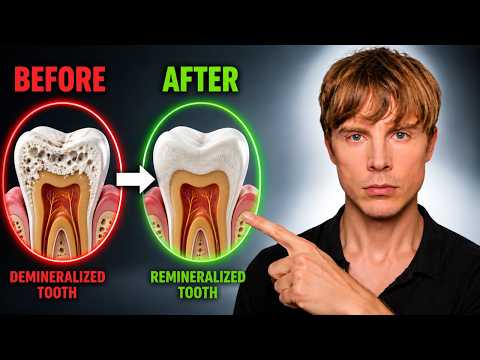
The Viral Toothpaste That Remineralizes Your Teeth
I. Executive Summary
Mature human tooth enamel is an acellular, highly organized crystalline matrix composed of approximately 96% carbonated hydroxyapatite by weight. Because ameloblasts undergo programmed apoptosis upon tooth eruption, adult enamel possesses no cellular machinery for biological self-repair or de novo structural regeneration once the prism architecture collapses. However, the physicochemical equilibrium between mineral dissolution (demineralization) and crystal precipitation (remineralization) remains dynamic. Prior to structural cavitation, subsurface porosities—such as incipient white spot lesions—are chemically reversible through exogenous mineral deposition.
Biomimetic nano-hydroxyapatite (nHAp) and microcrystalline hydroxyapatite (HAp) serve as bioactive calcium phosphate formulations designed to restore depleted enamel mineral density. Robust, double-blind randomized controlled trials (RCTs) in adult cohorts demonstrate that 10% HAp dentifrices achieve statistical non-inferiority compared to standard 1,450 ppm sodium fluoride controls in halting the progression of decayed, missing, and filled surfaces (DMFS) over 18 months. Similarly, pediatric RCTs confirm non-inferiority against age-appropriate fluoride regimens. In active white spot lesions, nHAp demonstrates superior microhardness recovery and lesion surface area reduction relative to fluoride alone.
For dentin hypersensitivity, systematic reviews and meta-analyses encompassing 44 human clinical trials establish that HAp outperforms both placebo (approximately 39.5% pain reduction) and conventional fluoride dentifrices (approximately 23% reduction). Mechanistically, nHAp particles (20–100 nm) penetrate and physically occlude open dentinal tubules, neutralizing intradental nerve mechanoreceptor activation per Brännström’s hydrodynamic theory. At the enamel surface, nHAp integrates into the acquired salivary pellicle, provides an epitaxial template for ion deposition, and acts as a sacrificial buffer releasing calcium (Ca2+) and phosphate (PO43−) during acid challenges.
Crucially, marketing claims asserting that nHAp “regrows lost enamel” are biologically false. Remineralization is strictly confined to filling microporosities within intact protein-mineral scaffolding; frank structural cavitation requires standard operative restoration. Systemically, preserving enamel integrity and preventing periodontal pocket formation eliminates chronic reservoirs of periodontal pathogens (e.g., Porphyromonas gingivalis), dampening systemic endotoxemia and downstream cardiovascular and neuroinflammatory cascades. From a biosafety standpoint, synthetic nHAp is non-toxic and non-fluorotic upon ingestion, provided particle morphology strictly adheres to rod-shaped dimensions (aspect ratio < 3) and avoids cytotoxic needle-shaped variants.
II. Insight Bullets
- Enamel is an acellular, non-regenerative tissue due to the total loss of ameloblasts following tooth eruption.
- Complete physical loss of enamel volume cannot be biological regrown; restorative intervention is required once structural cavitation occurs.
- Subsurface mineral depletion (demineralization) is a distinct, chemically reversible phase preceding macroscopic cavitation.
- White spot lesions represent early clinical manifestations of subsurface demineralization beneath an intact surface layer.
- Synthetic nano-hydroxyapatite (nHAp) chemically and structurally mimics native biological calcium apatite: Ca10(PO4)6(OH)2.
- In vitro demineralization models demonstrate that 10% nHAp recovers significantly greater enamel surface microhardness compared to fluoride or untreated controls.
- An 18-month double-blind adult RCT (Paszynska et al., 2023) established that 10% HAp toothpaste is statistically non-inferior to 1,450 ppm sodium fluoride in preventing DMFS index increase.
- In the 2023 adult trial, 89.3% of the HAp cohort and 87.4% of the fluoride cohort demonstrated zero progression of dental caries.
- A 48-week pediatric RCT in primary dentition (Paszynska et al., 2021) confirmed non-inferiority of microcrystalline HAp against 500 ppm fluoride for early childhood caries.
- In pediatric cohorts, roughly 73% of subjects in both HAp and fluoride groups exhibited some caries progression, emphasizing that dentifrice is secondary to dietary carbohydrate substrate control.
- A 2020 randomized clinical trial in orthodontic patients showed 6.7% nHAp toothpaste achieved significantly greater lesion shrinkage of post-orthodontic white spot lesions compared to fluoride.
- Systematic reviews and meta-analyses (Journal of Dentistry, 2024) confirm nHAp significantly enhances surface microhardness and mineral gain in initial enamel lesions.
- Dentin hypersensitivity arises when enamel erosion or gingival recession exposes open dentinal tubules to hydrodynamic fluid shifts.
- Fluid movement within patent dentinal tubules excites mechanoreceptors on intradental pulpal nerve fibers (A-beta and A-delta fibers).
- nHAp particles physically plug and occlude patent dentinal tubules within 2 to 8 weeks of daily application.
- A meta-analysis of 44 clinical trials (Limeback et al., 2023) demonstrated HAp reduces dentin hypersensitivity by 39.5% versus placebo and 23% versus fluoride.
- Synthetic nHAp achieves deep intratubular penetration due to particle sizing (20–100 nm) being smaller than dentinal tubule diameters (1–3 micrometers).
- nHAp deposits directly into microscopic surface fissures and inter-prismatic spaces via epitaxial crystal growth.
- Deposited nHAp serves as a localized, high-affinity reservoir of bioavailable Ca2+ and PO43− ions directly at the plaque-enamel interface.
- In the presence of bacterial organic acids (lactic acid), the outer synthetic HAp layer dissolves as a “sacrificial” barrier, buffering localized microenvironmental pH.
- Dissolution of the sacrificial HAp layer elevates local ion saturation, retarding the critical demineralization of the underlying endogenous tooth enamel.
- Clinical remineralization efficacy is verified at both 5% and 10% nHAp concentrations; dosages below 5% lack robust RCT backing.
- Brushing twice daily for a full two minutes is mandatory to provide adequate surface contact kinetics for crystal deposition.
- Post-brushing water rinsing washes away active nHAp nanoparticles; spitting without rinsing maximizes prolonged retention on enamel surfaces.
- Immediate mechanical tooth brushing following an acidic exposure (pH < 5.5 from coffee, soda, or gastric reflux) abrades softened enamel matrices.
- Salivary pellicle neutralization and natural buffering require at least 30 to 60 minutes following acute acid challenges before mechanical brushing.
- Chronic consumption of fermentable simple carbohydrates fuels bacterial glycolysis (Streptococcus mutans, Lactobacillus), sustaining acidogenic biofilm.
- Tooth decay and advanced periodontitis act as chronic sources of low-grade systemic bacteremia and pro-inflammatory signaling.
- Periodontal pathogens, specifically Porphyromonas gingivalis, secrete cytotoxic cysteine endopeptidases (gingipains) that degrade epithelial host defenses.
- Translocation of oral pathogens and lipopolysaccharides (LPS) into systemic circulation elevates high-sensitivity C-Reactive Protein (hs-CRP) and Interleukin-6 (IL-6).
- Chronic oral-derived systemic inflammation exacerbates vascular endothelial dysfunction, accelerates atheromatous plaque instability, and promotes microvascular cognitive impairment.
- P. gingivalis and its toxic proteases have been isolated post-mortem within Alzheimer’s disease cerebral parenchyma, correlating with neurofibrillary pathology.
- nHAp carries zero risk of dental fluorosis, making it fully biocompatible for pediatric populations and individuals with compromised swallowing reflexes.
- The European Scientific Committee on Consumer Safety (SCCS) strictly specifies that only non-coated, rod-shaped nHAp particles (aspect ratio < 3) are toxicologically safe for oral cosmetics.
- Needle-shaped nHAp nanoparticles exhibit cellular cytotoxicity and membrane disruption; consumer formulations must verify non-needle morphology.
III. Adversarial Claims & Evidence Table
| Specific Claim from Video | Speaker’s Evidence | Scientific Reality (Current Data) | Evidence Grade (A-E) | Verdict |
|---|---|---|---|---|
| Enamel cannot regrow, but mineral loss is reversible | Basic dental histology and structural definition of mature tooth enamel. | Biological reality: ameloblasts undergo apoptosis post-eruption, leaving enamel acellular. Subsurface remineralization occurs via chemical precipitation of Ca2+and PO43− (Cochrane Systematic Review, 2022). | Level A | Strong Support |
| 10% HAp toothpaste is non-inferior to 1,450 ppm Fluoride in adults | 2023 double-blind RCT with 189 adults over 18 months measuring DMFS index. | Confirmed via double-blind randomized clinical trial (Paszynska et al., 2023); no statistical difference in caries progression between 10% HAp (89.3% progression-free) and 1,450 ppm NaF (87.4% progression-free). | Level B | Strong Support |
| HAp is non-inferior to Fluoride in pediatric caries prevention | 48-week RCT in 214 children aged 3–7 evaluating ICDAS caries progression. | Confirmed via randomized controlled trial in primary dentition (Paszynska et al., 2021). Caries progression was 72.7% (HAp) vs 74.2% (fluoride), demonstrating clinical non-inferiority within the pre-set margin. | Level B | Strong Support |
| nHAp is superior for reversing early white spot lesions | 2020 6-month trial in 50 patients; 2024 meta-analysis on surface microhardness. | Systematic review and meta-analyses (Journal of Dentistry, 2024) demonstrate superior mineral gain, surface roughness reduction, and lesion area shrinkage for nHAp compared to standard fluoridated paste. | Level A | Strong Support |
| HAp significantly reduces dentin hypersensitivity vs Fluoride | 2023 systematic review & meta-analysis of 44 human clinical trials. | Validated by comprehensive meta-analysis (Limeback et al., 2023), demonstrating a 39.5% reduction in sensitivity vs placebo and 23.0% greater reduction vs fluoride dentifrices via tubule occlusion. | Level A | Strong Support |
| Immediate brushing post-acid exposure accelerates enamel damage | Warning to delay mechanical brushing following vomiting, coffee, or soda intake. | Acid challenges lower local pH below the critical point (5.5), demineralizing the outer few microns. Immediate mechanical abrasion strips the softened matrix before salivary calcium/phosphate remineralization can occur (Lussi et al., Clinical Oral Investigations). | Level B | Strong Support |
| Oral pathology directly fuels cardiovascular and cognitive decline | General claim linking tooth/gum inflammation to systemic and vascular pathology. | Human prospective cohorts establish that periodontitis and tooth loss elevate systemic hs-CRP/IL-6 and correlate with atherosclerotic CVD and cognitive decline (European Heart Journal / Alzheimer’s & Dementia); however, toothpaste choice alone is not a proven monotherapy to reduce systemic MACE. | Level C | Plausible |
| nHAp toothpaste “regrows” lost enamel structure | Explicitly debunked by speaker as social media marketing hype. | True biological regeneration of prismatic enamel architecture cannot occur; synthetic deposition only remineralizes existing porous matrices or forms a superficial synthetic layer (Biomimetics Review, 2023). | Level A | Unsupported (Debunked) |
IV. Actionable Protocol (Prioritized)
High Confidence Tier (Level A/B Evidence)
- Active Concentration Selection: Select oral dentifrices containing verified concentrations of 5% to 10% nano-hydroxyapatite (nHAp) or microcrystalline hydroxyapatite (HAp). Dosages below 5% exhibit insufficient crystal density for rapid tubule occlusion.
- Application Frequency & Contact Time: Brush twice daily for a minimum of 2 minutes with a soft-bristled or oscillating-rotating electric toothbrush.
- Post-Brushing Retention (No Rinse): Expectorate (spit out) excess foam thoroughly after brushing, but do not rinse with water immediately. Retaining residual nHAp in the salivary film prolongs surface contact, facilitating epitaxial crystal binding and ionic diffusion.
- Targeted Desensitization Protocol: For acute dentin hypersensitivity (exposed roots/cervical erosion), apply a pea-sized amount of 10% nHAp paste directly onto the symptomatic tooth with a clean fingertip, leaving it undisturbed for 2–3 minutes before bed.
Experimental Tier (Level C/D Evidence, High Safety Margin)
- Dual-Action Regimen (Hybrid Approach): For individuals with ultra-high caries risk, consider a alternating regimen (e.g., standard high-fluoride 5,000 ppm paste in the morning, 10% nHAp paste at night) to capitalize simultaneously on fluorapatite acid resistance and nHAp tubule-occluding/biomimetic remineralization.
- Post-Orthodontic White Spot Resolution: Apply a high-density nHAp paste via custom clear dental trays for 15 minutes daily following bracket de-bonding to accelerate subsurface mineral recovery in active white spot lesions.
- Zinc-Substituted Carbonate HAp: Utilize formulations combining nHAp with zinc-carbonate complexes to introduce localized bacteriostatic action against Streptococcus mutans alongside mineral deposition.
Red Flag Zone (Debunked or Safety Risk)
- Post-Acid Immediate Brushing: Never brush teeth within 30 to 60 minutes of an acute acid challenge (citrus juices, vinegar, sodas, black coffee, or gastric reflux). Rinse immediately with plain water or a sodium bicarbonate solution to accelerate pH buffering, then brush only after salivary pellicle remineralization has hardened the matrix.
- Unverified Nanoparticle Morphology: Discard and avoid nHAp products that fail to declare particle morphology or verify compliance with European Scientific Committee on Consumer Safety (SCCS) standards. Needle-shaped or high-aspect-ratio nHAp (>3) carries potential cytotoxicity and mucosal irritation risks. Only rod-shaped particles are certified safe.
- Relying on nHAp to Heal Cavitated Caries: Do not attempt to treat macroscopic, cavitated dentinal lesions with nHAp toothpastes. Once structural integrity is breached, the dentin-pulp complex requires formal clinical debridement and restorative material placement.
1 Like
Meaning, save your money.
1 Like
Even aside from “non-infiority” in prevention of cavities, it seems there could be other benefits?
But re: remineralization - does this do much more than fluoride already does? I see the claim that it does, but I don’t see convincing proof (yes, I saw the citation) that there’s a significant effect size difference between the two.
I wouldn’t be far off by claiming to never have NOT done this. 64 years and counting. (Dr Ellie would be very disappointed). Oops. My dentist says my teeth and gums are magnificent. …but I’ll work on this as my teeth have to last me a long time further. Thanks for the tip.
1 Like
Dr. Snyder focuses on what may be the 3 primary hallmarks of aging,with the requirement that the mitochondrial transplanation and stem cell replenishment has to be autologous.
“Dr Snyder proposes a tripartite theoretical blueprint for radical lifespan extension beyond the 120-year human barrier: autologous mitochondrial transplantation to replace organelles degraded by somatic mutations, targeted senolytic destruction of inflammatory senescent “zombie” cells, and continuous autologous pluripotent stem cell replenishment of failing organ niches”.
2 Likes
Stop Spinning Your Wheels: What Actually Took My Health From Top 15% to Top 1%
. Executive Summary
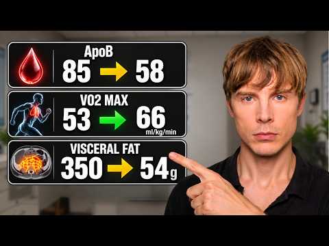
Optimizing long-term survivorship and metabolic longevity requires pivoting from generic “health behaviors” toward targeted, outcome-driven biomarker modification. Longevity is constrained by physiological bottlenecks rather than baseline strengths. Moving an individual from the 85th percentile of population health to the top 1st percentile demands ruthless prioritization of five core physiological levers: visceral adiposity reduction, cardiorespiratory fitness expansion, viscous and fermentable fiber intake, modulated protein consumption with dietary methionine-to-glycine balancing, and circadian sleep regularity.
Visceral adipose tissue functions as a pathogenic endocrine organ, driving chronic systemic inflammation, atherogenic dyslipidemia, and peripheral insulin resistance. Interventions that deplete visceral depots—primarily sustained caloric balance and zone-specific aerobic conditioning—demonstrate superior efficacy compared to resistance training alone for visceral fat mobilization. Concurrently, maximal oxygen uptake (VO2 max) demonstrates a continuous, graded, inverse relationship with all-cause mortality that extends past elite thresholds, eclipsing the longevity effect plateau observed in progressive muscular strength.
Nutritional interventions must prioritize functional endpoints over dietary dogma. High dietary fiber intake (approaching 50 g/day) delivers profound improvements in atherogenic particle clearance (ApoB/LDL-C reduction) and systemic metabolic stability. Concurrently, hyper-elevated protein consumption (exceeding 1.6 g/kg/day) offers diminishing lean mass gains while disproportionately increasing sulfur-containing methionine load. In translational models, excessive methionine intake in the absence of counterbalancing glycine accelerates metabolic dysfunction, whereas moderate protein intake coupled with glycine sufficiency preserves appendicular lean mass while enhancing insulin sensitivity.
Finally, circadian sleep consistency within a strict 30-minute window outpredicts gross sleep duration in reducing all-cause and cardiovascular mortality. Adequate sleep duration preserves lean tissue catabolism under caloric deficit conditions, preventing sarcopenic weight loss. By replacing unmeasured wellness routines with closed-loop biomarker tracking—measuring baseline parameters, deploying targeted protocols, and verifying multi-system adaptations—individuals systematically resolve rate-limiting physiological weaknesses to compress morbidity and decelerate biological aging.
II. Insight Bullets
- Bottleneck Constraint Principle: Human longevity and metabolic capacity are constrained by individual physiological bottlenecks rather than established physical strengths.
- Endocrine Pathology of Visceral Adipose Tissue: Visceral adipose tissue operates as a 24/7 pathogenic endocrine organ, releasing pro-inflammatory cytokines and driving hepatic insulin resistance.
- Visceral Adiposity Index and Cardiovascular Risk: A 2025 meta-analysis of prospective cohort data confirms that elevated visceral adiposity index significantly elevates risks of cardiovascular disease, stroke, and cardiovascular death, with risk increasing linearly per 0.5-unit increment (Wang et al., 2025).
- Calorie Restriction and Epigenetic Deceleration: Human caloric restriction trials, notably the two-year CALERIE Phase-2 RCT, confirm improvements in cardiometabolic markers and a 2% to 3% slowing in the pace of biological aging measured via DNA methylation algorithms like DunedinPACE (W. Daniel Belsky et al., 2023).
- Target Adiposity Thresholds: The lowest mortality risk across population data corresponds to maintaining body fat percentages below 15% for males and between 20% and 25% for females.
- Age-Stratified Visceral Fat Targets: Maintaining optimal cardiometabolic health requires suppressing absolute visceral fat mass into the lowest 10th percentile for age (e.g., approximately 100 g for young adults).
- Dietary Agnosticism in Body Composition: Diet composition functions strictly as a delivery vehicle; caloric balance and fat loss dictate the primary cardiometabolic benefits of dietary interventions rather than ideological dietary patterns.
- Resistance Training Longevity Ceiling: While muscular strength reduces all-cause mortality, its hazard-ratio reduction reaches an asymptote at moderate-to-advanced strength levels.
- Non-Plateauing Longevity Returns of VO2 max: Cardiorespiratory fitness (VO2 max) provides an unbroken dose-response curve with survival benefits continuing into elite fitness cohorts.
- Aerobic Superiority in Visceral Fat Mobilization: A systematic review and meta-analysis of 35 trials demonstrates that aerobic endurance exercise reduces visceral adipose tissue significantly, whereas progressive resistance training alone shows negligible isolated visceral fat reduction (Ismail et al., 2012).
- Cardiorespiratory and Muscular Synergy: Concurrent physical conditioning that maintains advanced muscular strength alongside elite cardiorespiratory fitness maximizes survival outcomes over single-modality specialization.
- Pragmatic Weekly Exercise Programming: A balanced longevity exercise framework prioritizes 3 to 4 progressive resistance training sessions alongside 1 to 3 targeted cardiovascular sessions (incorporating Zone 2 base and high-intensity intervals).
- High-Dose Dietary Fiber Utility: Daily dietary fiber intake scaled up to 50 g exerts profound systemic cardiometabolic effects, well beyond baseline population minimums.
- Atherogenic Lipoprotein Reduction via Fiber: Viscous and fermentable dietary fibers accelerate hepatic clearance of bile acids and downregulate circulating apolipoprotein B (ApoB) and low-density lipoprotein cholesterol (LDL-C).
- Colorectal Oncology Prevention: High intakes of dietary fiber (30 g to 50+ g daily) significantly reduce colorectal cancer incidence in population cohorts.
- Polyphenol-Driven Adipose Mobilization: Integrating polyphenol-dense whole foods enhances visceral fat oxidation compared to isocaloric, polyphenol-depleted nutritional regimes.
- Hypertrophy Protein Saturation Threshold: Meta-analyses show that lean mass accretion from dietary protein plateaus around 1.6 g/kg/day, rendering higher intakes metabolically superfluous for muscle retention in energy balance (Morton et al., 2018).
- Methionine-Induced Metabolic Strain: Excessive intake of the essential amino acid methionine drives hyperhomocysteinemia and metabolic derangements seen in rodent and preliminary translational models (Cavuoto & Fenech, 2012).
- Human Methionine Restriction Efficacy: Controlled human trials demonstrate that limiting methionine intake enhances systemic fat oxidation, clears hepatic lipid accumulation, and restores peripheral insulin sensitivity (Plummer et al., 2017).
- Glycine-to-Methionine Counterbalancing: Glycine acts as a biochemical mimetic of methionine restriction by enhancing clearance of methyl groups, buffering homocysteine synthesis, and mobilizing visceral lipid stores (McCarty et al., 2018).
- Circadian Sleep Consistency Superiority: Objectively measured day-to-day sleep regularity (going to bed and waking within a 30-minute window) reduces all-cause mortality by 20% to 48%, outpredicting raw sleep duration in epidemiological models (Windred et al., 2024).
- Sleep Deprivation Catabolic Partitioning: Caloric restriction conducted under sleep deprivation (5.5 hours vs. 8.5 hours in bed) shifts weight loss substrate utilization, inducing up to 80% lean mass loss and blunt fat mass oxidation (Nedeltcheva et al., 2010).
- End-State Optimization Over Activity Tracking: True health status is determined by downstream target biomarkers (ApoB, visceral fat volume, VO2 max, HbA1c, hs-CRP) rather than mere participation in “wellness” behaviors.
- Engineering Feedback Framework: Biological optimization requires applying an objective closed-loop feedback mechanism: quantify baseline biomarkers, deploy target interventions, systematically re-measure, and iterate.
- Top 1% Health Community: Siim Land manages the Top 1% Health Community (hosted on Skool), which provides biomarker assessments, coaching protocols, and peer tracking.
IV. Actionable Protocol (Prioritized)
High Confidence Tier (Level A/B Evidence)
-
Cardiorespiratory Optimization via Zone 2 & HIIT:
- Execute 150–200 minutes of weekly Zone 2 aerobic base training (lactate <2.0 mmol/L; conversational threshold) combined with one weekly high-intensity interval training (HIIT) session (e.g., 4x4 min at 90–95% max HR) to aggressively expand VO2 max and trigger visceral fat lipolysis (Ismail et al., 2012).
-
Hypertrophy Maintenance via Progressive Overload:
- Perform 3 to 4 full-body or upper/lower resistance training sessions per week focusing on compound multi-joint movements (6–12 reps at RPE 7–9) to maintain appendicular lean mass and bone mineral density without excessive systemic strain (Morton et al., 2018).
-
High-Viscosity Fiber Intake (40–50 g/day):
- Systematically scale intake of diverse soluble, insoluble, and prebiotic fibers to 40–50 g daily using whole-food sources (legumes, cruciferous vegetables, berries, chia seeds, root tubers) to bind biliary lipids, clear systemic ApoB, and improve glycemic control.
-
Circadian Anchoring & Sleep Consistency:
- Anchor daily sleep and wake times to a fixed window (±30 minutes, 7–8.5 hours total sleep) 7 days per week. Avoid severe sleep restriction during caloric deficits to prevent sarcopenic catabolism and sustain metabolic rate (Nedeltcheva et al., 2010; Windred et al., 2024).
-
Objective Target Biomarker Panel:
- Screen fasting blood panels quarterly: Target ApoB <60 mg/dL, hs-CRP <0.5 mg/L, fasting insulin <5 μIU/mL, and HbA1c <5.0%.
Experimental Tier (Level C/D Evidence, High Safety Margins)
-
Protein Titration & Methionine-Glycine Balancing:
- Cap total protein intake between 1.4 and 1.6 g/kg/day (deviating from 2.0+ g/kg bodybuilding dogmas) to minimize unnecessary sulfur amino acid load while saturating skeletal muscle protein synthesis.
- Supplement 5–10 g/day of pure glycine powder or collagen peptides alongside methionine-dense animal proteins (meat, poultry, eggs) to optimize the plasma glycine-to-methionine ratio and support hepatic transmethylation pathways (McCarty et al., 2018).
-
Polyphenol Densification for Adipose Remodeling:
- Consume polyphenol-rich foods daily (wild blueberries, pure cocoa, green tea, dark leafy vegetables) as adjuncts to accelerate visceral and hepatic lipid oxidation during caloric deficit or maintenance phases.
Red Flag Zone (Debunked Claims, Translational Gaps & Safety Risks)
-
Unmonitored Extreme Methionine Deprivation in Humans:
- Safety Data Absent: Extreme restriction of dietary methionine (<2 mg/kg/day) without clinical supervision risks negative nitrogen balance, anemia, muscle wasting, and hepatic dysfunction. Do not attempt synthetic amino-acid-stripped medical diets outside clinical trials.
-
“Health Halos” and Unvalidated Action Bias:
- Indulging in wellness practices (saunas, uncalibrated ice baths, indiscriminate longevity supplements, unregulated intermittent fasting regimes) without verifying downstream shifts in hard biomarkers (blood pressure, ApoB, DEXA visceral fat) is ineffective. Activity does not equal physiological adaptation.
-
Sacrificing Aerobic Conditioning Exclusively for Heavy Resistance Training:
- Relying solely on powerlifting or bodybuilding splits without structured cardiovascular endurance training elevates long-term all-cause mortality risk, blunts heart rate variability (HRV), and fails to clear visceral and pericardial lipid deposits.
Produced by Gemini 2.5 Pro
Target health metrics
Target health metrics for longevity require stratifying physiological capacity (such as cardiorespiratory fitness and adiposity distribution) by age decile, while holding systemic cardiometabolic biomarkers to age-invariant optimal standards. The data referenced from cohort analyses—including dual-energy X-ray absorptiometry (DEXA) normative databases of over 10,000 subjects (Ofenheimer et al., 2020) and the Cleveland Clinic cardiopulmonary cohort (Mandsager et al., 2018)—establish explicit targets to achieve top-tier health status across adulthood.
| Age Cohort | Visceral Adipose Tissue (VAT Mass) | Body Fat % (Men) | Body Fat % (Women) | VO2 Max: Elite (Men, ml/kg/min) | VO2 Max: Elite (Women, ml/kg/min) | Lean Mass (ALMI Target) |
|---|---|---|---|---|---|---|
| 20–29 | <100 g (Bottom 10th percentile) | 10–13% | 20–23% | ≥52–55+ | ≥43–46+ | Men: >8.5 kg/m² Women: >7.0 kg/m² |
| 30–39 | <150 g (Bottom 10th percentile) | 10–14% | 20–24% | ≥49–52+ | ≥40–43+ | Men: >8.5 kg/m² Women: >7.0 kg/m² |
| 40–49 | <220 g (Bottom 10th percentile) | 11–15% | 21–25% | ≥45–48+ | ≥36–40+ | Men: >8.3 kg/m² Women: >6.8 kg/m² |
| 50–59 | <320 g (Bottom 10th percentile) | 12–15% | 22–25% | ≥41–44+ | ≥33–37+ | Men: >8.0 kg/m² Women: >6.5 kg/m² |
| 60–69 | <420 g (Bottom 10th percentile) | 13–16% | 22–26% | ≥37–40+ | ≥30–34+ | Men: >7.8 kg/m² Women: >6.3 kg/m² |
| 70–80+ | <500 g (Absolute risk threshold) | 14–17% | 23–27% | ≥33–36+ | ≥27–31+ | Men: >7.5 kg/m² Women: >6.0 kg/m² |
Age-Stratified Physiological Metrics
Visceral Adipose Tissue (VAT)
- Normative Divergence vs. Longevity Target: In standard population cohorts, visceral fat accrues progressively from ~180 g in young adulthood to over 1,500 g past age 60. The video protocol mandates remaining in the lowest 10% for age rather than tracking population averages.
- Absolute Thresholds: Maintain DEXA visceral fat mass below 100 g during early adulthood, scaling to no more than 500 g at age 80. In cross-sectional imaging, VAT area should remain strictly below 100 cm² to avoid elevated cardiovascular event rates (Wang et al., 2025).
Cardiorespiratory Fitness (VO2 Max)
- The “Elite” Category Advantage: The landmark Cleveland Clinic study (Mandsager et al., 2018) showed that elite cardiorespiratory fitness (top 2.5% for age and sex) confers an approximate 5-fold reduction in all-cause mortality compared to the lowest quintile, with zero ceiling effect.
- Age Attenuation Rate: While sedentary adults experience an approximate 10% reduction in VO2 max per decade after age 30, structured endurance training (Zone 2 volume combined with interval protocols) limits this decline to roughly 5% per decade, allowing trained 60-year-olds to match the aerobic capacity of sedentary 30-year-olds.
Body Composition & Lean Mass Index
- Total Adiposity: Total body fat must remain below 15% for males and between 20% and 25% for females across middle age to avoid chronic low-grade inflammation. In adults over 65, lower thresholds adjust slightly upward (14–17% for men, 23–27% for women) to hedge against cachexia and frailty.
-
Appendicular Lean Mass Index (ALMI): Calculated as
Appendicular Lean Mass (kg) / Height (m)². The target is the top 10th to 20th percentile for age to prevent sarcopenia, supported by capping protein intake at 1.4 to 1.6 g/kg/day combined with progressive resistance training (Morton et al., 2018).
Age-Invariant Longevity Biomarkers
Unlike functional capacity, clinical targets for atherogenic particles, glycemic control, and systemic inflammation must not be adjusted upward with advancing age. Longevity medicine targets remain uniform across all adult cohorts:
- Apolipoprotein B (ApoB): <60 mg/dL (optimally 50–60 mg/dL) to arrest the progression of coronary atheroma.
- Low-Density Lipoprotein Cholesterol (LDL-C): <70 mg/dL (optimally 50–60 mg/dL), mediated via high intake of soluble/viscous fiber (40–50 g/day).
- Fasting Insulin: <5.0 uIU/mL, serving as an early indicator of peripheral insulin sensitivity before changes manifest in fasting glucose.
- Hemoglobin A1c (HbA1c): 4.5% to 5.0%, maintained via caloric management and circadian sleep anchoring.
- Total Plasma Homocysteine: <7.0 umol/L (optimally 5.0–6.0 umol/L), modulated by balancing dietary methionine with 5–10 g/day supplemental glycine or collagen peptides (McCarty et al., 2018).
- High-Sensitivity C-Reactive Protein (hs-CRP): <0.5 mg/L, reflecting the suppression of active visceral adiposity cytokine secretion.
- Sleep Regularity Index: Daily sleep onset and wake variance locked within a ±30-minute window, which delivers a 30% to 48% reduction in all-cause mortality regardless of baseline duration (Windred et al., 2024).
Produced by Gemini 2.5 Pro
1 Like
Critical Marker Insights
Inflammation: hsCRP Should Approach Zero
Healthy individuals maintain hsCRP levels near 0.1 mg/L. Levels between 0.5-1.5 mg/L suggest excess body fat or compromised metabolic health.
Lower is unequivocally better when measuring chronic systemic inflammation.
Kidney Function: Cystatin C Over Creatinine
Creatinine fluctuates with muscle mass and exercise intensity, making it unreliable for true kidney assessment. Cystatin C remains stable regardless of muscle tissue, providing accurate kidney function data.
If you want to measure your kidney function, measure cystatin C. Creatinine can give you some additional information, but in the absence of cystatin C, it can be misleading.
Liver Enzymes: Context Matters
Lower GGT, ALT, and ALP generally indicate reduced liver stress. However, extremely low values in sedentary individuals may coincide with poor body composition.
Exercise volume influences these markers—reduced training frequency can lower liver enzymes independent of other health changes.
4 Likes
Unfortunately, Increasing VO2 Max Really Is This Simple

I. Executive Summary
Sprint Interval Training (SIT) serves as a time-efficient exercise modality capable of eliciting rapid improvements in maximal oxygen uptake (VO2 max) and peripheral cardiometabolic parameters, particularly in untrained and moderately trained populations. The primary physiological premise challenges the conventional paradigm that progressive aerobic adaptations require multiple hours of weekly low-to-moderate-intensity continuous training (MICT). A comprehensive meta-analysis of 34 studies encompassing 418 participants (Vollaard et al., 2017) demonstrated that SIT protocols yield an average ~8% improvement in VO2 max over training periods lasting up to 12 weeks. Most notably, the meta-analytic data establish a non-linear dose-response relationship: expanding sprint repetitions per session (e.g., 8–10 repetitions) does not confer superior cardiorespiratory adaptations compared to minimalist dosing of 2–3 supramaximal repetitions.
Controlled randomized trials confirm that minimalist protocols—such as three 20-second all-out cycling sprints separated by 2 minutes of active recovery, performed three times weekly—induce robust VO2 max gains between 10% and 19% across 6 to 12 weeks (Gillen et al., 2016). This equates to merely 2 to 3 minutes of cumulative supramaximal workload per week within sub-10-minute training sessions. The primary cellular mechanism driving these adaptations involves acute intracellular ATP and phosphocreatine depletion coupled with rapid calcium, AMPK, and p38 MAPK pathway signaling, stimulating skeletal muscle mitochondrial biogenesis (e.g., citrate synthase activity) comparable to prolonged endurance exercise.
From an epidemiologic perspective, cardiorespiratory fitness provides potent, dose-dependent risk reduction against all-cause mortality. Observational data across 750,302 veterans (Kokkinos et al., 2022) demonstrate that mortality risk reduction reaches an optimal nadir at approximately 14.0–14.3 metabolic equivalents (METs), corresponding to a VO2 max of approximately 49–50 mL/kg/min. While low-volume SIT provides an efficient ramp to transition sedentary individuals into this cardioprotective fitness bracket, it cannot fully replace high-volume polarized training for athletes targeting elite thresholds (>60 mL/kg/min), where central hemodynamic factors like eccentric left ventricular hypertrophy and plasma volume expansion become rate-limiting.
II. Insight Bullets
- Sprint interval training (SIT) stimulates rapid improvements in cardiorespiratory fitness while requiring substantially less time than traditional continuous endurance training.
- A meta-analysis of 34 trials with 418 participants showed that SIT interventions increase VO2 max by an average of 8% over periods up to 12 weeks (Vollaard et al., 2017).
- The training studies examined in the meta-analysis primarily utilized cycle ergometers with supramaximal sprints lasting 10 to 30 seconds separated by 2-minute recovery bouts.
- Increasing the number of sprint repetitions per workout does not produce greater cardiorespiratory fitness adaptations.
- Performing as few as two 20-second all-out sprints three times per week increased VO2 max by 10% to 13% over a 6-week intervention.
- A 12-week protocol consisting of three 20-second sprints three times per week elicited a 19% VO2 max increase in previously sedentary participants (Gillen et al., 2016).
- The cumulative net duration of maximal sprint exertion required to achieve these cardiorespiratory gains was limited to two to three minutes per week.
- Protocols employing high repetition volumes of 8 to 10 sprints generated comparable or potentially attenuated adaptations compared to lower-volume protocols.
- Baseline aerobic capacity dictates the relative magnitude of improvement, with sedentary subjects showing the greatest relative percentage increases.
- Individuals with moderate baseline VO2 max scores in the 40s to low 50s mL/kg/min still achieved measurable fitness increases from low-volume sprint protocols.
- Advanced individuals with baseline VO2 max values exceeding 55 to 60 mL/kg/min cannot rely solely on minimalist sprinting and require higher overall training volume to progress.
- A retrospective cohort study of 750,302 U.S. veterans demonstrated an inverse, graded association between cardiorespiratory fitness and all-cause mortality (Kokkinos et al., 2022).
- The lowest mortality hazard in the veteran cohort was observed at approximately 14.0 to 14.3 METs, corresponding to a VO2 max of approximately 49 mL/kg/min.
- Maintaining a VO2 max at or above 50 mL/kg/min captures the vast majority of all-cause mortality risk reduction without requiring elite aerobic thresholds.
- The core SIT protocol comprises a 2-minute low-intensity warm-up, followed by three 20-second all-out sprints interspersed with 2-minute recovery periods.
- The total session time for a complete low-volume sprint workout remains under 10 minutes when factoring in warm-up and active recovery intervals.
- The sprint protocol can be executed via stationary cycling or overground running, though cycling eliminates eccentric musculoskeletal impact.
- Maximizing cardiorespiratory adaptations requires participants to deliver genuine supramaximal, all-out effort during each sprint repetition.
- Shortening recovery intervals below two minutes impairs intracellular energy replenishment, dropping exercise intensity from supramaximal to submaximal thresholds.
- Skipping the pre-sprint warm-up substantially increases the incidence of acute musculoskeletal injury during supramaximal exertion.
- Minimalist sprint training is clinically sufficient to raise cardiorespiratory fitness to the protective 50 mL/kg/min benchmark in non-athletic populations.
- Personalized multimodal training plans and metabolic biomarker tracking are offered through the creator’s Top 1% Health community.
IV. Actionable Protocol (Prioritized)
High Confidence Tier (Level A/B Evidence)
| Domain | Clinical Action | Dosing / Target | Primary Endpoint |
|---|---|---|---|
| Sprint Interval Training (SIT) | Execute low-volume supramaximal cycle ergometer intervals | 3 x 20-sec all-out sprints (>100% VO2 max), 2-min active recovery, 3 days/wk (Gillen et al., 2016; Vollaard et al., 2017) | Increase VO2 max by 8–19% and upregulate skeletal muscle mitochondrial enzymes |
| Cardiorespiratory Target | Attain and sustain protective cardiorespiratory fitness threshold | ≥14.0 METs (VO2 max ≥ 49–50 mL/kg/min) verified via cardiopulmonary exercise testing (CPET) (Kokkinos et al., 2022) | Maximize reduction in all-cause mortality risk hazard ratios |
| Zone 2 Aerobic Base | Pair supramaximal training with polarized low-intensity endurance | 150–300 min/wk at 65–75% HRmax (below first lactate threshold) (Bull et al., 2020) | Drive eccentric cardiac remodeling, mitochondrial capillary density, and fat oxidation capacity |
Experimental Tier (Level C/D Evidence - High Safety Margin)
- Overground Sprint Progression: Modality transition from stationary cycling to resisted hill sprinting, performing 3 x 15–20 second hill sprints on a 6–8% grade twice weekly. The inclined plane reduces hip hyperextension forces and eccentric hamstring strain compared to flat sprints while preserving supramaximal cardiovascular strain. Monitor for posterior chain soreness and Achilles tendon tension.
- Micro-Volume Exercise Snacking: Protocol dosing of single 20-second maximal stair climb or cycle sprints dispersed 2 to 3 times throughout the day, separated by 1 to 4 hours of inactivity. This mechanism leverages acute glycogen depletion and rapid AMPK activation without dedicated training blocks. Monitor resting blood pressure and autonomic recovery via morning HRV.
- Concurrent Creatine Monohydrate Loading: Compound dosing of 3–5 g/day creatine monohydrate. Mechanisms include enhanced intramuscular phosphocreatine stores, accelerating ATP resynthesis during repeat supramaximal bouts to prevent power output decay across successive 20-second sprints. Monitor baseline serum creatinine and cystatin C to distinguish benign creatinine elevations from renal dysfunction.
Red Flag Zone (Debunked, High-Risk, or Contraindicated)
Cold Unconditioned Overground Sprinting: Initiating flat maximal track or turf sprints without prior aerobic conditioning or dynamic warm-up. In unconditioned adults over age 30, explosive maximal deceleration and high eccentric biceps femoris strain carry high risks of acute hamstring muscle tear, Achilles tendinopathy, and plantar fascial rupture. Stationary cycle ergometry or uphill grades should precede flat running.
Truncating Inter-Set Recovery (<60–90 Seconds): Compressing rest periods between supramaximal sprints under the mistaken assumption that higher density increases aerobic gains. Insufficient recovery impairs phosphocreatine resynthesis and intracellular pH buffering, degrading power output by >30% and turning a supramaximal neuromuscular stimulus into submaximal glycolytic fatigue.
Sole Reliance on SIT for Comprehensive Cardiovascular Remodeling: Substituting all low-intensity steady-state endurance training exclusively with 3 minutes of weekly sprinting. While SIT induces peripheral skeletal muscle mitochondrial adaptations (citrate synthase upregulation) equivalent to continuous training, it lacks sufficient sustained venous return and end-diastolic filling duration to stimulate left ventricular eccentric hypertrophy and large-scale plasma volume expansion.
Produced by Gemini 2.5 Flash
2 Likes