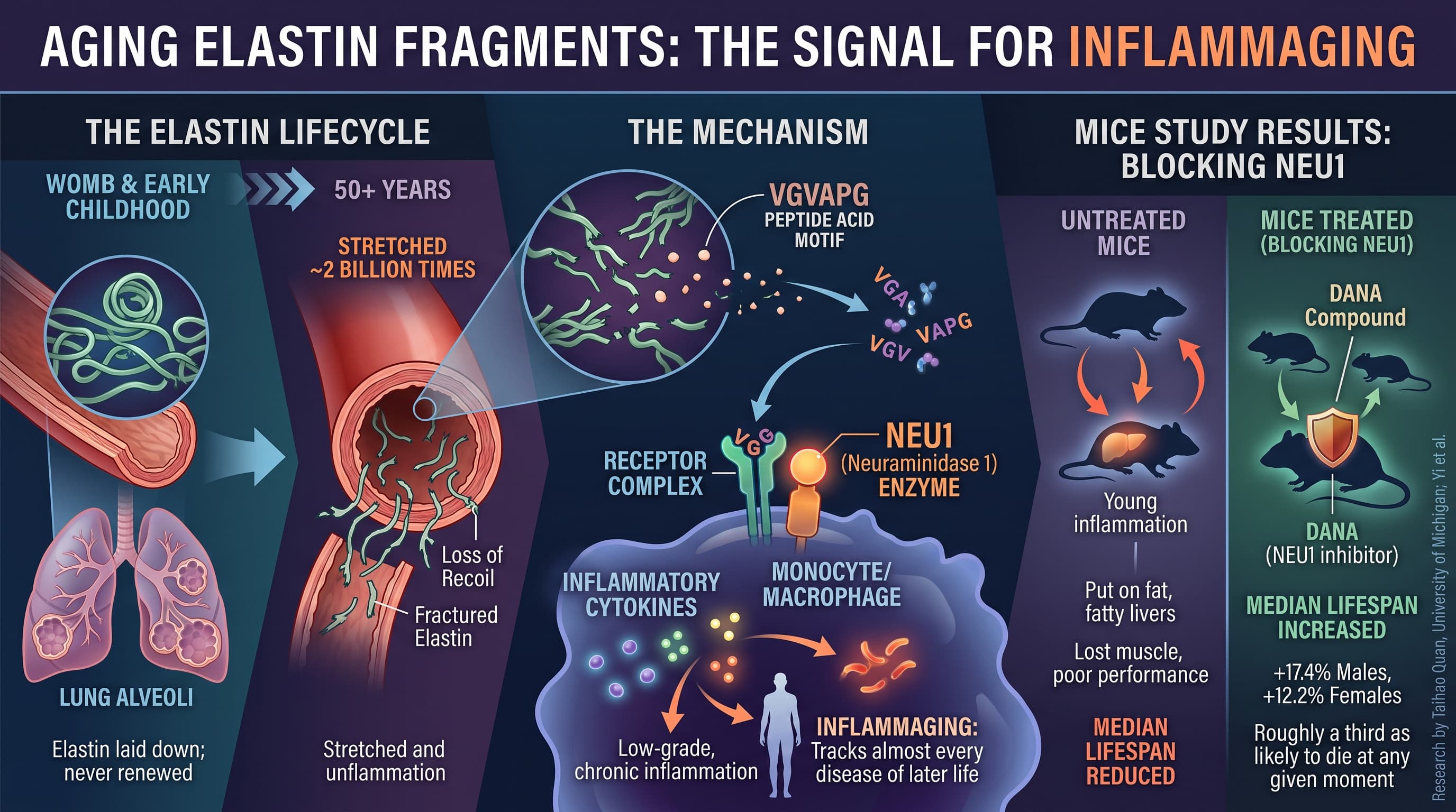
Elastin is the rubber-band protein that lets your aorta recoil with every heartbeat, your lungs spring back with every breath, and your skin snap back when pinched. You make almost all of it before you finish growing, and you never meaningfully replace it. Over decades of mechanical fatigue, sunlight, and enzyme attack, it shatters into short peptides that leak into the bloodstream. This review argues that those fragments are not passive debris but active hormone-like signals that switch on immune cells and drive chronic, body-wide inflammation. The author, a dermatology researcher at the University of Michigan, sets out to answer a specific question: which organs are actually producing this circulating debris? His answer is that three tissues dominate, the aorta, the lungs, and the sun-damaged dermis, each with its own signature set of protein-cutting enzymes. The practical payoff he proposes is source-resolved biomarkers, meaning a future blood test that could tell a clinician whether a given patient’s inflammation is coming mainly from their arteries, their lungs, or their skin, and therefore which tissue to treat.
There is a protein in your body that is almost as old as you are. Elastin, the elastic fibre that gives arteries their recoil and lungs their spring, is laid down in the womb and in early childhood and then, for practical purposes, never renewed. By the time you are fifty, the elastin in your aorta is fifty years old and has been stretched roughly two billion times.
That longevity is the problem. A new mini-review from Taihao Quan at the University of Michigan pulls together an emerging and genuinely provocative idea: as elastin finally fatigues and fractures, the pieces it sheds are not inert rubbish. They are signalling molecules. One short sequence in particular, a six-amino-acid motif written VGVAPG, docks onto a receptor complex on the surface of monocytes and macrophages and tells them to start producing inflammatory cytokines. The result is inflammaging, the smouldering low-grade inflammation that tracks almost every disease of later life.
The evidence that pushed this from curiosity to serious hypothesis came in 2025, when a team led by Yi and colleagues injected these fragments into young mice at concentrations matching those found in elderly humans. The young animals put on fat, developed fatty livers, lost muscle, performed worse on treadmills, and died sooner. Blocking the receptor’s key enzyme, an enzyme called NEU1, did the reverse: median lifespan rose by around 17 per cent in males.
Quan’s contribution is to ask a question that sounds pedantic but is not. If these fragments matter, where are they coming from? Elastin, unlike collagen, is not everywhere. It is concentrated in exactly three places that take a beating: the aorta and large arteries, where it makes up 30 to 50 per cent of the vessel wall’s dry weight; the lung, whose alveoli inflate and deflate about 15,000 times a day in a bath of inhaled oxidants; and the dermis, which absorbs a lifetime of ultraviolet radiation. Each of these tissues degrades its elastin using a slightly different mix of enzymes.
The implication is a shift in how we might think about anti-inflammatory medicine. Rather than blanket immunosuppression, you could read the precise chemical signature of a patient’s circulating fragments, identify which organ is the dominant source, and intervene there. Someone whose fragments carry the fingerprint of macrophage elastase is telling you about their lungs. Someone whose fragments look neutrophil-cut may be telling you about sun-damaged skin.
That test does not exist yet. Neither does a usable drug. But the framing is new, and it is testable.
Actionable Insights
Be clear about what this review can and cannot do for you. It is a hypothesis paper with no human intervention data, so there is no supplement, dose, or protocol to take away from it. What it offers is a mechanistic reason to take three unglamorous behaviours more seriously, plus one number.
The number is this: in mice, blocking the receptor enzyme NEU1 with a compound called DANA raised median lifespan by 17.4 per cent in males and 12.2 per cent in females. Reconstructing the strength of that result from the reported statistics, a treated mouse was roughly a third as likely to die at any given moment as an untreated one. That is a large effect by animal-study standards, large enough that it should be treated with suspicion until someone repeats it. DANA is not a drug you can obtain, and nothing equivalent has been tested in a person.
The behaviours the mechanism supports are sun protection, not smoking, and blood pressure control. Each reduces the mechanical or chemical load on one of the three elastin reservoirs. None has a measured effect on circulating elastin fragments in humans.
Context and Source
- Paywalled Paper: Circulating elastin fragments drive systemic aging: Where do pro-aging elastin fragments come from? A mini-review
- Author: Taihao Quan, sole author
- Institution: Department of Dermatology, University of Michigan Medical School, Ann Arbor, Michigan
- Country: United States
- Journal: Mechanisms of Ageing and Development, volume 233 (2026), article 112218, published by Elsevier
- Impact evaluation: The impact score of this journal is 6.5 (Journal Impact Factor, Web of Science, most recent release), evaluated against a typical high-end range of 0 to 60+ for top general science, therefore this is a Medium impact journal.