Senolytic Therapy: What are you doing?

If the 12 Hallmarks of Aging have meaning, then one aspect is to evaluate how they interact with each other. Which Hallmarks have the highest level of interactions? If that matters, then addressing the Hallmarks in order of interaction may be a interesting idea.

  1. Senescence - 8 interactions
  2. Inflammaging - 7 interactions
  3. mitochondrial disfunction - 6 interactions
  4. genomic instability - 6 interactions
  5. stem cell exhaustion - 5 interactions
    on down the line

This is just an idea as some of the Hallmarks that have fewer interactions may have greater harm/benefit if addressed.

Interactions_of_the_12_Hallmarks

https://pubs.acs.org/doi/10.1021/acschemneuro.3c00531

1 Like

@RapAdmin Sorry, late to this party. Not sure how I missed it :woman_shrugging:

Testing requires phlebotomy collection, then shipped back in a special cooler we provide. No dry ice, postage to and from included in the price.

And yes, @DrFraser has access. I also did a podcast a week or so ago that goes over what we test and why. Dr Fraser will post the link in the near future

1 Like

What is your cost?

I thought I would provide some of the key papers that persuaded me of my view with probably the majority of senescence.

This paper:

https://www.nature.com/articles/s43587-021-00105-8

for which I have done my standard chatGPT prompt:

Paper

Pouikli et al., “Chromatin remodeling due to degradation of citrate carrier impairs osteogenesis of aged mesenchymal stem cells,” Nature Aging, 2021.

Summary

This paper argues that ageing impairs the osteogenic capacity of bone-marrow mesenchymal stem cells by disrupting a mitochondrial–nuclear acetyl-CoA pathway.

The authors isolate bone-marrow-derived MSCs from young and old mice. Aged MSCs retain MSC identity and trilineage potential, but show reduced colony proliferation and a shift away from osteogenesis toward adipogenic behaviour, matching the known ageing phenotype of poorer bone formation. They then show that aged MSCs have reduced chromatin accessibility, especially around promoters, increased promoter nucleosome density, reduced nascent transcription, and lower global H3 and H4 acetylation.

At the locus level, ageing redistributes histone marks on lineage genes. H3K27ac is not globally reduced, but it is lost from putative enhancers linked to MSC differentiation, chondrogenesis and skeletal development, including loci near genes such as Postn and Wnt16. H3K27me3 increases globally and across gene bodies, supporting a less transcriptionally permissive chromatin state.

The central mechanistic finding is counterintuitive: aged MSCs have higher total acetyl-CoA, yet lower histone acetylation. The authors interpret this as a compartmentation problem. Acetyl-lysine staining shifts from the nucleus toward mitochondria in aged cells, suggesting mitochondrial trapping of acetyl-CoA. They identify reduced levels of the mitochondrial citrate carrier CiC, encoded by Slc25a1, as the key bottleneck. CiC normally exports mitochondrial citrate, which ACLY converts to cytosolic/nuclear acetyl-CoA. Loss of CiC would therefore reduce the acetyl-CoA pool available for histone acetylation even if total cellular acetyl-CoA is high.

The rescue experiments are strong. Lentiviral overexpression of CiC in aged MSCs restores acetyl-CoA export, increases nuclear acetylation and improves osteogenic differentiation. Conversely, inhibiting CiC in young MSCs with BTA shifts acetyl-lysine signal toward mitochondria and impairs osteogenesis. Sodium acetate, which can be converted to acetyl-CoA in the cytosol by ACS, restores H3 acetylation genome-wide and rescues osteogenic capacity of aged MSCs.

Finally, the authors propose that aged MSCs lose CiC through enhanced lysosomal degradation, apparently involving mitochondrial-derived vesicles rather than general mitophagy. Slc25a1 mRNA and promoter accessibility are not significantly reduced, mitochondrial protease markers are not changed, Bafilomycin A1 does not restore CiC, but the lysosomal inhibitor E64d does. CiC colocalises more with LAMP2 after E64d treatment, and electron microscopy shows more MDV-like vesicles in aged MSCs.

Main conclusion

The paper proposes this causal chain:

ageing → mild mitochondrial stress / MDV formation → lysosomal degradation of CiC → impaired citrate export → reduced cytosolic/nuclear acetyl-CoA → histone hypoacetylation → chromatin compaction at osteogenic regulatory regions → impaired osteoblast differentiation.

This is highly relevant to your citrate/acetyl-CoA model because it directly links mitochondrial citrate export, ACLY-dependent nuclear acetyl-CoA supply, histone acetylation and age-related failure of osteogenesis.

Novelty

The novelty is not simply that metabolism affects chromatin; that was already known. The novelty is the specific ageing mechanism:

  1. Compartmental acetyl-CoA matters more than total acetyl-CoA. Aged cells have more total acetyl-CoA but less histone acetylation, implying that the relevant variable is nuclear/cytosolic acetyl-CoA availability, not whole-cell abundance.

  2. CiC/SLC25A1 is placed at the centre of stem-cell ageing. The paper identifies the mitochondrial citrate carrier as a causal link between mitochondrial quality control and chromatin state.

  3. MDV–lysosome degradation of a mitochondrial carrier is connected to epigenetic ageing. The authors suggest that a mitochondrial quality-control pathway can selectively remove CiC, thereby altering nuclear chromatin.

  4. Functional rescue is demonstrated. CiC overexpression and acetate supplementation both rescue histone acetylation and osteogenic differentiation, moving the work beyond correlation.

  5. The osteogenesis defect is framed as an acetylation/chromatin plasticity problem. Rather than simply saying old MSCs are metabolically impaired, the paper links metabolism to lineage-specific enhancer accessibility and differentiation potential.

Strengths

The study is mechanistically coherent and uses several independent layers of evidence: ATAC-seq, CUT&RUN, RNA-seq, immunofluorescence, metabolomics, lipid staining, electron microscopy, pharmacological inhibition, acetate supplementation and CiC overexpression.

The rescue experiments are particularly persuasive. Showing that aged MSCs can regain osteogenic capacity after CiC overexpression or acetate supplementation supports causality rather than mere association. The BTA inhibition experiment in young cells also gives a useful reverse perturbation: blocking CiC mimics aspects of ageing.

The paper also avoids a common mistake in metabolism–epigenetics papers: it does not assume that total metabolite abundance equals functional nuclear availability. The observation that aged MSCs accumulate acetyl-CoA while being histone-hypoacetylated is an important conceptual point.

Critique and limitations

The main limitation is that much of the work is ex vivo. The MSCs are isolated, expanded in culture, sorted and kept for several passages under 2% oxygen. The authors are careful about maintaining niche-like oxygen and MSC identity, but culture expansion can still reshape metabolism, chromatin and differentiation behaviour. The paper shows consistency with old-mouse bone quality, but it does not fully prove that the same CiC degradation mechanism is operating in vivo at sufficient magnitude to drive skeletal ageing.

The second limitation is cell-type specificity. The mechanism is shown in mouse bone-marrow MSCs. It may be relevant to other ageing tissues, but that is not demonstrated. Different cell types may have different reliance on citrate export, acetate metabolism, ACLY, ACSS2, fatty-acid oxidation or direct mitochondrial–nuclear signalling.

Third, the MDV mechanism is suggestive but not fully nailed down. E64d restoring CiC and CiC–LAMP2 colocalisation support lysosomal degradation, and EM shows more MDV-like structures, but the paper does not genetically block MDV formation or identify the cargo-selection machinery that specifically targets CiC. Therefore, the MDV claim is plausible but less directly causal than the CiC/acetylation/osteogenesis rescue experiments.

Fourth, sodium acetate rescue is informative but not necessarily physiologically specific. Acetate can feed cytosolic acetyl-CoA through ACS enzymes, but it may also have broader metabolic and signalling effects. The acetate experiment supports “cytosolic acetyl-CoA availability matters,” but it does not by itself prove that citrate is the only relevant source.

Fifth, the study does not deeply examine splicing, despite the strong relevance of histone acetylation and chromatin accessibility to co-transcriptional RNA processing. Given your interest, this is a missed opportunity: reduced promoter/enhancer acetylation and lower transcriptional output could plausibly affect isoform choice, exon inclusion and differentiation programmes, but the paper mainly interprets the phenotype through gene expression and chromatin accessibility rather than alternative splicing.

Finally, the paper would be stronger with human validation. It cites prior human MSC/citrate work, but this study’s central ageing mechanism—age-related CiC loss by lysosomal/MDV degradation and rescue of osteogenesis—needs confirmation in aged human MSCs and ideally in vivo bone-regeneration models.

Overall assessment

This is a strong and important paper. Its best-supported claim is that aged mouse MSCs suffer from a functional shortage of nuclear/cytosolic acetyl-CoA despite increased total acetyl-CoA, and that restoring this pool can reopen chromatin and improve osteogenesis. The weakest part is the precise upstream mechanism of CiC degradation via MDVs, which is plausible but not as causally established as the downstream CiC–acetylation–osteogenesis axis.

For your model, this paper is highly supportive: it provides a direct experimental example of mitochondrial citrate export failure causing histone hypoacetylation and age-related stem-cell dysfunction.

This paper looks at causality. I discussed it with a professor at Birmingham who suggested it would not get through peer review because IL-10 is anti-inflammatory. That to me is a flaw with peer review. It probably means getting funding for further research would be hard.

Summary

This is a 2017 MSc thesis, “Decoding the role of IL-10 in aging”, examining whether sustained over-expression of the anti-inflammatory cytokine IL-10 can itself drive ageing-like phenotypes. The central model is the pMT-10 mouse, in which IL-10 over-expression is induced by zinc administration. The motivation is that IL-10 is often framed as protective against inflammation, but its long-term systemic consequences are not well understood.

The thesis tests 75 days of sustained IL-10 over-expression. The main findings are:

  1. Premature mortality and weight effects
    pMT-10 mice over-expressing IL-10 died prematurely. The cause of death was not established; the author speculates that abnormal haematopoiesis may contribute. IL-10 over-expression also prevented normal weight increase over time, but gut histology did not suggest malabsorption, leaving reduced food intake as a possibility.

  2. A skin- and hair-ageing phenotype
    IL-10 over-expression caused hair depigmentation, especially in males, and produced skin changes reminiscent of ageing: thinner dermis, reduced subcutaneous fat, altered hair follicle length/frequency, and reduced hair follicle melanin.

  3. IL-10 receptor dependence
    The phenotype was not seen in pMT-10 mice lacking IL-10Rα, supporting the interpretation that the effects are mediated by IL-10 signalling rather than simply by zinc administration or transgene background.

  4. Delayed wound repair
    Wound healing was impaired, especially after longer IL-10 induction. However, the wound-healing experiments also raise a confounding issue: zinc itself may contribute to the early wound-healing delay, and the author notes a possible IL-10–zinc interaction.

  5. Fibroblast defects and senescence-like changes
    Fibroblasts isolated from IL-10-exposed mice showed increased mitotic duration, longer cell-cycle duration, and more cytokinesis failure, suggesting reduced mitotic fidelity. In vitro, recombinant IL-10 exposure increased senescence-associated features in mouse adult fibroblasts, including more 53BP1 DNA-damage foci after 7 days.

  6. Not a complete progeroid syndrome
    The mice did not show several broader progeroid features within the 75-day window, such as cataracts, lordokyphosis, or kidney histological ageing. The author therefore suggests that IL-10 may particularly affect skin ageing, or that longer follow-up is needed to see broader systemic ageing.

Novelty

The novel element is the inversion of the usual IL-10 story. IL-10 is commonly viewed as anti-inflammatory and potentially protective, but this work suggests that chronic, sustained IL-10 signalling can promote ageing-like tissue dysfunction, at least in skin and fibroblasts. The thesis therefore reframes ageing not simply as “too much inflammation”, but as a problem of immune imbalance, where excessive anti-inflammatory signalling may also be harmful.

More specifically, the novelty lies in:

  • using an inducible IL-10 over-expression mouse model to study long-term systemic effects;
  • linking IL-10 over-expression to hair depigmentation, skin thinning, impaired wound repair, and fibroblast mitotic defects;
  • showing receptor dependence using IL-10Rα-deficient pMT-10 mice;
  • proposing IL-10 as a possible regulator of cellular senescence and premature skin ageing.

Critique

The work is interesting and hypothesis-generating, but it is not yet conclusive evidence that IL-10 is a general driver of ageing.

The strongest part is the phenotypic consistency: premature mortality, hair depigmentation, skin thinning, wound-healing delay, fibroblast cell-cycle defects, and in vitro senescence-like changes all point in the same direction. The IL-10Rα knockout experiment is especially important because it argues that the skin phenotype is genuinely IL-10 receptor mediated.

The main weakness is that the model is an over-expression model, not physiological ageing. Sustained high IL-10 may produce pathology that resembles ageing without being part of normal ageing. The thesis would be stronger if it showed that endogenous IL-10 rises in ageing skin, that blocking IL-10 signalling rescues age-related skin defects, or that naturally aged mice share the same IL-10-dependent mechanisms.

A second limitation is the zinc-induction confounder. The author controls for this with BL/6 + zinc and pMT-10 without zinc, but the wound-healing data explicitly suggest that zinc may itself affect repair, and possibly interact with IL-10. That does not invalidate the skin findings, but it makes the wound-healing conclusion less clean.

A third issue is sex dependence and variability. Hair depigmentation was stronger in males, not universal even among males, and did not correlate clearly with serum IL-10 levels. This is biologically interesting, but it also means the phenotype may depend on hormonal status, water/zinc intake, timing of sampling, or other unmeasured variables.

Fourth, the senescence evidence is suggestive but incomplete. The thesis uses p21 and 53BP1 as markers and discusses future work on p16, p21, IL-6, Mmp3, ROS and SA-β-gal. That means the core claim “IL-10 induces cellular senescence” is plausible but under-characterised. A robust senescence claim would need multiple markers, functional growth arrest, SASP profiling, reversibility tests, and ideally rescue by blocking IL-10R/STAT3 signalling.

Finally, the paper does not yet show a mechanism. It does not establish whether IL-10 acts directly on fibroblasts through STAT3, indirectly through immune remodelling, through stem/progenitor exhaustion, through mitochondrial dysfunction-associated senescence, or through haematopoietic pathology. The author’s own future-work section recognises this and proposes broader kinetic, tissue, and molecular profiling.

Overall assessment

This is a valuable thesis because it challenges the simplistic idea that anti-inflammatory cytokines are necessarily anti-ageing. The best-supported conclusion is:

Chronic IL-10 over-expression in pMT-10 mice produces a receptor-dependent, skin-focused premature ageing-like phenotype, with fibroblast mitotic defects and senescence-like changes.

The broader claim that IL-10 is a general regulator or driver of organismal ageing remains promising but not yet proven.

chatGPT prompt: Going back to the original paper: what happens to the osteoblasts which fail to differentiate?

In this paper, they are not really osteoblasts that fail to differentiate; they are aged MSCs/progenitors that fail to commit efficiently to the osteoblast lineage.

What seems to happen is:

  1. They remain less osteogenic / poorly committed

    The aged MSCs have reduced chromatin accessibility and reduced histone acetylation at promoters/enhancers of osteogenic genes. So they cannot properly activate the osteoblast differentiation programme. The paper’s model is that acetyl-CoA is trapped in mitochondria because CiC/SLC25A1 is degraded, leaving too little cytosolic/nuclear acetyl-CoA for histone acetylation and chromatin opening at osteogenic genes.

  2. They are biased toward adipogenesis instead

    The authors explicitly say aged MSCs show “skewed adipogenic differentiation … at the expense of osteogenesis,” which matches the known increase in marrow fat with age and osteoporosis risk.

  3. They do not appear simply to die

    The paper does not present the failed osteogenic cells as undergoing apoptosis or being eliminated. The emphasis is on a lineage-choice defect: aged MSCs are still multipotent, but their chromatin state makes osteoblast differentiation harder and favours the adipocyte route.

  4. Osteoblast differentiation requires more chromatin remodelling than adipogenesis

    This is an important point. The authors note that osteoblast differentiation required a longer induction period than adipogenesis, and they interpret this as evidence that becoming an osteoblast needs a more advanced level of chromatin remodelling. So when histone acetylation is insufficient, osteogenesis is especially vulnerable.

So the likely fate is: many aged MSCs fail to enter the osteoblast programme efficiently and are instead diverted toward adipocyte-like differentiation or remain poorly differentiated progenitors, contributing to less bone formation and more marrow adiposity. The paper does not prove a specific terminal fate for every failed cell.

Also: They resemble senescent cells in the broad sense of being aged, dysfunctional and poorly regenerative, but the evidence points more specifically to a reversible metabolic–epigenetic lineage-bias state, not classical cellular senescence.

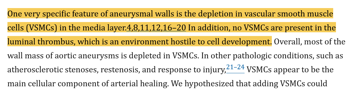
Another prompt: is there any other research on osteoporosis which finds senescent adipocytes replacing osteoblasts?

Yes — there is related evidence, but the precise wording needs care.

The evidence does not usually say that “senescent adipocytes replace osteoblasts” directly. More often it says:

ageing / osteoporosis → BMSCs become senescent or dysfunctional → they preferentially form marrow adipocytes rather than osteoblasts → bone formation falls and marrow fat rises.

There is also newer evidence that bone marrow adipocytes themselves can become senescent and then impair osteoblast formation through SASP-like signalling.

A few relevant strands:

1. Senescent or aged BMSCs shift toward adipocytes rather than osteoblasts.
Reviews of osteoporosis and skeletal senescence describe the key pattern: senescent BMSCs differentiate into adipocytes more than osteoblasts, reducing bone formation. (Springer Link) This matches the Nature Aging paper you uploaded, except that Pouikli et al. did not prove classical senescence; they showed a reversible CiC/acetyl-CoA/chromatin block.

2. Clearing senescent cells in old mice reduces marrow fat and increases osteoblast activity.
A major mouse study, Farr et al. 2017, found that targeting senescent cells prevents age-related bone loss. Later reviews summarise that clearing p16-positive senescent cells in old mice preserved trabecular and cortical bone, reduced osteoclasts, increased osteoblasts, improved bone formation rates, and reduced bone marrow adipocytes. (PubMed) (PMC) That is strong evidence that senescent cells help drive the osteoblast–adipocyte imbalance.

3. Senolytics improve aged BMSC osteogenesis.
A 2021 npj Regenerative Medicine paper reported that dasatinib plus quercetin improved the osteogenic capacity of aged mouse BMSCs both in vitro and in vivo. The authors interpreted the durable effect after short exposure as more consistent with eliminating a harmful senescent subpopulation than simply transient pathway modulation. (Nature)

4. Bone marrow adipocyte senescence is now an active research area.
A 2024 review says there is preliminary evidence that at least a subset of bone marrow adipocytes can undergo cellular senescence, remaining viable and metabolically active while acquiring senescence features. It also notes that senolysis in mice can modulate the marrow niche to increase osteoblast numbers and reduce marrow adipocytes. (PMC)

5. A 2024 Bone Research paper gives direct evidence for senescent marrow adipocytes impairing bone.
Xie et al. reported that macrophage-derived PCLAF induced senescence in BMSC-derived adipocytes, with higher p16, p21 and SA-β-gal. In vivo, recombinant PCLAF induced senescence markers in bone marrow adipocytes; conditioned medium from these senescent adipocytes impaired osteogenic differentiation, increased p16/p21 in osteoblast-lineage cells, reduced mineralisation and enhanced adipogenic differentiation. Transplanting PCLAF-treated marrow adipocytes caused bone loss and lower mineral apposition in recipient mice. (Nature)

So the best synthesis is:

Yes, osteoporosis research supports a senescence-linked replacement pattern: osteoblast production declines while marrow adiposity rises. Some of this is due to senescent/dysfunctional MSCs being biased toward adipogenesis, and some newer work suggests senescent marrow adipocytes can themselves worsen the niche and suppress osteogenesis.

But it is probably not literally that mature osteoblasts turn into senescent adipocytes. It is more likely a lineage allocation and niche effect: shared MSC progenitors increasingly choose or are pushed into adipocyte fate, while senescent cells and senescent marrow adipocytes secrete factors that further inhibit osteoblast differentiation.

If it looks like a duck, walks like a duck, and quacks like a duck, then it’s probably a duck.

For D&Q or Foxo4-DRI?

Your total monthly cost for your posted protocol.

About $250 CAD.

I think I have resolved this issue in my detailed post recently. If you think anything is outstanding please give me evidence as to what is outstanding.

I do recognise that senescence can be caused in a number of ways and that I am only looking at one of those routes. However, I think it is an important route that causes most of the harm (and most of the senescence).

The whole senescent cell field needs a complete overhaul - it’s based on a giant error in a critical tool:

3 Likes

I disagree with your broad overgeneralization here as discussed in the other thread you linked to. I’m not a fan-boy of senolytics, but it’s also not accurate to make claims that it’s “based on a giant error in a critical tool.” There’s a lot of research in senolytics of varying quality, and the recent P-16 scandal makes a total mess of sorting it all out. Very unfortunate. But it’s not helpful to paint the entire senolytics field as “based on a giant error in a critical tool.” That’s simply not an accurate portrayal of the situation, which is far more nuanced.

2 Likes

You said you only mean Fisetin in the other thread. You should just maybe delete this message.

Hardly. Try again. I said the field needs overhaul, not elimination. Fisetin in the other hand…

And holy moses, is it that controversial to recommend an overhaul when you have a systemic error of that magnitude? What are the alternatives? I mean, really. Wonders never cease.

Meanwhile fisetin has failed, the ITP for one. Looking at the literature and the weight of evidence, I think it’s done. Of course, if someone feels there’s a benefit in their situation, more power to them.

My position remains unchanged and my post stands until contrary evidence emerges, at which point I’ll gladly adjust my framework. YMMV.

3 Likes

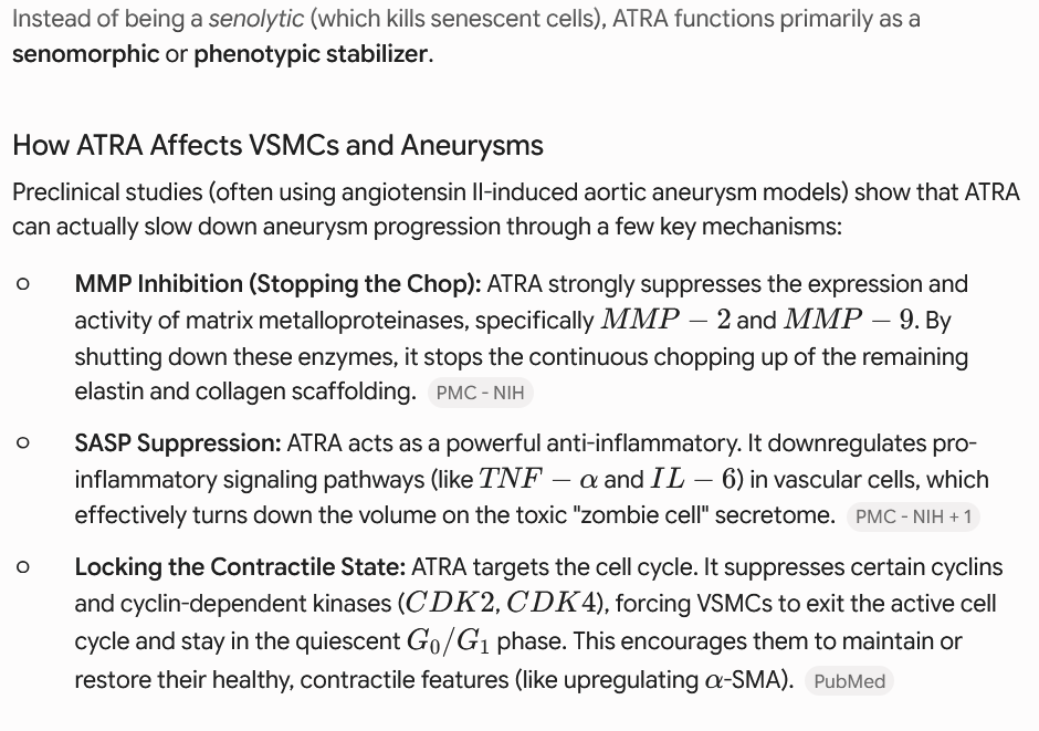
Fisetin also appears to have failed the Mayo Clinic human trials. Senolytics also failed in Aubrey DeGreys mouse trials - they reduced life expectancy of the mice! I dabbled with them and they gave me very bad diarrhea. I think they are only marginally useful for the very old. Stay away from them if you’re young and stick to senomorphics IMHO! Mouse trials with senomorphics vs D+Q found senomorphics like Rapamycin and Taurine to be much more effective at reducing senescent cells load!

3 Likes

I agree. I think senomorphics are a far more fruitful direction. I’m not going to say the whole field of senolytics is worthless, but it seems to me a relatively minor tributary in the anti-aging field, where we want to prevent senescence altogether rather than just dealing with the consequences. Senescent cells avoiding cancer strategy is a recognized modality and elimination of such cells needs a lot more research. Wound healing is another pathway and senolytics there might be actively undesirable. Regardless, benefits need to be demonstrated and the available senolytic agents seem subpar and blunt instruments at this point in time - perhaps better can be had in the future. YMMV.

2 Likes

Michael P Lisanti

Said this (fisetin) was not good as a senolytics several year ago.

I’m not a fan of fisetin (which is a senomorphic btw), but to claim there is no benefit from D&Q or other senolytics is pushing it a bit.
But yea, too many liars in the world, including science, where it’s publish or perish. Maybe in Utopia we can all enjoy pursuits which we enjoy, but we are not there.

So saying Fisetin isn’t a worthy senolytic makes sense to me. But suggesting D+Q or other FOXO4-DRI maybe? idk, there’s a lot of research to be done still, and n=1 experiments included.

Fiestin is not a senolytic. It was incorrectly assumed to be due to a mistake in measuring a similarly named antibody. This effect was confirmed by the ITP.

2 Likes

If you look closely at the Fisetin data that you’re dismissing, you’ll find conflicting evidence related to varying protocols.

For example, you cite the ITP research, but ITP also did continuous Fisetin at a specific dosage. Critics claim the dosage was both insufficient and should be applied hit-and-run to produce the desired systemic effects.

Alternative, earlier research that produced meaningful results did much larger dosages in a hit-and-run protocol.

Similarly, there’s some excellent research with strong effect sizes using D+Q.

Again, this stuff is nuanced, and you’re painting with a broad brush.

Even your overall position on senolytics doesn’t make sense (to my mind, at least…)

The research shows our senescent cell population is roughly balanced until age 40 at the first aging inflection point. A balanced senescent cell population is the healthy goal, not total eradication.

After age 40, the senscent cell population enters geometric growth curve (not arithmetically, but geometrically - a key point) with aging. Just as with financial geometric growth curves, the early compounding is not that significant. But as we approach 60 the numbers get more dramatic. This geometric growth in senescent cells correlates (correlation, not causation, but there is a clear potential mechanism of action) with a roughly similar pattern in inflammaging.

Inflammaging, in turn, is correlated with a wide variety of aging related health issues, including cardiovascular disease.

There’s clear logic to occasional hit-and-run senolytic treatments to revert the geometric population growth to the level of normal health. You’re not trying to kill all senscent cells. You’re simply trying to offset the geometric growth equation that’s likely related to multiple downstream negative health impacts.

This view I’m sharing is very different from the one you’re advocating. I’m surprised you’re seeing this topic the way you discuss, because you’re view doesn’t even seem connected to any of the relevant research.

I’ve had similar confusion with others who have chimed in against senolytics, but then they are in alignment with taking multiple prescription medications to lower LDL to unnaturally low levels. I get that there’s no research showing there’s no floor to LDL that shows negative health impacts (right now), but we all know that today’s science is tomorrow’s myth. It’s fairly safe to say that there is an issue with artificially low LDL through medication, but we just haven’t figured out what it is.

The inconsistencies in cardiovascular disease research make clear that disease progression is a function of multiple co-morbidities, not just LDL, or any other single factor. It’s a systemic response to reduced endothelial health, poor metabolic health, and (most important for this thread) chronic inflammation.

Having someone get on the low LDL bandwagon with multiple meds, but ignore senescent cells/inflammaging and various ways to preserve endothelial health makes no sense. It’s a system, so taking parts in isolation is oversimplified thinking.

You completely dismiss senolytics, yet I personally went from massive debilitating chronic joint pain to now being a 65 year old pain free distance runner using D+Q once per quarter. Yes, that’s an N=1, but that was the change in my protocol that opened the door. That life-changing difference is the reason I study this stuff.

Similarly, I have run chronically elevated LDL my entire life, and yet my CCTA shows zero soft plaque and a hard plaque score of 9 (and declining with each test, so approaching zero). Related is my hsCRP which doesn’t even register on the measurement scale (likely related to the senolytics treatments), a HOMA-IR of .5-.7 indicating excellent metabolic health, healthy blood pressure, excellent Triglyceride/HDL ratio, and so on.

My point is how I’m surprised that very knowledgeable and vocal members of this community are on the bandwagon of artificially lowering LDL to extreme low levels in the name of cardiovascular health, but then they’re equally dismissive of senolytics when cardiovascular research clearly shows the problem is a function of comorbidities.

I think senolytics deserves a seat at the table and is clearly relevant. Dismissing this topic as “hype” and then reverting your position to “anti-Fisetin” isn’t aligning.

Again, I’m not trying to take you personally to task with this comment. I’m merely using your comment as related to other positions taken in this forum to ask the question of these inconsistent polarizing positions.

I hope that’s helpful.

4 Likes

All these studies are compromised (maybe). Because researchers used another testing compound for a decade! And have measured another P16!)

Its kinda scandal) And maybe it compromise all senolytic’s theme

2 Likes

Congratulations on your fisetin experience - I hope I’ve made it clear that I applaud anyone’s experience with any intervention that they feel is helpful to them… I’ve said that in pretty much every post regarding this topic (and other topics too). I don’t necessarily think n=1 or personal testimonials proves anything about MOA or is going to be true for someone else, but who cares - even if it’s a placebo effect, if it works, more power to you and it’s not important what anyone else (including me) thinks about fisetin.

Wrt. the senolytic field and my low enthusiasm for it, I think we are starting to go in circles at this point, so I’m not going to write a novel here. I don’t feel like repeating myself, so in a nutshell, I put greater stock in anti-aging interventions further upstream. Dealing with damage that results from cell aging is like dealing with the consequences of a fire that burns down a building - I’m more enthusiastic about preventing the fire in the first place. Regarding the LDL analogy you brought up - I guess again I see it differently. To me, trying to bring down ApoB/LDL is attempting to go upstream from a state of atherosclerosis before endothelial damage is done and the MACE cascade sets in. In other words, it’s trying to prevent the fire in the building. Now, there is an entirely legitimate discussion to be had about whether extremely low ApoB/LDL is a good target and an even more fraught question as to whether the tools currently employed (LLT - statins etc.) are optimal or even helpful. But it still is an attempt to get ahead of the problem. To me senolytics in your analogy would be like focusing on how to limit damage once atherosclerosis has set in, or heart failure or other downstream consequences. Legitimate insofar as it goes - which is why I never dismissed the field of senolytics entirely - but of less interest to me than trying to get ahead of the issue by addressing upstream factors. I have greater enthusiasm for fixing the issues even further upstream. If there are gene therapies (as we’ve seen with recent trials) which can take the issue of atherosclerosis entirely off the table that’s even better, because that’s moving further upstream from even LLT therapies and you don’t have to deal with side effects of things like statins. Of course as always, we need to validate that this gene therapy is a global good without side effects etc. Anyhow, that’s my explanation of why I (others may have different motivations) am more onboard with LLT compared to senolytics - not only are current LLTs more clinically validated compared to something like fisetin with a much greater knowledge base around the MOA, physiological impact and side effects, but it is more of a preventative upstream intervention (this is also true on a personal level my CAC at 65 was zero, and I’m hoping to keep it that way). Whereas I look at the fisetin literature and don’t know what to do with it - like I said before since cells enter senescence for a variety of reasons, it’s not clear to me how a single senolytic agent addresses all these… even in cases like wound healing (see paper below*), not to mention cancer avoidance. Now somebody says “just take fisetin” or “just take X” - like I wrote before, it strikes me as an exceptionally blunt instrument unlikely to address the complexities of cell senescence. Anyhow it’s not in my personal drug stack, but if it works for someone else, I heartily congratulate them.

Sorry if I’m not addressing the other points you make, but I’m not sensing we’re making progress here if we feel the need to repeat ourselves - perhaps our perspectives are different as a result of a different vantage point.

  • Role of Senescent Cells in Cutaneous Wound Healing
2 Likes