From a mechanistic perspective, however, the real time changes are very interesting.
Looking, for example, at the differentiation of stem cells. We start with a cell which has complex 1 inhibited and depends upon pyruvate to maintain redox status. Then (and there is some link here to coming out of a hypoxic environment) it moves towards differentiation, NF kappa B is turned right up creating a substantial citrate flux from the mitochondria and large scale acetylation occurs to enable differentiation. It is at this point that differentiation can fail and the cell enter senescence. It would be nice to understand this at a lower level, but I don’t think it can be measured properly.
Acetyl-CoA is also part of the cell division process and I would like to fully understand the process whereby cells move from a single stem cell to a daughter cell and continuing stem cell.
chatGPT has given me a bit of an answer on this.
Acetyl-CoA is more than a metabolic intermediate; during proliferation it acts as a three-way hub that couples carbon availability to the nuclear, cytosolic and membrane-building events that let a cell duplicate itself.
Pillar
What happens
Why acetyl-CoA is indispensable
1. Epigenetic ignition of the cell-cycle engine
The first wave of genes needed for G1/S entry—including the yeast G1 cyclin CLN3 and hundreds of “growth” genes—are switched on only when their promoters become hyper-acetylated. That burst of acetyl marks is limited by how much acetyl-CoA is present in the nucleus; boosting acetyl-CoA drives histone acetylation and cell-cycle entry, while starving it keeps chromatin closed and the cycle off.
• Acetyl-CoA directly fuels GCN5/CBP/p300 histone acetyl-transferases. • Local nuclear production by ATP-citrate lyase (ACLY) or acetyl-CoA synthetase (ACSS2) ensures that histone acetylation can spike even when cytosolic levels are low. (PubMed, Nature)
2. Building blocks for two daughters
Every round of division roughly doubles plasma-membrane area, ER and Golgi surface, and the lipid sleeve that envelopes segregating chromosomes. All of that membrane is built from fatty acids and sterols that start as cytosolic acetyl-CoA.
• Cytosolic acetyl-CoA (mainly from ACLY) is converted by ACC → FASN → elongases/desaturases into phospholipids and neutral lipids. • Knocking down ACLY, blocking ACC, or inhibiting FASN stalls cells in G1 or kills them because membrane mass cannot keep pace; a 2024 study even uncovered a “lipid checkpoint” in early G1 that arrests the cycle when fatty-acid synthesis (and thus acetyl-CoA consumption) is inadequate. (PubMed, Nature)
3. Acetylation as a molecular timer
Many cell-cycle regulators themselves are acetylated. For example, Cyclin A must be acetylated at specific lysines before the APC/C can ubiquitinate and destroy it, ensuring orderly mitotic exit. Tubulin and several CDKs are similarly controlled. Because acetyl-CoA is the acetyl donor, its concentration influences how quickly these timers tick.
• When acetyl-CoA is limiting, cyclin acetylation and degradation slow, lengthening mitosis; excess acetyl-CoA has the opposite effect. (PubMed)
How the three arms are coordinated
Nutrient sensing → acetyl-CoA synthesis. Growth-factor signalling (PI3K–mTOR) up-regulates ACLY and ACLY’s citrate supply; AMPK and PGC-1α fine-tune ACSS2 and mitochondrial export, ensuring that carbon flows to acetyl-CoA only when energy is adequate.
Spatial pools matter. Nuclear ACLY/ACSS2 complexes generate an “on-site” pool for chromatin, while cytosolic ACLY feeds ACC/FASN for lipids. Compartmentalisation lets a cell prioritise gene activation even when membrane synthesis must pause, and vice-versa.
The G1 lipid checkpoint senses incomplete fatty-acid synthesis (a proxy for low acetyl-CoA flux) and activates PERK–ATF4 → p21, blocking CDK activity until lipid supply recovers.
Big-picture view
Signal Acetyl-CoA levels rise in response to carbon and growth signals.
Set chromatin Local nuclear acetyl-CoA spikes open proliferation genes.
Supply biomass Cytosolic acetyl-CoA is channelled into fatty-acid/cholesterol synthesis so the cell can physically double.
Synchronise timing Acetyl-CoA-dependent acetylation of cyclins/CDKs/microtubules ensures each phase ends only when the previous one is metabolically “paid for.”
Because all three roles depend on the same metabolite, perturbing acetyl-CoA synthesis or utilisation (e.g., with ACLY inhibitors) simultaneously starves chromatin remodelling, lipid supply and cell-cycle timing—explaining why such enzymes are promising anti-proliferative drug targets in cancer and why acetate supplementation can sometimes rescue dividing cells under metabolic stress.
In short: acetyl-CoA acts as the metabolic throttle of cell division, translating carbon abundance into the chromatin state, macromolecular building blocks and molecular timers that collectively allow one cell to become two.
I thought I would ask chatGPT for a bit more on the process of differentiation of stem cells. I suppose it is not really relevant to this thread as C Elegans adult cells don’t divide.
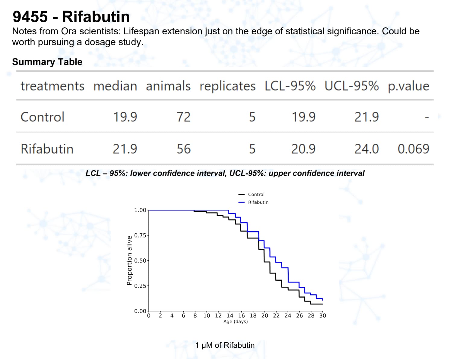
Also (as I think John is alluding to): null results are not that significant. But we should care about a significant life extension or shortening and move these compounds to larger animal models.
a) What has been found to extend life in mammals but not in c elegans
then
b) Do table
I also worry personally about the food question. Hence something that helps E Coli can harm C Elegans lifespan. SImilarly if you kill E coli you extend C Elegans life.
What I think is that the experiments are easy, fast and cheap to do. As a general principle things that are easy, fast and cheap are not good quality and the information we get is not good quality. It is, however, better than having no information at all.
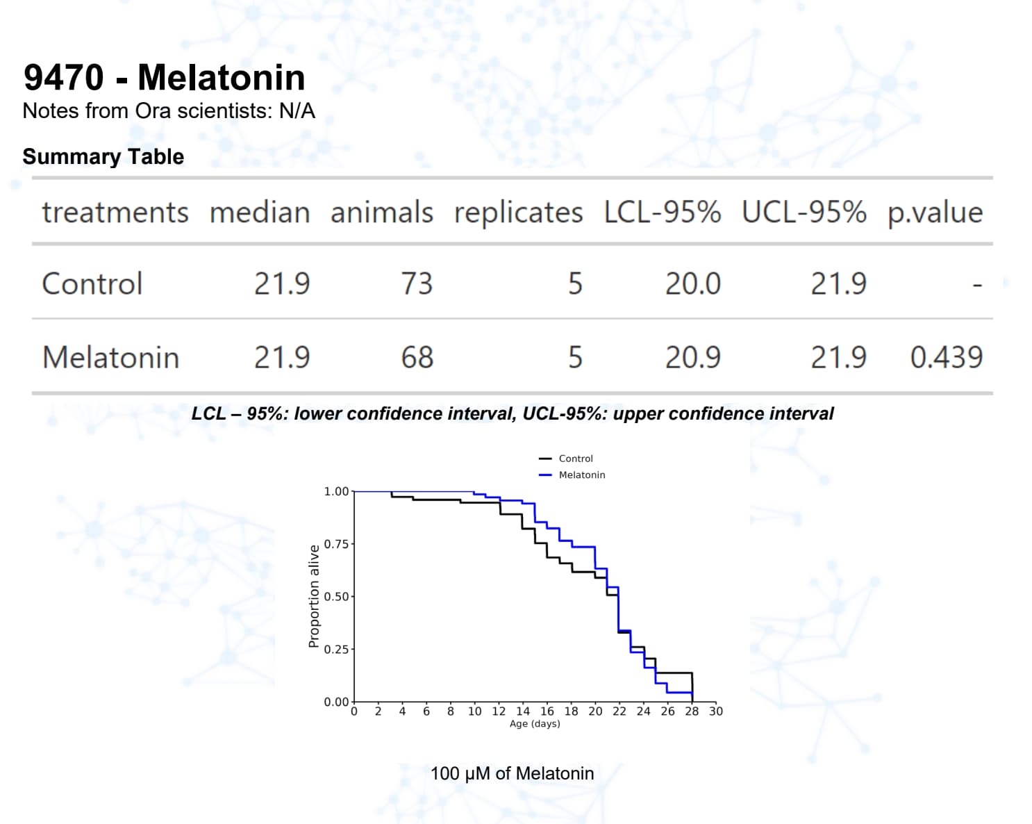
Melatonin, for example, has a good track record in mice.
Citrate, has a good track record in drosophila.
If I get a bit of time I intend reading up on the difference between mtDNA mutations in C Elegans and other species.
I think the relative absence of PINK1/Parkin is probably a key element in the short lifespan.
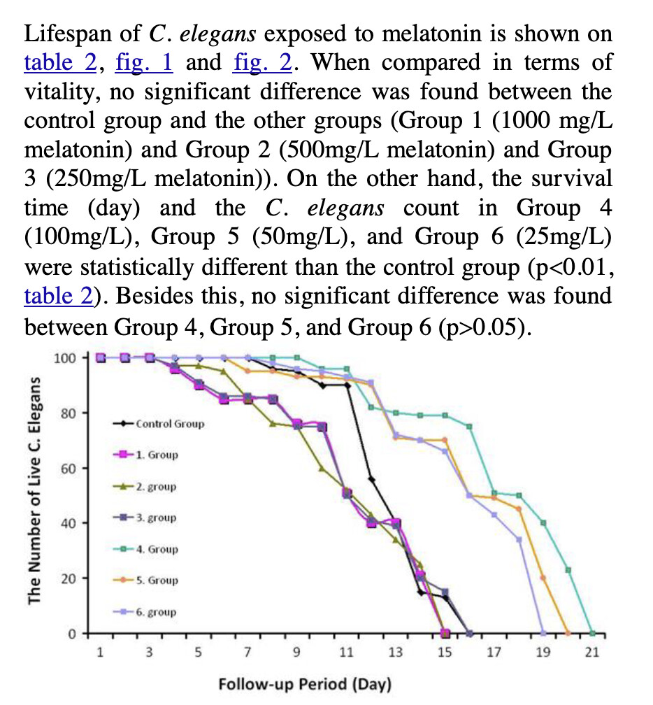
Group 6 = 25 mg/dL = 108 µM. I tried 100 µM so should have found the same result. The best is Group 4 = 100 mg/dL = 430 µM. I’ll ask Ora and maybe try 430 µM.
Melatonin will reduce ROS. It may be that too much melatonin removes too much of the ROS signalling. However, carrying forward dosing issues from C Elegans to humans is a really hard thing to do. I would not really try.
I personally continue with my weekly blood tests and taking between 1 and 2g of melatonin per night. I do think the timing matters although sometimes I take some during the day.
I think what probably happens with the worms is that the damage done to the mitochondria is such that even if you skew splicing decisions to a higher energy state it has no effect on the organism. They basically don’t have senescent cells.