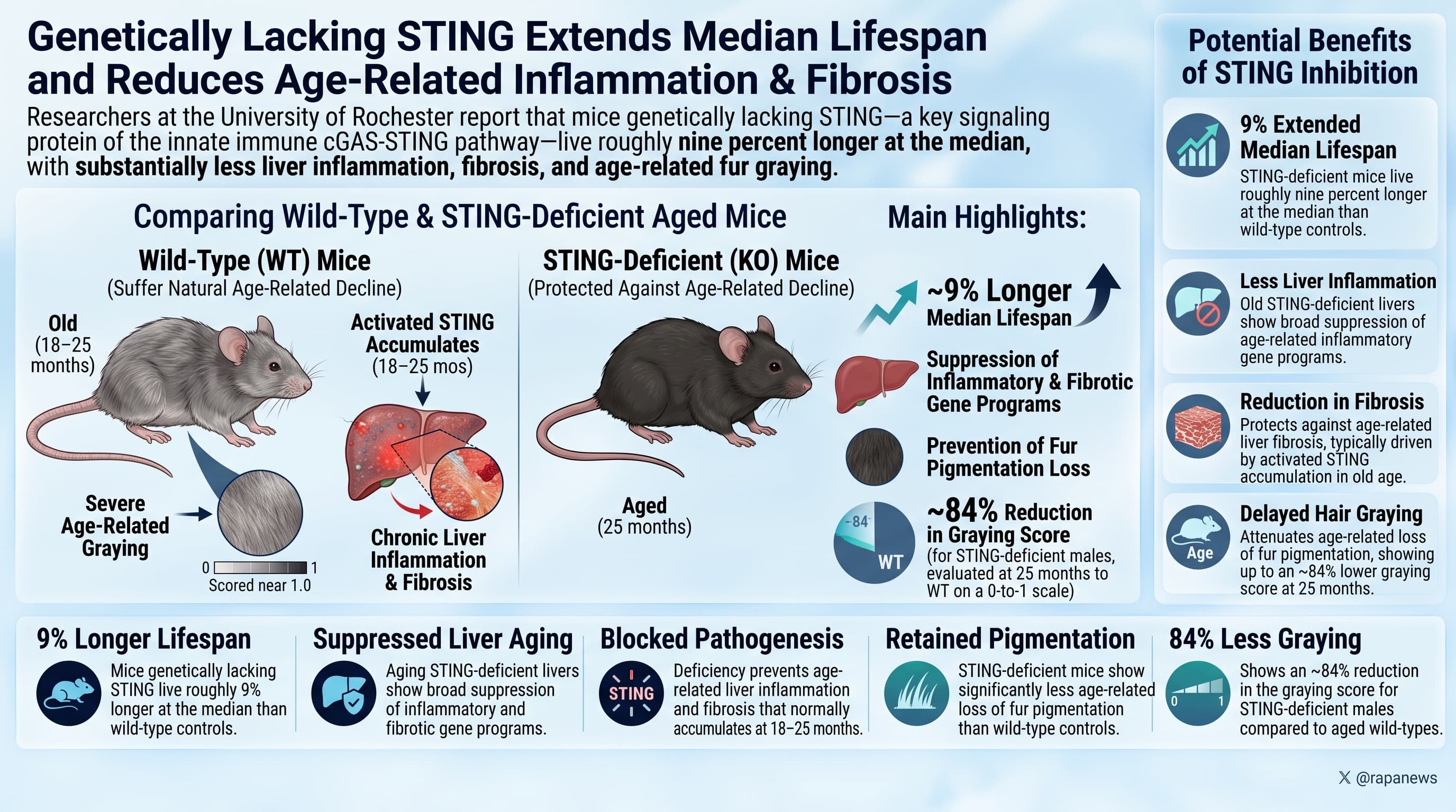
Researchers at the University of Rochester report that mice genetically lacking STING, a central signaling protein of the innate immune cGAS-STING pathway, live roughly nine percent longer at the median than wild-type controls and show substantially less age-related liver inflammation and fibrosis. Activated STING accumulates in wild-type mouse livers between 18 and 25 months of age, and livers from old STING-deficient mice show broad suppression of inflammatory and fibrotic gene programs. STING-deficient mice showed less age-related loss of fur pigmentation than wild-type controls, scored at 25 months an approximately 84 percent reduction in the graying score. The result contrasts sharply with the same group’s earlier finding that deleting cGAS, the upstream sensor in the same pathway, shortens lifespan.
Aging comes with a low, persistent burn of inflammation that no infection explains. One of the best characterized sources of that burn is a cellular alarm system built to detect loose DNA in the cytoplasm. The sensor, cGAS, finds the DNA. The adaptor, STING, carries the signal forward and turns on interferon responses. In young animals this is antiviral defense. In old ones, the DNA leaking out of damaged mitochondria, derepressed transposable elements, and crumbling nuclei trips the same alarm, over and over, with nothing to fight.
The obvious move is to shut the alarm off. The problem has been knowing where to cut the wire. Gorbunova and Seluanov’s lab at Rochester previously deleted cGAS in mice and got the opposite of what they expected: shorter lives, more inflammation, faster tissue damage. Their explanation is that cGAS has a second job in the nucleus, organizing chromatin, that has nothing to do with immune signaling, and that losing it costs more than the inflammation it prevents.
This new preprint tests the other end of the wire. Mice lacking STING, but with cGAS intact, reached a median survival of 773 days against 712 days for wild-type animals, a gain of 61 days. Where the effect shows up most clearly is the liver. Across heart, lung, liver, kidney, and pancreas, four of the five tissues looked largely the same between groups. The liver did not. Old wild-type livers were studded with inflammatory cell clusters and laced with collagen scarring by three independent stains. Old STING-deficient livers were comparatively clean, with scarring reduced by roughly 65 to 88 percent depending on the stain.
The transcriptional data line up with the histology. Sequencing 25-month-old livers turned up 907 genes expressed at lower levels in the knockouts against 240 higher, and the suppressed programs are the expected ones: inflammasome assembly, leukocyte migration, cytokine production, interferon signaling, collagen metabolism, tissue remodeling. Staining for the activated, phosphorylated form of STING showed it is nearly absent in 18-month-old livers and abundant by 25 months, which makes the protein a plausible driver rather than a bystander.
The big idea is that inflammaging has specific addresses, and that the pathway diagram matters more than the pathway name. Two proteins in the same cascade, deleted separately, produce opposite lifespan outcomes. That is an argument for molecular precision in geroscience and a warning against treating “block inflammation” as a strategy.
It is also a result that should be held loosely. The control animals were short-lived by field standards. The survival benefit rests on a statistical test that favors early separation of the curves. The maximum lifespan does not appear to have moved. And a 2026 paper from Tufts found that the same STING deletion makes the aging brain worse, with a leaky blood-brain barrier and neuromotor decline. Longer life, better liver, possibly worse brain, is a genuinely unresolved trade.
Actionable Insights
Nothing here is directly actionable today. This is a germline gene deletion present from conception, not a drug, and no STING inhibitor has yet been tested for human safety or efficacy in slowing aging.
What the numbers say, in plain terms. Median survival rose 61 days, from 712 to 773 days, which is 8.6 percent and not the “nearly 10 percent” the authors write. Translated to a human analogy, that is like moving a median from 78 to 85 years, but only if the starting point were a fair one, and here it is not. The control mice died about 190 days earlier than well-housed mice of this strain typically do. Interventions that look good against sickly controls frequently evaporate against healthy ones.
The liver findings are stronger. Fibrosis by Sirius red fell about 85 percent, with a standardized effect size near 2.8. An effect size of 0.8 is conventionally “large,” so this is very large, though measured in only four animals per group, which inflates it. Inflammatory cell clusters fell about 83 percent, effect size near 1.5, measured in 18 animals, which is more trustworthy.
Practical read: the liver is an underrated organ-specific aging priority, and cytosolic DNA driven inflammation is a defensible mechanism. That is a research direction, not a protocol.
Context and Source
- Open Access Paper: STING deficiency extends median lifespan and mitigates age-related liver pathology in mice
- Institution: Department of Biology and Department of Medicine, University of Rochester, New York, with a contributing author at Brown University, Rhode Island.
- Country: United States
-
Venue: bioRxiv, posted October 19, 2026.
Impact Evaluation: The impact score of this journal is N/A, because bioRxiv is a preprint