Lost in Translation: Why Your Cells Mis-Read Their Own Genes As You Age — And How Rapamycin May Fix It

Mining transcriptomes from over 16,000 human and thousands of mouse samples, researchers find that aging corrupts the cell’s RNA-editing machinery in a consistent, damaging way they call “splicing degeneration” — and that this corruption is partially reversed by rapamycin and calorie restriction, positioning it as a candidate new hallmark of aging.

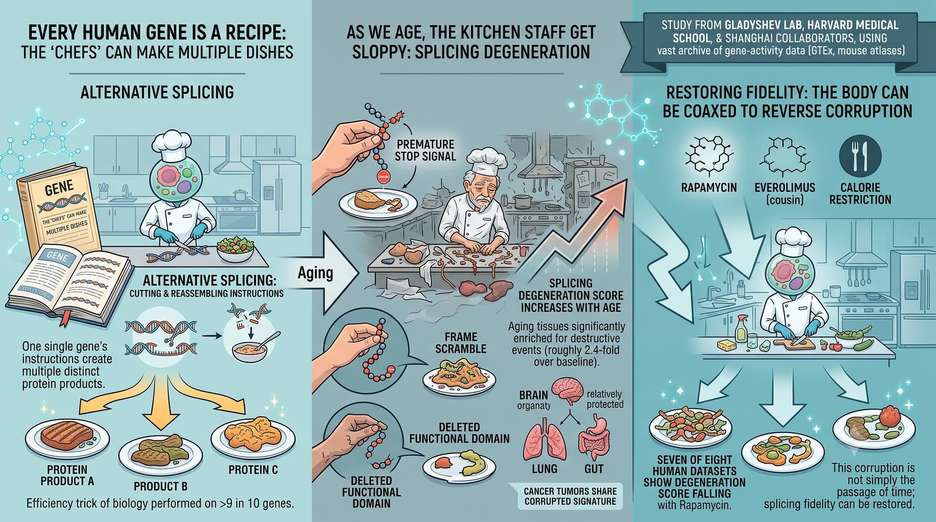
Every human gene is a recipe that can be cooked several ways. Through a process called alternative splicing, a single gene’s instructions are cut and reassembled into multiple distinct protein products — a feat performed on more than nine in ten of our genes. It is one of the great efficiency tricks of biology. This new study argues that, as we age, the kitchen staff get sloppy.

Researchers from Vadim Gladyshev’s lab at Harvard Medical School, working with collaborators in Shanghai, scoured a vast archive of gene-activity data: 16,627 human tissue samples from the GTEx project plus large mouse atlases. Rather than simply cataloguing which splicing patterns shift with age — which others have done — they asked a sharper question: do these shifts actually damage the resulting proteins?

Their answer is a fairly emphatic yes. The team built a classifier that flags splicing changes as “damaging” when they introduce a premature stop signal, scramble the protein’s reading frame, or delete a functional protein domain. Aging tissues, they found, are significantly enriched for exactly these destructive events (roughly 2.4-fold over baseline). They distilled this into a single “splicing degeneration” score that climbs steadily with age across most tissues. Intriguingly, the brain appears relatively protected, while the lung and gut are hit hard. Tumours, notably, show the same corrupted signature — hinting at shared machinery between aging and cancer.

The headline-grabbing part: the corruption is not simply the passage of time made visible. When the team examined cells and mice treated with rapamycin — the gold-standard lifespan-extending drug — seven of eight human datasets showed the degeneration score falling. Everolimus (a rapamycin cousin) and calorie restriction pointed the same direction. This suggests splicing fidelity is something the body can, in principle, be coaxed to restore.

Mechanistically, the authors finger specific “splicing factor” proteins — the regulators that decide how RNA gets cut — as culprits. Silencing many of these factors made cells look transcriptomically older on the lab’s own aging clocks, with the splicing-degeneration regulators producing the largest effect.

The big idea is to promote alternative splicing from a footnote in aging biology to a potential hallmark and therapeutic target in its own right. If splicing fidelity can be measured as a biomarker and nudged back toward youthfulness pharmacologically, it joins a growing list of aging processes that are, at least on paper, addressable. The caveat — and it is a large one — is that this entire edifice rests on correlation and computational inference, not on a single experiment showing that fixing splicing makes an animal live longer.

Actionable Insights

This is a biomarker-and-mechanism paper, not a trial. The “intervention” data are reanalyses of rapamycin, everolimus, calorie restriction, and metformin datasets that already informed longevity practice — the paper adds a new readout (splicing score), not a new intervention.

The take-home with the most evidential weight is that rapamycin’s benefit signal extends to a previously unmeasured layer of cellular fidelity — reinforcing its standing in a longevity stack. Effect-size reality check: the splicing-degeneration changes are statistically detectable but tiny in absolute terms. Treated-vs-control score shifts in the figures live in the third decimal place (e.g. ~0.235 → ~0.230), i.e. low-single-digit percent relative reductions. The strongest quantitative signal in the whole paper is the baseline enrichment of damaging splicing in old tissue (odds ratio 2.4), not the magnitude of any intervention’s reversal.

Practical translation for the stack-builder: nothing changes your protocol today. Rapamycin remains as a key supported lever; metformin and S6K1 deletion gave non-significant trends (p = 0.12 and 0.35). Do not treat “splicing degeneration” as a measurable personal biomarker — there is no assay you can order.

Source:

  • Open Access Paper: Mammalian aging involves genome-wide splicing degeneration leading to functional decline, Posted June 29, 2026.
  • Institutions: Division of Genetics, Brigham and Women’s Hospital / Harvard Medical School (Boston, USA); Shanghai Institute of Nutrition and Health, Chinese Academy of Sciences (Shanghai, China)
  • Country: USA / China (collaboration)
  • Journal: bioRxiv — preprint server. Posted June 29, 2026. Not peer reviewed
Intervention effect sizes — the critical weakness:

The authors report p-values and vote-counts, not standardized effect sizes, and the absolute magnitudes are minuscule. Representative figures (Fig 4), with my relative-change estimates:

  • Rapamycin in vivo (12-mo mice): ~0.235 → ~0.230, ≈2% relative reduction, p = 0.017.
  • Various human-cell rapamycin panels: shifts of ~0.005 on a 0.2–0.3 scale, ≈1.5–3% relative, p = 0.012–0.049.
  • Rapamycin overall: 7/8 datasets directionally consistent (vote-count, not effect size).
  • Everolimus: 3/4 datasets, one panel p = 0.0028.
  • Calorie restriction: significant only in young mice (p = 0.05); aged mice p = 0.4 (NS).
  • Metformin: p = 0.12 (NS). S6K1 deletion: p = 0.35 (NS).

I cannot compute Cohen’s d for these because the figures report neither SDs nor n per panel in a usable form. But the visual spread relative to the between-group difference suggests small-to-moderate d at best, with very wide uncertainty given n ≈ 2–4 per group in reused datasets. A ~2% shift in a composite proportion score, with no clinical or functional anchor, is of uncertain practical significance.

1 Like

https://www.biorxiv.org/content/10.64898/2026.06.26.734787v1

Obviously not news to anyone who reads this forum

chatGPT(5.5paid):

Summary

The paper argues that mammalian aging is accompanied by a genome-wide decline in the fidelity of alternative splicing, which the authors call “splicing degeneration.” Rather than simply cataloguing age-associated splice changes, they try to distinguish splice changes likely to damage protein function from those that may be neutral or adaptive.

The authors analyse large RNA-seq datasets from humans and mice, especially GTEx human tissues. They calculate PSI values — percent-spliced-in — for alternative splicing events, then compare older samples with younger reference samples. They find that the number of differential splicing events rises with age across many tissues. They also build splicing-based aging clocks, showing that splicing patterns can predict chronological age reasonably well.

A central step is their classification of splicing events as potentially damaging if they:

  1. introduce a premature termination codon, especially through intron retention;
  2. cause a frameshift;
  3. alter a region overlapping a protein domain.

Using this framework, they find that age-associated splicing events are enriched for damaging consequences. These events occur disproportionately in conserved genes and in genes involved in RNA metabolism, RNA splicing, antigen presentation and related processes.

They then define a quantitative splicing degeneration score: the proportion of damaging alternative-splicing events present in a sample. This score increases with age in many human and mouse tissues. Lung, spleen and small intestine appear among tissues with relatively consistent age-associated increases.

The paper also claims that splicing degeneration is modifiable. In reanalysed datasets, rapamycin, everolimus and calorie restriction tend to reduce splicing degeneration. Metformin and S6K1 deletion show weaker or non-significant effects. This is used to argue that splicing degeneration is not merely a passive correlate of chronological time, but may be linked to modifiable aging biology.

Mechanistically, the authors focus on splicing factors. They identify splicing factors whose expression or own splicing changes with age, including members of the SRSF and hnRNP families. They propose a “self-regulation” model: age-related mis-splicing of splicing factors contributes to broader splicing degeneration. They combine GTEx correlations, ENCODE knockdown RNA-seq, and eCLIP binding data to identify candidate regulators, including SRSF1, SRSF5, U2AF1, PTBP1, QKI, PUF60, SF3B1, HNRNPA2B1, HNRNPC, PRPF8 and others.

Finally, they use transcriptomic aging clocks to show that knockdown of many splicing factors increases predicted transcriptomic age. This supports the idea that splicing factor dysfunction can push gene-expression states toward an older profile.

Claimed novelty

The main novelty is not the observation that splicing changes with age; that is already known. The novelty is the attempt to define a functional, genome-wide measure of age-related splice damage.

The paper’s key novel contributions are:

1. “Splicing degeneration” as a formal aging phenotype
The authors move from “alternative splicing changes with age” to “damaging isoforms accumulate with age.” That is a stronger conceptual claim.

2. A quantitative degeneration score
They propose a score based on damaging splice outcomes: premature stops, frameshifts and domain-disrupting events. This makes splicing degeneration measurable across datasets, tissues and interventions.

3. Cross-species and multi-tissue integration
They analyse human GTEx, mouse datasets, Tabula Muris, cancer datasets and intervention datasets. This breadth strengthens the case that the phenomenon is general rather than tissue-specific.

4. Link to longevity interventions
The claim that rapamycin, everolimus and calorie restriction reduce splicing degeneration is particularly interesting, because it links splice fidelity to interventions already known to affect aging pathways.

5. Splicing factors as both victims and drivers
The “self-regulation” model is useful: splicing factors themselves become mis-spliced or mis-expressed with age, which may amplify downstream splicing disorder.

6. Connection to transcriptomic age
Showing that splicing-factor knockdowns increase transcriptomic age connects splice regulation to broader aging signatures rather than treating it as an isolated molecular defect.

Critique

The paper is important and highly relevant to aging biology, but several claims need caution.

First, “damaging” is computationally inferred, not experimentally proven. A frameshift, premature stop or domain overlap is plausibly damaging, but not all such events will produce stable protein products. Many may be degraded by nonsense-mediated decay or may occur at low abundance. Conversely, some splice changes classified as damaging could be regulated, adaptive or tissue-specific.

Second, bulk RNA-seq is vulnerable to cell-composition effects. Aging tissues often change in immune infiltration, fibrosis, senescent-cell burden and cell-type proportions. Some apparent splicing degeneration may reflect a different mixture of cells rather than splice failure within the same cells. The authors acknowledge this, but it remains a major limitation.

Third, GTEx is cross-sectional and post-mortem. GTEx samples differ in cause of death, ischemic time, tissue quality, RNA integrity and donor health. The model adjusts for some variables, but residual confounding is likely. Age-associated splice changes in GTEx may partly reflect illness, agonal state or sample handling.

Fourth, the PSI thresholds are somewhat arbitrary. The score uses PSI > 0.3 for damaging intron retention and PSI < 0.7 for damaging exon/skipping-related events. The authors test cutoffs, but the biological meaning of these thresholds is not obvious. A small change in a highly expressed essential gene may matter more than a large change in a low-expression gene.

Fifth, protein-level validation is limited or absent. The argument is about functional decline of protein products, but most evidence is transcriptomic. Ribosome profiling, proteomics, long-read RNA-seq, isoform-specific protein detection, or functional rescue experiments would make the case much stronger.

Sixth, the intervention analysis is suggestive but not definitive. Rapamycin-associated reductions in splicing degeneration are interesting, but these are reanalyses of heterogeneous datasets. Differences in cell type, dose, duration, sequencing method and baseline state could influence the result. It does not prove that rapamycin extends lifespan by restoring splicing fidelity.

Seventh, Mendelian randomisation is a bold addition, but interpretation should be cautious. Splicing QTLs can have pleiotropic effects on expression, chromatin or nearby genes. Inferring that particular splicing events causally influence Horvath age or lifespan is plausible but not settled.

Eighth, the “splicing degeneration” label may be partly value-loaded. Some age-associated splice changes may be adaptive stress responses, tumour-suppressive shifts, immune remodeling, or compensatory changes. The term “degeneration” is appropriate for the damaging subset, but perhaps too broad if applied to all age-related AS change.

Overall assessment

This is a strong and conceptually useful paper. Its main value is that it reframes age-related alternative splicing as a measurable form of molecular damage, rather than just another transcriptomic correlate of age. The link with rapamycin and calorie restriction is especially interesting.

The paper is best viewed as a large-scale computational hypothesis-generating study. It makes a persuasive case that splice fidelity declines with age and may be modifiable, but it does not yet prove that splicing degeneration is a primary driver of aging or functional decline.

The next decisive experiments would be:

  1. single-cell or sorted-cell validation to separate cell-composition effects from within-cell splice decline;
  2. long-read RNA-seq to confirm full-length isoform changes;
  3. proteomic or ribosome-profiling evidence that damaging isoforms alter protein output;
  4. intervention experiments where specific splicing factors are restored in aged tissues;
  5. tests of whether correcting selected age-damaging splice events improves cellular or organismal function.

For your broader acetylation/splicing hypothesis, the paper is quite supportive: it strengthens the idea that aging involves a genome-wide deterioration in isoform control, especially affecting conserved and functionally important genes. It does not directly prove an acetyl-CoA or histone-acetylation mechanism, but it fits well with a model in which age-related transcriptional and chromatin changes impair co-transcriptional splicing fidelity.

The interesting thing to me about splicing having read up on this over a number of years is that the acetylation of the histone itself has a bigger effect on splicing than the acetylation of splicing factors (or so it seems). That is because the electrostatic effects enable the recruitment of the bromodomain protein.

An interesting reference paper (of the above preprint) for this forum is this one:

Rapamycin is a naturally occurring macrolide whose target is at the core of nutrient and stress regulation in a wide range of species. Despite well-established roles as an inhibitor of cap-dependent mRNA translation, relatively little is known about its effects on other modes of RNA processing. Here, we characterize the landscape of rapamycin-induced post-transcriptional gene regulation. Transcriptome analysis of rapamycin-treated cells reveals genome-wide changes in alternative mRNA splicing and pronounced changes in NMD-sensitive isoforms. We demonstrate that despite well-documented attenuation of cap-dependent mRNA translation, rapamycin can augment NMD of certain transcripts. Rapamycin-treatment significantly reduces the levels of both endogenous and exogenous Premature Termination Codon (PTC)-containing mRNA isoforms and its effects are dose-, UPF1- and 4EBP-dependent. The PTC-containing SRSF6 transcript exhibits a shorter half-life upon rapamycin-treatment as compared to the non-PTC isoform. Rapamycin-treatment also causes depletion of PTC-containing mRNA isoforms from polyribosomes, underscoring the functional relationship between translation and NMD. Enhanced NMD activity also correlates with an enrichment of the nuclear Cap Binding Complex (CBC) in rapamycin-treated cells. Our data demonstrate that rapamycin modulates global RNA homeostasis by NMD.

1 Like

Also:

1 Like