For how long to wait? I tested last October (4 mo ago).
I agree about units. However, the units seem wrong. 39ng/dl is 3.9ng/ml which would be surprisingly low. In SI it would be 9.75 nmol/l
2 Likes
Actually thinking about this i am out by a factor of ten. ie (0.39/0.975)
1 Like
6 months after a change is plenty. Time to steady state seems variable in my experience but measured in months not days or weeks like the other Bs
1 Like
I have weekly results for B12, B9 and 25OHD. That goes back for about 3 years 9 months. I have done specific experimentation with cholecalciferol, calcifediol and seeing what the serum 25OHD (calcifediol) results are. I have not experimented with B12 and B9, but I have varied the inputs and seen outputs changing and my experience is that they change moreso over a period of a week or less. I don’t do daily blood tests so I cannot say much about that.
“Vitamin B12 and D supplementation was reported to be associated with accelerated aging.”
Source: Association Between Vitamins and Slower Biological Aging
1 Like
Yet again, I want to point out that FORM MATTERS. Are we talking b12 as methylcobalamin or cyanocobalamin? Are we talking D as D2? D3? No findings matter without specifying form, and I didn’t see any of that in the source article.
3 Likes
“Daily intakes of 11 vitamins were estimated using the multiple source method to account for within-person variation from two 24 -h recalls, incorporating both food and supplement contributions. Total vitamin intake was calculated as their sum.”
I couldn’t understand so much of the methodology of this paper so it doesn’t really help much to me.
So many potential confounders that aren’t really explained. Sort of p hacking also.
2 Likes
My latest vitamin D test results are in: 69 (up from 42 a year ago) after taking calcifediol 30 mcg (3 pills of vitamoreD) per day for 2 months. I was previously taking 2 pills (20mcg) per day for 10 months. (no test in between; just a hunch). I’ll stay with the higher dose and recheck in a few months to see if that’s my dose or I need to back off.
5 Likes
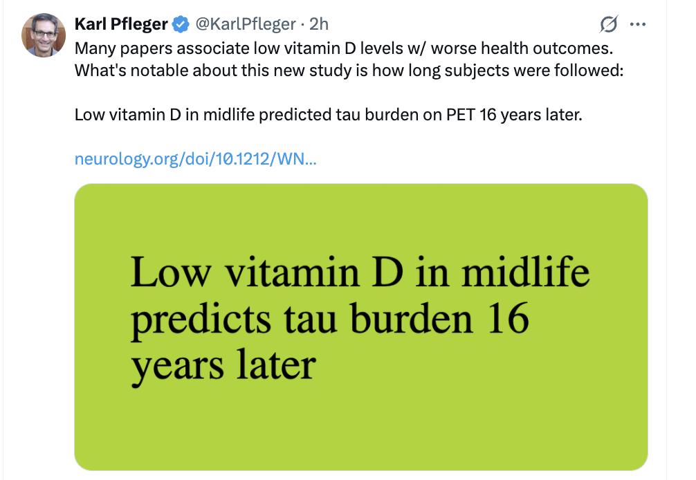
Association of Circulating Vitamin D in Midlife With Increased Tau-PET Burden in Dementia-Free Adults
Paper: https://www.neurology.org/doi/10.1212/WN9.0000000000000057
5 Likes
I am doing some experiments at the moment trying to work out whether cholecalciferol taken whilst fasting creates more serum 25OHD or not.
However, regardless I tend to think I want to be at least 100 nmol/l (40ng/ml).
4 Likes
Among patients with previously untreated mCRC, addition of high-dose vitamin D3, vs standard-dose vitamin D3, to standard chemotherapy plus bevacizumab did not improve PFS.
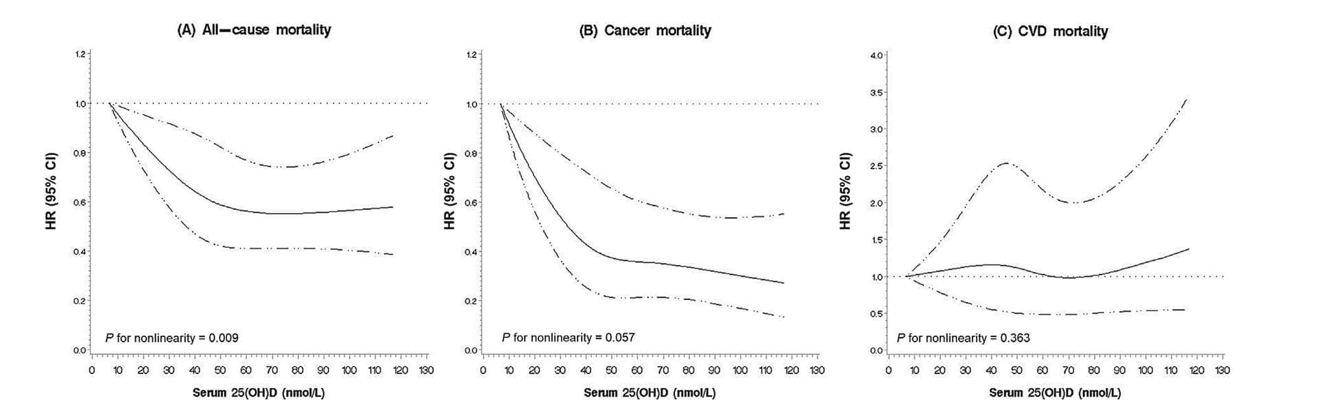
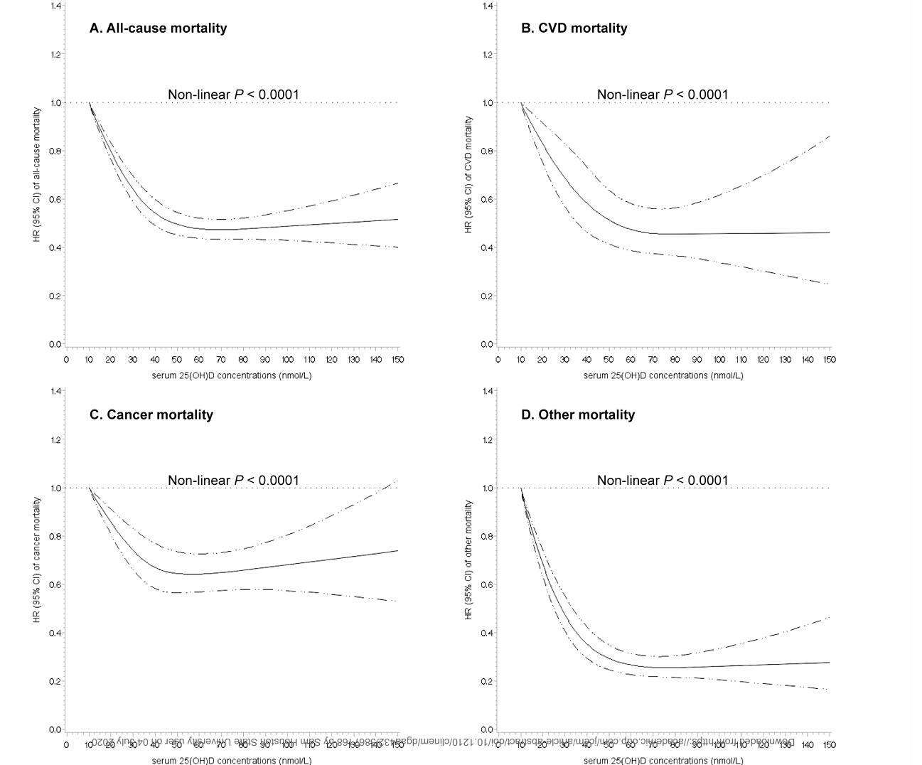
Judging by metrics like ACM, the optimal 25(OH)D level seems to be around 75 nmol/L. But with long-term supplementation, even just 2,000 IU/day of Vitamin D is enough to push serum levels above 110 nmol/L. Meanwhile, most over-the-counter supplements sold today are 5,000 IU. It’s hard to believe the sweet spot for VD dosage is actually this narrow.
What bothers me is that here we are in 2026, yet most clinical trials are still stuck using 2,000 IU or 4,000 IU daily. And if baseline levels are already around 30 ng/mL, adding another 2,000–4,000 IU shows virtually no benefit. I personally suspect the issue might be that these trial doses are simply too small, but mainstream medicine remains very resistant to long-term high doses like 10,000–20,000 IU daily.
@CronosTempi , what’s your take on this?
The problem is that the relationship between daily cholecalciferol and calcifediol (25OHD) is not that easy to map. There is a limit as to how much can be converted each day. This varies by individual.
I like to keep my calcifediol quite high at least 150 (uk units nmol/l). I feel a bit manky when i go as low as 90.
I have a mechanistic theory that if there are say 1,000 or so genes which are expressed from the VDR (calcitriol receptor) then I am inclined to have more of them functioning.
Still looking at those charts (and it is not clear where the source is) there would be arguably a merit of increasing up to say 75 (uk units) 30 (us units). However, not necessarily a demerit of increasing beyond there.
Using calcifediol to supplement I did get as high as 400.
I don’t think there is a good argument to megadose with cholecalciferol mainly because it does not convert reliably and secondly I think unprocessed cholecalciferol is mildly toxic (it disrupted my sleep when I did the experiments).
In itself, that is an argument against bolus doses of cholecalciferol. If people want a loading dose it should be calcifediol.
1 Like
As a matter of curiosity here are my results since February 22, I went weekly in May 22
90 168 116 419 337 209 359 257 225 198 185 174.55 185 120.7 120.08 160 170.2 3000+10xded 185.35 odd result maybe sun 147 123 108 less sun basic 3000iu 184 15 drops 25OHD each day 165.69 139.81 144.78 odd result given no 25OHD 143.98 123.83 needs some 25OHD 209.93 upper 220 174 147.18 153 147 195.29 finished off a couple of bottles of dedrogyl no quantities 205.58 220.15 137.7 235.64 230.76 206.5 191 216.11 195.14 183.55 181 178 196 179.7 207 191 170 249 211 216 253 247 214 201 195 206 176 184 140 is this an error? Or varioxia 171 170 213 202 202 186 196 193 189 182 161 204 167 187 205 184 194 196.89 198 245 220 230.84 188 197 188 182 181 200 216 66 looks like someone else’s sample 169 166 190 175 200 now on 6000iu 179 166 161 167.93 may stabilise at this level 112 didn’t stabilise 101 still going down 176 10 x dedrogyl daily going up quite a bit 193 still 10xded but now moving to 5xded 194 5xded 140 5xded 194 154 seem to have used a lot 238 153 only 3000iu 201 144 215 quite a bit of dedrogyl 209 231 210 171 187 182 189 194 169 164 297 175 207 222 146 208 189 203.05 220 188 180 188 189 >241 7 196 160 225 138 150 170 140 181 179 178 223 204 184 194 152 145 146 141 123 141 100 84 233 223 222 202 205 208 197 187 164 176 172 181 182 168 163 148 264 204 197 186 176 174 145 132 142 130 143 187 257 238 213 204 194 238 213 173 156 182 194 170 159 162 140 147 131 146 131 128 136 122 110 131 123 129 124 183 212 212 273 258 208 208
Obviously without the full analysis of what I was eating, supplementing, walking around in the sun etc there is not a lot that can be read into this.
I normally supplement with 3,000iu of cholecalciferol.
I also don’t know how accurate the test is.
Once serum 25(OH)D exceeds 75 nmol/L, we see a sharp rise in either CVD mortality or cancer mortality, alongside an uptick in overall all-cause mortality. Personally, I suspect this spike at higher concentrations might just be an artifact of smaller sample sizes at those levels, leading to a strange upward curve.
However, if that right-hand rise in the U-curve is actually real, then the 2,000 IU or 5,000 IU supplements on the market are surprisingly unsafe, and 400 IU to 600 IU might turn out to be the true sweet spot.
Once serum 25(OH)D exceeds 75 nmol/L, we see a sharp rise in either CVD mortality or cancer mortality, alongside an uptick in overall all-cause mortality.
Can you please give me a link to the source for this data?
However, if that right-hand rise in the U-curve is actually real, then the 2,000 IU or 5,000 IU supplements on the market are surprisingly unsafe, and 400 IU to 600 IU might turn out to be the true sweet spot.
The problem is that 3,000 iu struggled in the past with me to maintain 90 (uk units).
1 Like
I actually am not a big vitamin D enthusiast. There’s been consistent hype from certain quarters to push for high serum levels and supplementation. But the evidence simply is not there, sorry. I see all sorts of graphs, but the CI is usually crazy wide. Incidentally, this controversy is as old as the hills. Already back in the early 2000’s on the CRSociety site there were vigorous discussions about optimal vitamin D serum levels. From what I remember, the conclusion from all the evidence available at the time (as expressed by Michael Ray) was that 25ng/mL (65 nmol/L) is good enough, and no reason to go much beyond. However, if you look at the literature the range is super wide. I think you can go as high as 100 nmol/L and be fine, but there really is no good reason to go higher.
Supplementation is a pointless discussion. Why? Because the individual differences in translation to serum levels are exceptionally broad. Some people barely look at a pill and their levels jump, others gobble up 10K IU and can’t get it to budge. There really is only one way: measuring blood levels - this incidentally is a point made frequently by Matt Kaeberlein, who regards discussions of supplement doses as pointless, you need to “dose + measure”. Don’t fall below 20ng/mL though.
I’m at 38ng/mL, so just above 90nmol/L and OK with that level which I hit with a small amount of sun exposure and 1000 IU daily D3 supp. I see no reason to go higher - no convincing evidence or study.
1 Like
I think this is the key point we should have no real problem getting to a consensus on.
One reason why I tend to go for highish serum levels is that there is an argument that in certain circumstances it encourages autophagy.
I will give a response from chatGPT (5.5paid) on this:
Yes. There is reasonably strong mechanistic evidence that higher availability of 25-hydroxyvitamin D [25(OH)D, calcifediol] can increase autophagy, but the important qualification is that much of the effect appears to occur after intracellular conversion of 25(OH)D to 1,25(OH)₂D (calcitriol). The strongest evidence comes from macrophages and antimicrobial autophagy.
The clearest chain is:
higher extracellular 25(OH)D → more substrate for CYP27B1 → increased intracellular 1,25(OH)₂D → VDR activation → cathelicidin/LL-37 → autophagy
1. Macrophage experiments directly implicate 25(OH)D availability
Campbell and Spector’s work on human macrophages is particularly relevant. Macrophages express CYP27B1, allowing them to convert circulating 25(OH)D into calcitriol locally. They found that stimulation of macrophages induces CYP27B1 and that vitamin-D signalling then increases CAMP/cathelicidin and autophagic flux. Importantly, they explicitly argue that because macrophage CYP27B1 is not regulated in the same way as renal CYP27B1, its production of calcitriol can be substantially substrate-dependent on the concentration of 25(OH)D available to the cell. (PubMed Central (PMC))
They describe the pathway roughly as:
25(OH)D₃
→ CYP27B1
→ 1,25(OH)₂D₃
→ VDR
→ CAMP/LL-37
→ autophagosome formation / autophagic flux.
In vitamin-D-deficient serum, macrophages fail to generate the same response. (PubMed Central (PMC))
2. A striking threshold/substrate experiment
One of the more interesting findings concerns macrophages cultured in human serum.
When serum contained low 25(OH)D, macrophage antimicrobial responses were impaired. Replacing it with vitamin-D-sufficient serum restored the response in a 25(OH)D-concentration-dependent fashion. A review summarising these experiments notes that serum above about 30 ng/mL (75 nmol/L) could rescue the macrophage vitamin-D-dependent pathway. (Frontiers)
This isn’t proof that 75 nmol/L is an optimal systemic autophagy threshold, but it is unusually direct evidence that availability of 25(OH)D can be rate-limiting for a cellular pathway involving autophagy.
3. Mycobacterial TLR stimulation provides a detailed mechanism
Shin et al. studied human primary monocytes exposed to a mycobacterial TLR2/1 ligand. They found:
TLR2/1 activation
→ AMPK/p38 signalling
→ increased CYP27B1
→ conversion of 25(OH)D → 1,25(OH)₂D
→ VDR activation
→ cathelicidin expression
→ autophagy activation.
Blocking parts of this pathway impaired the autophagic response. (PubMed Central (PMC))
This is particularly convincing because it connects the availability of 25(OH)D to a fairly well-defined autophagic signalling pathway rather than merely finding an association.
4. Calcitriol itself clearly stimulates autophagy
There is stronger experimental evidence for calcitriol itself.
Physiologically relevant concentrations of 1,25(OH)₂D induced autophagy in human macrophages through pathways requiring ATG5, Beclin-1 and PI3K, and reduced HIV replication. (PubMed)
Related work showed that vitamin-D-induced autophagy could suppress both M. tuberculosis and HIV and was dependent on cathelicidin and autophagic maturation. (PubMed Central (PMC))
Thus the biological plausibility of the second half of the chain:
calcitriol → VDR → autophagy
is quite strong.
5. What higher serum 25(OH)D probably does
The implication is slightly different from saying that 25(OH)D itself is an autophagy agonist.
I would describe the evidence as:
25(OH)D acts as a substrate whose availability can determine how much locally produced calcitriol a CYP27B1-expressing cell can make, and thereby how strongly vitamin-D-dependent autophagy can operate.
This may be particularly important in cells expressing inducible CYP27B1, including macrophages.
There is evidence that very low 25(OH)D becomes limiting. Campbell et al. cite failure of macrophage responses at approximately <25–45 nmol/L, depending on the activating stimulus. (PubMed Central (PMC))
So a plausible relationship is:
| Serum 25(OH)D | Likely consequence for this pathway |
|---|---|
| <25 nmol/L | Strong substrate limitation |
| 25–50 nmol/L | Potentially limited intracrine calcitriol production |
| 50–75 nmol/L | Increasingly adequate substrate |
| ≥75 nmol/L | Macrophage pathway appears largely rescued in some experiments |
| Much higher levels | Little evidence that autophagy continues increasing proportionally |
That final point is important. I don’t think the literature currently establishes a monotonic relationship such as 75 → 125 → 175 nmol/L producing progressively more systemic autophagy.
A useful counterexample
A human osteoarthritis study gave 40,000 IU/week vitamin D₂ for six months and measured circulating LC3A. Surprisingly, supplementation reduced serum LC3A, despite raising vitamin-D status. (PubMed Central (PMC))
That result doesn’t necessarily mean autophagy fell: circulating LC3A isn’t a clean measure of tissue autophagic flux, and pathological accumulation of LC3 can reflect blocked rather than enhanced autophagy. But it illustrates why serum biomarker studies shouldn’t simply be interpreted as “more vitamin D = more autophagy.”
Bottom line
I think the evidence supports this statement quite well:
Raising low 25(OH)D into the sufficient range can enable or increase autophagy in CYP27B1-expressing cells by providing substrate for local calcitriol synthesis.
The evidence is much weaker for:
raising an already sufficient 25(OH)D from, say, 75 to 150 nmol/L causes substantially greater whole-body autophagy.
The macrophage work is especially interesting because it suggests that serum 25(OH)D itself can be the rate-limiting reservoir, while serum calcitriol may tell you relatively little about this intracrine pathway.
If useful, I can next look specifically for experiments that plot autophagic flux against 25(OH)D concentration, to see whether there is evidence for a dose-response curve and where it saturates.
and
I found very little evidence that directly plots true autophagic flux against a range of 25(OH)D concentrations. Most papers either use one 25(OH)D dose or generate a dose-response using 1,25(OH)₂D (calcitriol) instead. That means there is not currently a convincing experimentally determined “25(OH)D saturation concentration for autophagy.”
The most relevant studies are these:
| Study/model | Vitamin D species and concentrations | Autophagy measurement | Dose-response/saturation information |
|---|---|---|---|
| Sustova et al., C2C12 muscle cells | 25(OH)D₃, 100 nM | LC3 with/without chloroquine; LC3–LAMP2 colocalisation | Direct evidence that 100 nM increases flux, but only one 25(OH)D concentration tested for flux |
| Cervical SiHa cells | 25, 60, 250, 2500 nM 25(OH)D₃ | LC3-II flow cytometry | No significant increase over the entire range; not a rigorous flux assay |
| Tian et al., intestinal cells | 25(OH)D₃ | Beclin-1, p62, LC3 plus inhibitors | Supports increased autophagic maturation, but does not provide a useful graded concentration-response curve |
| Campbell/Spector, human macrophages | 1,25(OH)₂D₃ dose range | LC3B-II + lysosomal inhibition | Clear dose response; response approaches maximum around 1 nM calcitriol |
| Macrophage intracrine studies | extracellular 25(OH)D | CYP27B1 → calcitriol → CAMP/autophagy | Suggest deficiency becomes limiting below roughly 25–45 nM, but do not directly plot flux versus 25(OH)D |
The strongest direct 25(OH)D result: 100 nM
Sustova et al. treated differentiated C2C12 muscle cells with 100 nM 25(OH)D₃ for 24 hours. Importantly, they did not simply measure LC3-II; they used chloroquine to block autophagosome clearance.
25(OH)D produced strong LC3 accumulation only when chloroquine was present. That is the pattern expected when basal autophagosomes are being rapidly cleared and therefore indicates increased autophagic flux rather than blocked autophagy. LC3/LAMP2 colocalisation gave consistent evidence of increased autophagolysosome formation. (PubMed)
So we can say fairly confidently:
100 nM 25(OH)D₃ can stimulate autophagic flux in skeletal-muscle cells.
But the experiment unfortunately does not tell us whether:
50 nM < 75 nM < 100 nM < 150 nM
in terms of autophagy.
A concentration-series experiment actually produced a negative result
The SiHa cervical-cell study is useful because it tested a wide range:
25 → 60 → 250 → 2500 nM 25(OH)D₃
and measured LC3-II. There was no significant increase in LC3-II across the concentration range. (PubMed Central (PMC))
There are two important caveats. First, this was measuring autophagy-associated cell death rather than carefully measuring basal autophagic flux. Second, LC3-II alone can be misleading because rapid autophagic clearance can actually lower steady-state LC3-II.
Nevertheless, this demonstrates something important: 25(OH)D does not universally produce a concentration-dependent increase in autophagy in every cell type.
The macrophage data suggest a lower threshold rather than a high optimum
The human macrophage literature provides a useful indirect clue.
TLR-activated macrophages increase CYP27B1 and convert:
25(OH)D → 1,25(OH)₂D → VDR → cathelicidin → autophagy.
Campbell and Spector note that TLR2/1-mediated responses fail when 25(OH)D concentrations are below approximately 25 nM, while IFN-γ-dependent responses can become impaired below approximately 45 nM. (PubMed Central (PMC))
That suggests something more like:
very low 25(OH)D → substrate limited
followed by
adequate 25(OH)D → sufficient local calcitriol → effective autophagy
rather than evidence for unlimited enhancement at progressively higher concentrations.
We do have a saturation curve for calcitriol
This is where the evidence becomes much better.
Campbell and Spector exposed human macrophages to increasing concentrations of 1,25(OH)₂D₃ and measured LC3B-associated autophagosomes. The percentage of LC3-positive cells rose significantly with calcitriol dose, and they describe 1,000 pM = 1 nM calcitriol as sufficient to produce the maximal response. True flux was confirmed using lysosomal inhibition. (PubMed Central (PMC))
The response was already biologically strong at much lower concentrations: around 50 pM produced substantial effects, and 100 pM was strongly active. (PubMed Central (PMC))
So there is a genuine saturating response approximately of the form:
calcitriol 0 → 50 pM → 100 pM → 1,000 pM
low → increasing → strong → approximately maximal autophagic response
But one cannot directly translate that into serum 25(OH)D because intracellular calcitriol generation depends on CYP27B1, cell type, activation state, vitamin-D-binding protein, CYP24A1 and other factors.
What concentration of 25(OH)D would I predict gives near-maximal effect?
Based on these data—not as a directly measured fact—I would currently expect something roughly like:
| 25(OH)D | Likely effect in CYP27B1-responsive cells |
|---|---|
| <25 nM | Clearly substrate-limited in some immune-cell systems |
| 25–50 nM | Increasing substrate availability; some pathways still limited |
| 50–75 nM | Probably substantial restoration |
| 75–100 nM | Plausibly near the plateau for many intracrine vitamin-D responses |
| 100–150 nM | Possible additional effect in some tissues, but direct evidence for additional autophagic flux is lacking |
| >150 nM | No convincing evidence that autophagy continues increasing |
The 100 nM concentration is particularly interesting, because that is exactly the concentration at which Sustova et al. directly demonstrated increased autophagic flux in muscle cells. (ResearchGate)
But that should not be interpreted as evidence that serum 25(OH)D of 100 nM is an experimentally established autophagy optimum.
One result makes the biology even more interesting
The Sustova study found something rather unexpected: 25(OH)D increased autophagic flux, whereas 1,25(OH)₂D could actually block it in these muscle cells. The authors linked the opposing effects partly to differential induction of CYP24A1 and production of 24-hydroxylated metabolites. (PubMed)
So the simple model
more 25(OH)D → more calcitriol → more autophagy
is probably incomplete outside macrophages.
My current reading of the evidence is therefore that 25(OH)D around 75–100 nM is biologically plausible as a range in which vitamin-D-dependent autophagy is no longer strongly substrate-limited, but I have not found an experiment demonstrating that autophagic flux rises progressively from 50 → 75 → 100 → 125 nM and then plateaus.
The experiment that would really answer the question would expose primary human cells to, say, 10, 25, 50, 75, 100, 150 and 250 nM 25(OH)D, measure flux using LC3-II ± bafilomycin/chloroquine or tandem mCherry-GFP-LC3, and simultaneously measure intracellular 1,25(OH)₂D and 24,25(OH)₂D. I cannot find a published study that has done that cleanly.
JH: If it only soups up the macrophages that is good enough for me.
1 Like