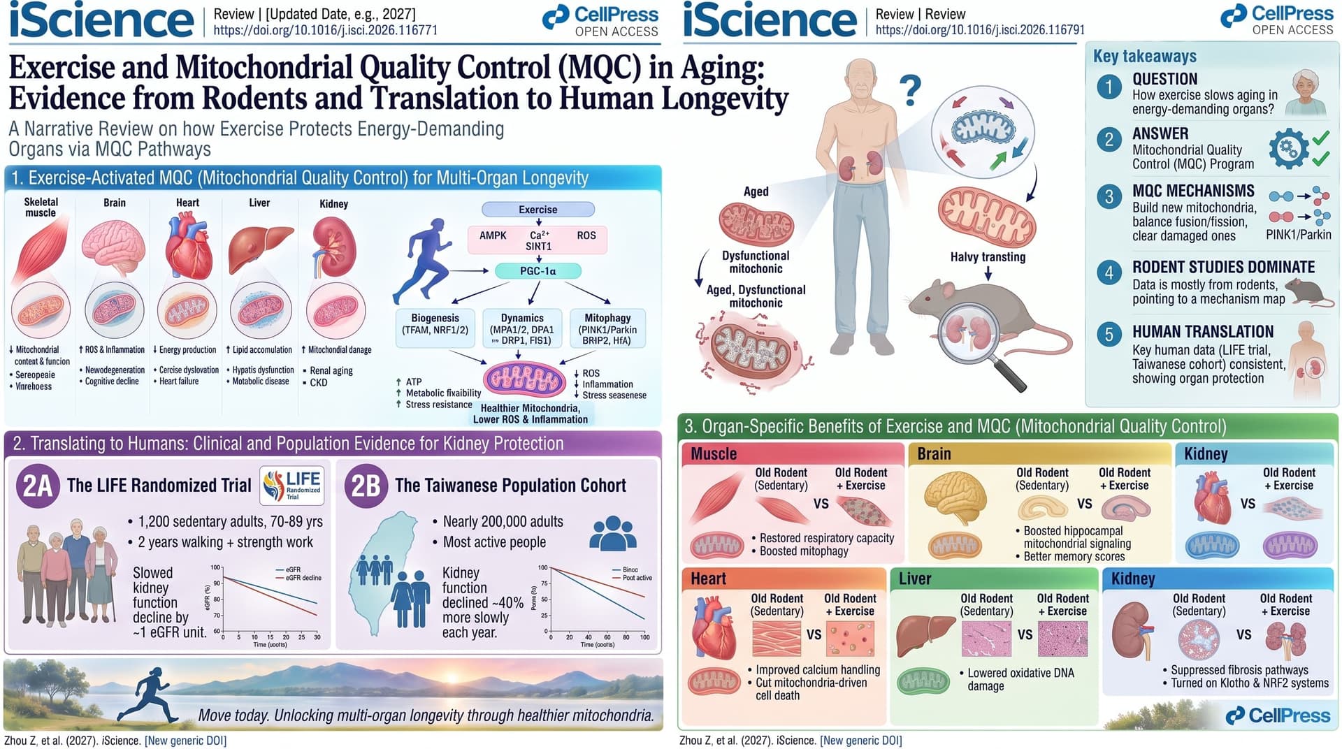
This narrative review from Chinese researchers asks one question: how does exercise slow aging in the organs that burn the most energy? Those are skeletal muscle, liver, brain, kidney and heart. The authors argue that the answer is mitochondrial quality control. Exercise switches on the AMPK and PGC-1alpha signals that build new mitochondria. It balances fusion and fission, clears out damaged mitochondria through mitophagy, and limits the oxidative, inflammatory and apoptotic signals that come from failing mitochondria. The review is a map of possible mechanisms, drawn mostly from rodent studies. Where human data exist, they point the same way.
Narrative
Your liver burns about 200 kilocalories per kilogram of tissue each day. Your heart and kidneys burn about 440, and your brain about 240. Resting skeletal muscle burns only about 13. These organs run almost entirely on mitochondria, which makes them among the first to suffer when mitochondria wear out. A new review in iScience argues that exercise is still the most reliable tool we have to slow that wear, and that it acts on the same basic machinery in every organ.
The shared mechanism goes like this. Hard muscular work drains ATP, and the rising ratio of AMP to ATP switches on AMPK, the cell’s fuel gauge. AMPK activates PGC-1alpha, a master regulator that turns on the genes for building new mitochondria and copying their DNA. Exercise also changes mitochondrial shape. Endurance work tends to favor fusion, which joins mitochondria into networks that share healthy components. Resistance work and high intensity tend to favor fission, which splits damaged pieces off so the cell can destroy them through mitophagy. The result is more mitochondria, and better ones.
The authors then go through five organs. In aged rodent muscle, training restores respiratory capacity and mitophagy. In the liver, eight weeks of treadmill running in 21-month-old rats lowered oxidative DNA damage to levels seen in rats half their age. In the brain, swimming and resistance programs in old rats restored hippocampal mitochondrial signaling alongside better memory scores. In the kidney, exercise suppressed fibrosis pathways and turned on the protective Klotho and NRF2 systems. In the heart, it improved calcium handling and cut mitochondria-driven cell death.
Human data are thinner but consistent. In the LIFE randomized trial, which followed about 1,200 sedentary adults aged 70 to 89, two years of walking plus strength work slowed kidney function decline by roughly 1 unit of eGFR. In a Taiwanese cohort of nearly 200,000 adults, the most active people lost kidney function about 40 percent more slowly each year than the least active.
The review is also candid about the limits. Too much exercise backfires. Exhaustive or high-intensity bouts in old animals raised oxidative damage in the heart and triggered brain inflammation. Some studies found no benefit at all, including one on old rats with kidney disease. The gains also fade quickly. Older adults lost their training-induced mitochondrial enzyme activity faster than young adults after they stopped. Forced exercise in rodents generally did worse than voluntary running, and sex, hormones and baseline fitness all changed the results.
The review’s value is its cross-organ framing. Aging does not strike one organ at a time, and exercise does not act on one organ at a time either. Its weakness is that it catalogues associations without weighing them. It does not rank studies by quality, run a meta-analysis, or say which pathway matters most. For the field, the message is not that exercise works. We knew that. The message is that mitochondrial quality control may be the common currency, and that dosing by age, and possibly by organ, is the unsolved problem.
Actionable Insights
The best-supported human protocols in the review are modest. Around 150 minutes of brisk walking per week plus light strength work, which was the LIFE trial design, cut the share of older adults with rapid kidney decline from 32.2 percent to 25.9 percent. That is about 6 fewer people with fast decline per 100 people exercising, roughly a 20 percent relative reduction. Put another way, about 16 people need to exercise for two years to spare one person from rapid decline. Average kidney function was 0.96 eGFR units better after two years. That is real but small, about one year’s worth of normal age-related loss.
In the population data, highly active adults lost 0.27 eGFR units a year versus 0.46 for the least active, and they had about 9 percent lower risk of developing chronic kidney disease. Because this was observational, part of that gap may reflect healthier people choosing to exercise.
For muscle, 10 weeks of full-body resistance training twice weekly (3 sets of 10 to 12 reps) raised mitochondrial respiratory complex levels in adults around 59. The practical rules are these: combine aerobic and resistance training, avoid chronic exhaustive sessions after middle age, and never stop. Older muscle loses its mitochondrial gains faster once training ends.
Context and Source
- Open Access paper: Exercise-induced mitochondrial remodeling in energy-demanding organs during aging
- Institutions: Anhui University of Chinese Medicine (Hefei) and Wuhan Sports University (Wuhan)
- Country: China
- Journal: iScience (Cell Press), published August 21, 2026
- Impact evaluation: The impact score of this journal is 4.5 (JIF), evaluated against a typical high-end range of 0 to 60+ for top general science, therefore this is a Medium impact journal.