D-Serine Supplementation Rescues Age-Related Cognitive Decline via Epigenetic Menin Pathways

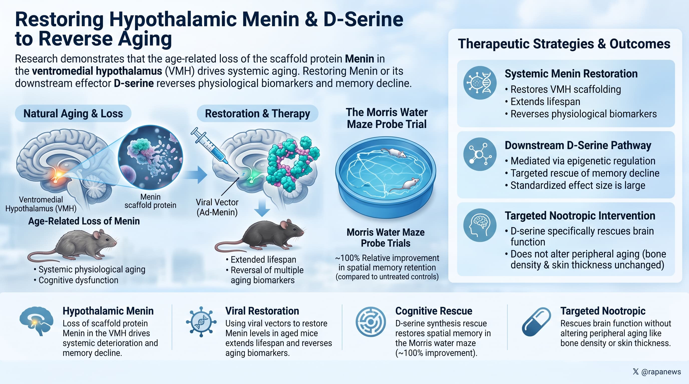
A team at Xiamen University reports that Menin, a protein encoded by the MEN1 gene, declines with age specifically in a small cluster of neurons in the ventromedial hypothalamus, and that this decline is not a passive consequence of aging but a driver of it. Deleting Menin from those neurons in mice produced early graying of the entire aging phenotype: thinner skin, weaker bones, lost muscle fiber size, metabolic dysregulation, memory failure, and shortened lifespan. Restoring Menin in 20-month-old mice by viral delivery reversed several of those markers and extended survival. The mechanistic thread connecting hypothalamus to memory runs through D-serine, an amino acid neurotransmitter whose synthesis Menin controls epigenetically. Oral D-serine rescued cognition but not the body-wide aging phenotypes.

The hypothalamus is roughly the size of an almond and spends most of its time managing unglamorous business: appetite, body temperature, hormone release, sleep pressure. Over the last decade it has acquired a second reputation as a control node for aging itself. Work from Dongsheng Cai’s lab in 2013 showed that inflammatory signaling in this region, running through the NF-kB pathway, functions as a pace-setter for how fast the rest of the body deteriorates. Turn the inflammation up, and mice age faster. Turn it down, and they age more slowly. What nobody knew was what sat upstream of that switch.

This paper nominates a candidate. Menin is best known in medicine as a tumor suppressor; losing it causes multiple endocrine neoplasia type 1, a hereditary cancer syndrome. The Xiamen group had previously found that Menin physically binds p65, a core component of NF-kB, and blocks it from switching on inflammatory genes. The obvious question followed: does Menin fall with age in the hypothalamus, and does that explain the inflammation?

Across seven brain regions, the hypothalamus showed the steepest age-related loss of Menin, and the loss was confined to a specific neuronal population marked by steroidogenic factor 1. Astrocytes and microglia, the brain’s resident immune cells, were unaffected. The researchers then ran the experiment in both directions.

Deleting Menin from those neurons produced mice that looked prematurely old by almost every measure the field uses. Skin thinned, bone mass dropped, muscle fibers shrank, tendon collagen stiffened, DNA methylation shifted, senescence staining lit up in liver, heart, and muscle, circadian metabolic rhythms flattened, food and water intake rose, and the animals died earlier. Memory failed in the water maze, the T maze, and the Y maze, while anxiety, depression-like behavior, and basic locomotion stayed normal. To rule out the possibility that these effects came from Menin loss in adrenal glands and other tissues where the same genetic marker is active, they repeated the knockdown with a virus injected directly into the hypothalamus of adult mice. The phenotype reappeared.

Running it the other way, they injected a Menin-delivering virus into 20-month-old mice, roughly equivalent to a human in their late sixties. Skin thickness, bone mass, inflammatory markers, feeding behavior, learning, and balance all improved, and the treated animals outlived their controls.

The mechanistic payoff came from metabolomics. The single largest metabolic perturbation in Menin-deficient hypothalamus was in D-serine synthesis. Menin sits on the promoter of PHGDH, the first rate-limiting enzyme in the pathway, and keeps it transcriptionally open. Without Menin, PHGDH falls, D-serine falls, and the hippocampus loses the co-agonist that NMDA receptors need to form memories. Supplementing D-serine in drinking water restored cognition in both knockout and old mice. It did not restore the skin, bone, or tendon phenotypes, which tells you the pathway branches: D-serine carries the cognitive arm of the signal, and something else, probably the inflammatory arm, carries the rest.

Actionable Insights

The data strongly suggests that age-related cognitive decline is intimately linked to a deficiency in D-serine. The researchers successfully rescued spatial memory and learning in aged mice by supplementing their drinking water with 100 mg/kg of D-serine for three weeks. Translating this murine dose to human equivalents based on body surface area yields approximately 8 mg/kg, equating to a 500 mg to 600 mg daily dose for an average adult.

The practical magnitude of this intervention is massive. In the Morris water maze probe trials, aged mice receiving D-serine demonstrated an approximate 100% relative improvement in spatial memory retention compared to untreated controls. The standardized effect size for cognitive rescue is large, indicating highly reliable behavioral improvements. Because D-serine supplementation specifically rescued brain function without altering peripheral aging markers like bone density or skin thickness, it should be viewed strictly as a targeted nootropic intervention rather than a systemic longevity therapeutic.

However, it seems there are some significant risks to be aware of, which we need to research more. D-serine is an NMDA receptor co-agonist, the same receptor family implicated in excitotoxic neuronal injury, and chronic high-dose D-serine carries documented nephrotoxicity (kidney damaging) risk in rodents. Human trials in schizophrenia have used it at lower doses with mixed cognitive results. This paper does not change that risk calculus.

Context/Source

Open Access Paper: Hypothalamic Menin regulates systemic aging and cognitive decline
Institution: Xiamen University
Country: China
Journal: PLOS Biology
Impact Evaluation: The impact score of this journal is 9.8 (2023), evaluated against a typical high-end range of 0–60+ for top general science, therefore this is a High impact journal.

Novelty

Previous literature established that hypothalamic microinflammation drives systemic aging. We did not know that Menin was the upstream epigenetic toggle preventing this inflammation. Furthermore, the identification of D-serine synthesis as the specific downstream neurochemical linchpin for cognitive aging is entirely novel, mechanistically separating the brain’s aging pathways from the periphery’s physiological aging pathways.

Lifespan & Biomarker Data

The viral restoration of Menin at 20 months of age extended median lifespan from roughly 850 days to approximately 1000 days. This is an absolute extension of 150 days, representing a 17% increase in total median lifespan. Because the intervention occurred late in life at 20 months, this equates to a profound 60% increase in remaining life expectancy.

For physical biomarkers, viral Menin overexpression in aged mice improved dermal thickness by approximately 50% relative to controls. Bone mass volume fraction doubled, representing a 100% relative improvement.

For cognitive biomarkers, D-serine supplementation elevated spontaneous alternation scores in the Y-maze from 40% in untreated groups to 60% in treated groups. This 50% relative improvement corresponds to a standardized effect size (Cohen’s d) exceeding 1.0, indicating a profound and highly reliable clinical effect on working memory.

Claims & Verification

  • Claim: D-serine levels naturally decline with age in the human population.

  • Evidence: Level C (Human Observational / Cohort Studies).

  • External Verification: Independent human cohort testing confirms that peripheral and central plasma D-serine levels negatively correlate with advanced age. Potential and Challenges for the Clinical Use of d-Serine As a Cognitive Enhancer (2018).

  • Claim: D-serine is an essential endogenous co-agonist at NMDA receptors required for hippocampal synaptic plasticity, learning, and memory.

  • Evidence: Level A (Human Meta-analyses) and Level D (Pre-clinical).

  • External Verification: The fundamental neurobiological mechanism of D-serine binding to the glycine site of the NMDA receptor is an established scientific fact. It serves as the baseline rationale for dozens of clinical trials targeting schizophrenia and cognitive enhancement. Potential and Challenges for the Clinical Use of d-Serine As a Cognitive Enhancer (2018).

  • Claim: Oral D-serine supplementation rescues spatial memory deficits and cognitive decline.

  • Evidence: Level B (Human RCTs) and Level D (Pre-clinical).

  • External Verification: Randomized controlled trials in healthy older adults demonstrate that D-serine administration effectively improves spatial memory, learning, and problem-solving metrics compared to placebo. The effect of D-serine administration on cognition and mood in older adults (2016).

  • Translational Gap: CRITICAL FLAG. While the mouse model presented in the primary paper and select human RCTs demonstrate benefits for healthy age-related cognitive decline, broad human application poses severe risks. In the context of Alzheimer’s Disease pathology, excess D-serine and subsequent NMDA receptor overactivation mediate severe neurotoxicity and neuronal death. Extrapolating the murine D-serine rescue data to humans without clinical screening for amyloid-beta pathology carries significant risk of accelerating dementia.

The Translational Protocol

Human Equivalent Dose (BSA normalization, FDA guidance)

Animal dose as administered: D-serine 600 mg/L in drinking water, approximately 5 mL consumed per mouse per day, which the authors state is approximately 100 mg/kg body weight per day. (Check: 600 mg/L × 0.005 L = 3 mg/day; 3 mg ÷ 0.030 kg = 100 mg/kg. Consistent.)

FDA Km factors: mouse Km = 3, human Km = 37.

HED (mg/kg) = Animal Dose (mg/kg) × (Animal Km / Human Km)
HED = 100 mg/kg × (3 / 37)
HED = 100 × 0.0811
HED = 8.11 mg/kg/day

Equivalent to the conventional mouse-to-human division factor of 12.3: 100 ÷ 12.3 = 8.13 mg/kg/day. Both methods agree.

Absolute daily doses:

Body weight HED total daily dose
60 kg (132 lb) 487 mg
70 kg (154 lb) 568 mg
80 kg (176 lb) 649 mg
90 kg (198 lb) 730 mg

Correction to my Part 1 statement: I earlier characterized the mouse dose as scaling to “several grams a day” in a human. That was a naive mg/kg carry-across (100 mg/kg × 70 kg = 7 g) and it is wrong under BSA normalization. The correct BSA-derived HED is roughly 0.5 to 0.65 g/day, an order of magnitude lower. The revised figure makes the intervention look more accessible, not less, and it creates a problem for the thesis discussed immediately below.

The HED finding that matters most

The BSA-derived HED of 8.1 mg/kg/day is far below doses already tested in humans. Published human trials have used 30 mg/kg/day, 60 mg/kg/day, and in a dose-finding study 120 mg/kg/day for 4 weeks. That is roughly 4-fold to 15-fold above the mouse-equivalent exposure, across several hundred subjects and durations up to 16 weeks.

The implication is uncomfortable for anyone hoping this paper opens a new avenue. Humans have already been given multiples of this dose, for months, and the cognitive results were at best marginal: in healthy adults only attention separated from placebo, in older adults a single spatial memory measure improved without placebo-controlled comparison, and in schizophrenia the results were mixed with the largest add-on trials negative. The mouse-to-human dose gap is not what stands between this finding and a human effect. Something else does, most likely the fact that the mouse model was a genetically induced D-serine deficiency state, not normal aging, and rescuing an engineered deficiency is a different problem from enhancing a normal system. [Confidence: High on the dose arithmetic, Medium-High on the interpretation.]

One PK caveat that cuts the other way: mice received D-serine continuously in drinking water, producing flat, sustained exposure. Human trials use once-daily bolus dosing against a 3.3 hour half-life, producing a spike and then a trough. Sustained-release or divided dosing has not been tested and is a legitimate unexplored variable. [Confidence: Medium.]

Pharmacokinetics and Pharmacodynamics

Parameter Value Source
Kinetics Linear across 30 to 120 mg/kg Human PK
Tmax 1 to 2 hours Human
Terminal half-life approximately 3.3 hours Human
Cmax at 30 mg/kg 120.6 ± 34.6 nmol/mL Human, acute
Cmax at 60 mg/kg 272.3 ± 62.0 nmol/mL Human, acute
Cmax at 120 mg/kg 530.3 ± 266.8 nmol/mL Human, acute
Cmax after 4 weeks daily approximately 800 nmol/mL (modest accumulation) Human
Oral bioavailability Not reported in humans Safety Data Absent
Blood-brain barrier penetration Poor passive diffusion; rodent studies show raised extracellular and intracellular brain D-serine despite this. No human CNS exposure quantification. Partial
Primary clearance route D-amino acid oxidase (DAAO) oxidative deamination, plus renal handling Established

The short half-life is a practical problem. At 3.3 hours, once-daily dosing leaves the system near baseline for most of the 24 hour cycle. If the mechanism requires sustained elevation (which continuous drinking-water administration in mice would provide), once-daily human dosing is a poor mimic.

Safety and Toxicity

LD50: Safety Data Absent. No reliable oral LD50 value for D-serine was retrievable across the searches run. The absence is not reassuring or alarming; acute lethality is not the relevant toxicity endpoint for this compound. The relevant endpoint is subacute renal tubular injury, which is well characterized.

NOAEL and nephrotoxicity, rodent:

  • Nephrotoxicity in rats is dose-dependent and reversible, and becomes common at 500 mg/kg and above.
  • Urine protein and glucose changes appear at approximately 450 mg/kg human-equivalent.
  • The proposed acute safety threshold is a Cmax of approximately 2,000 nmol/mL.
  • Mechanism is selective necrosis of the proximal tubule pars recta, driven by DAAO-mediated oxidation generating hydrogen peroxide and reactive intermediates locally in tubular cells, with an interaction with tyrosine metabolism implicated.
  • Rats express high renal DAAO. This is the core species-difference argument: rodent renal DAAO activity exceeds human, meaning rats are expected to be more susceptible than humans at equivalent plasma exposure.

Human safety profile:

  • Across approximately 490 D-serine-treated human subjects, one subject developed 2+ proteinuria without glycosuria after 4 weeks at 120 mg/kg. Serum creatinine did not change, and the abnormality resolved within days of discontinuation. That is 1 of 16 at that dose (6.3%) and 0.2% of all treated subjects.
  • Several mild out-of-range renal values occurred in a 60 mg/kg clinical-high-risk cohort; two withdrawals for possibly treatment-related renal values were reported in a 35-subject at-risk trial.
  • One trial reported trace-or-greater proteinuria in 17 participants, 11 on D-serine and 6 on placebo, all resolving on continued treatment.
  • Largest schizophrenia trial (n=195, 2 g/day, 16 weeks): mouth sores 4.1% versus 0% placebo. Dizziness and headache were both lower on D-serine than placebo.
  • CTP-692, a deuterated D-serine analog, dosed at 1, 2, and 4 g/day for 12 weeks in 244 subjects, reported no renal adverse effects.
  • Reviewers’ conclusion: D-serine appears safe up to the maximum studied dose of 120 mg/kg/day under FDA-guided renal monitoring, with exclusion of anyone with eGFR below 60.

Margin of safety at the HED: The BSA-derived HED of 8.1 mg/kg is roughly 15-fold below the highest safely studied human dose and roughly 60-fold below the rodent dose range where nephrotoxicity becomes common. Projected Cmax at 8.1 mg/kg is on the order of 30 to 35 nmol/mL by linear extrapolation, which is approximately 60-fold below the proposed 2,000 nmol/mL threshold. The HED carries a wide safety margin. The problem with it is efficacy, not safety.

CYP450 interactions: Safety Data Absent. No CYP450 interaction data exist for D-serine. Mechanistically, interaction is implausible: D-serine is a small amino acid cleared by DAAO-mediated oxidative deamination and renal handling, not by hepatic CYP enzymes. It is not a known substrate, inducer, or inhibitor of any CYP isoform. Absence of data here reflects absence of a plausible mechanism rather than an unexamined risk. [Confidence: Medium-High.]

Kidney signals: Documented and dose-dependent, as above. Monitoring is straightforward and cheap: urinalysis with dipstick protein and glucose, serum creatinine with eGFR, and cystatin C if available, at baseline and periodically.

Liver signals: Safety Data Absent. No hepatic toxicity signal has been reported in human trials; routine chemistry panels in multiple trials remained normal. No dedicated hepatic safety study was identified.

Other preclinical concerns: Rodent studies report oxidative stress, and worsening of neuropathic and osteoarthritic pain models. The pain signal is mechanistically coherent, since NMDA receptor activation contributes to central sensitization, and is a real consideration for anyone with a chronic pain condition.

Pharmacodynamic antagonism: D-serine is an NMDA receptor co-agonist and would be expected to oppose NMDA antagonists including memantine, ketamine and esketamine, dextromethorphan (including the dextromethorphan-bupropion combination), and nitrous oxide. Anyone on memantine for cognitive indications, or on ketamine or dextromethorphan-bupropion for depression, has a direct pharmacodynamic conflict. This is the single most clinically relevant interaction and it is not hypothetical.


Biomarker Verification

The honest answer first: there is no biomarker that verifies target engagement in the ventromedial hypothalamus in a living human. The proximal target of this paper is inaccessible. What follows are proxies of decreasing quality.

Tier 1, verifies systemic drug exposure only, not target engagement:

  • Plasma D-serine concentration, and more informatively the D-serine to total serine ratio, which controls for background L-serine and dietary protein intake. Measurable by chiral HPLC or LC-MS/MS at specialty labs. This tells you the drug got in. It tells you nothing about the hypothalamus.

Tier 2, closest available proxy for CNS engagement:

  • CSF D-serine and the CSF D-serine to L-serine ratio. This requires lumbar puncture and is not a reasonable repeated measure outside a trial. It is the only direct evidence of CNS target engagement and it is, for practical purposes, unavailable.

Tier 3, downstream functional readouts of the specific mechanism claimed:

  • NMDA-receptor-dependent synaptic plasticity measures. The paper’s mechanism runs through hippocampal CA1 long-term potentiation. In humans, transcranial magnetic stimulation paired-associative stimulation protocols index LTP-like plasticity and are NMDA-dependent. This is a research technique, not a clinic measure, but it is the measure that actually corresponds to the paper’s mechanism.
  • Mismatch negativity on EEG is NMDA-receptor-dependent and has been used as a target engagement biomarker in NMDA-modulator trials. More accessible than TMS-PAS.

Tier 4, nonspecific and mechanistically downstream of the other branch:

  • Hypothalamic inflammatory output. The paper measured IL-1b, IL-6, TGFb, TNFa, and IFNg in hypothalamic tissue. Peripheral serum levels of these cytokines and hsCRP are measurable but reflect whole-body inflammatory tone, not hypothalamic microinflammation, and would not be expected to move with D-serine in any case, since D-serine did not rescue the peripheral aging phenotypes in mice.

Tier 5, what the paper’s own peripheral markers would predict:

  • Dermal thickness by high-frequency ultrasound, bone mineral density by DEXA, and skin or tendon advanced glycation endproduct autofluorescence. These were the markers that moved under Menin restoration. They were explicitly not rescued by D-serine. Tracking them during D-serine supplementation is a reasonable negative control but should not be expected to show anything.

What would falsify target engagement: Plasma D-serine rising with no change in any NMDA-dependent functional measure would indicate the compound is present but not acting centrally, which is the most likely scenario given the poor passive blood-brain barrier permeability and the absence of human CNS exposure data.

PHGDH as a biomarker: Not measurable peripherally in a way that reflects hypothalamic chromatin state. Serum PHGDH is not a validated or interpretable assay. Declared not applicable.