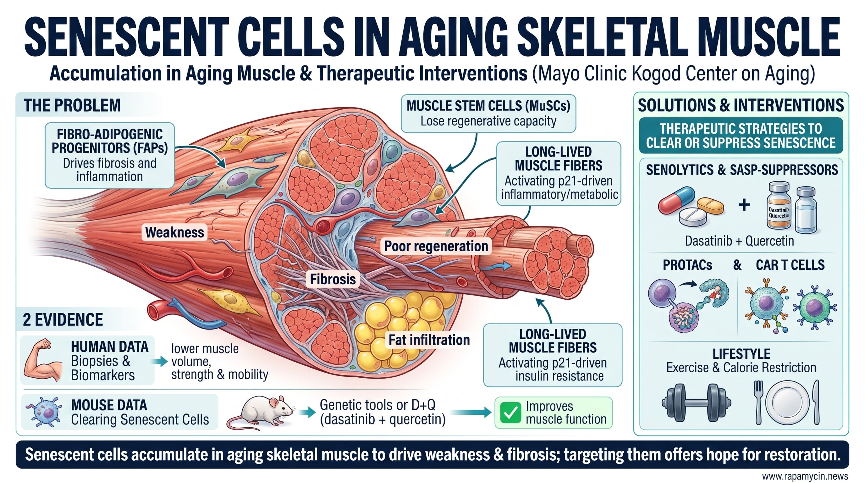
This invited review from the Mayo Clinic’s Kogod Center on Aging summarizes evidence that senescent cells accumulate in aging skeletal muscle and contribute to weakness, poor regeneration, fibrosis, fat infiltration and insulin resistance. The authors describe three main senescent populations: fibro-adipogenic progenitors that drive fibrosis and inflammation, muscle stem cells that lose regenerative capacity, and long-lived muscle fibers that switch on a p21-driven inflammatory and metabolic program. Human data from muscle biopsies and blood biomarkers link higher senescence burden to lower muscle volume, strength and mobility. In mice, clearing senescent cells with genetic tools or drugs such as dasatinib plus quercetin improves muscle function. The review also covers emerging senolytics, SASP-suppressing drugs, PROTACs and CAR T cells, along with exercise and calorie restriction.
Every adult loses muscle with age. After about 50, mass and strength decline steadily, and for some people the loss becomes sarcopenia and then frailty. Despite decades of research, no drug is approved to prevent or treat age-related muscle loss. A new review in the Annual Review of Physiology argues that a key piece of the puzzle may be senescent cells, sometimes called zombie cells because they stop dividing but refuse to die.
Senescence is a protective response to damage. When a cell’s DNA, telomeres or mitochondria are badly injured, it can lock itself into permanent growth arrest rather than risk becoming cancerous. The problem is that senescent cells do not sit quietly. They secrete a cocktail of inflammatory molecules, enzymes and growth factors known as the senescence-associated secretory phenotype, or SASP. Over time, that cocktail disrupts the tissue around them.
The authors, led by Nathan LeBrasseur, describe how senescence shows up across the cell types that make up muscle. Fibro-adipogenic progenitors, which normally help repair injured muscle, become senescent and pump out inflammatory signals that encourage scarring and fat build-up. Muscle stem cells, the reserve that rebuilds damaged fibers, slip from a reversible resting state into permanent senescence and lose their ability to regenerate tissue. Even the muscle fibers themselves, which never divide, switch on a protein called p21 and begin behaving like senescent cells, with inflammatory output and impaired responses to insulin.
The human evidence comes mainly from large studies of older adults. In the Study of Muscle, Mobility and Aging, muscle biopsies from more than 700 people showed that those with higher senescence signatures had smaller, fattier, weaker muscles and worse physical function. Blood levels of SASP proteins such as GDF-15 and activin A track with frailty and predict who will lose the ability to walk a quarter mile.
Animal studies go further. Clearing senescent cells in mice with genetic tools or with the drug combination dasatinib plus quercetin improves grip strength, endurance and muscle regeneration. Forcing muscle to overexpress p21 does the reverse, causing atrophy and weakness.
The review is strong on biology and candid about gaps. Nobody has yet cleared senescent cells specifically from muscle to test whether they drive sarcopenia directly. Senescence markers are notoriously unreliable, and some cells that look senescent, such as certain macrophages, may simply be activated. Muscle fibers are particularly hard to classify, and the p21 program in fibers overlaps with stress pathways that drive atrophy for other reasons.
The translational picture is less rosy than the review implies. Human trials of senolytics so far have been small, short and mostly focused on other conditions, with mixed results, and none has measured muscle as a primary outcome. One much-publicized natural senolytic, fisetin, failed to reduce senescent cells or extend lifespan in the rigorous NIH Interventions Testing Program.
The big idea is that muscle aging may be driven not only by wear and tear in the fibers but by a small population of dysfunctional cells that poison the neighborhood. For now, the best-supported ways to keep those cells in check are the oldest ones: exercise, especially resistance training, and avoiding excess body fat.
Actionable Insights
This is a review of mechanisms, largely from mouse studies. No senolytic has been shown to improve muscle in humans.
- Exercise first. In the authors’ own human studies, structured exercise lowered senescence-related transcripts in blood immune cells and reduced circulating SASP markers. Resistance training reduced senescent cells in fat tissue. Exercise is also the only intervention proven to build muscle in older adults.
- Manage body fat. Senescence biomarkers rise stepwise from normal weight to overweight to obesity.
- Modest calorie restriction lowered SASP markers in the CALERIE trial, which used about 12 percent restriction for two years. Older adults should weigh this against the risk of losing muscle.
- Senolytic drugs are still a work in progress. Human trials of dasatinib plus quercetin have been small, and a 2024 randomized trial in postmenopausal women missed its primary bone endpoint.
How big are the effects? In old mice, senescent cells are cleared about four times more slowly than in young mice. In one key mouse study, dasatinib plus quercetin extended remaining median lifespan by roughly a third, but that result has not been replicated in more rigorous lifespan testing programs. Safety Data Absent for senolytic use in sarcopenia.
Context and Source
- Paywalled Paper: Cellular Senescence in Skeletal Muscle: Molecular Mechanisms, Physiological Consequences, and Therapeutic Opportunities, published 2026 Oct 21.
- Institution: Robert and Arlene Kogod Center on Aging; Paul F. Glenn Center for Biology of Aging Research; Department of Physical Medicine and Rehabilitation; Mayo Clinic Graduate School of Biomedical Sciences, Rochester, Minnesota
- Country: United States
- Journal: Annual Review of Physiology (Annual Reviews)
- Impact evaluation: The impact score of this journal is 20.3 (2025 Journal Impact Factor; second of 89 in Physiology), evaluated against a typical high-end range of 0 to 60+ for top general science, therefore this is a High impact journal.