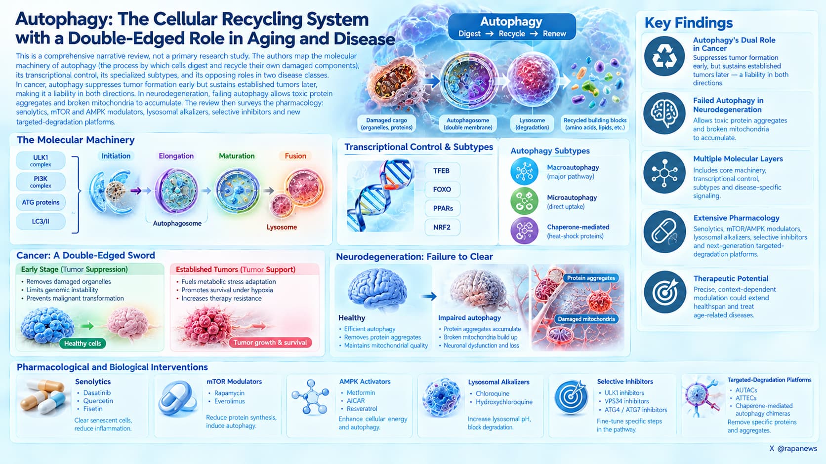
This is a comprehensive narrative review, not a primary research study. The authors map the molecular machinery of autophagy (the process by which cells digest and recycle their own damaged components), its transcriptional control, its specialized subtypes, and its opposing roles in two disease classes. In cancer, autophagy suppresses tumor formation early but sustains established tumors later, making it a liability in both directions. In neurodegeneration, failing autophagy allows toxic protein aggregates and broken mitochondria to accumulate. The review then surveys the pharmacology: senolytics such as dasatinib, quercetin and fisetin, mTOR and AMPK modulators, lysosomal alkalizers such as chloroquine, selective inhibitors of ULK1, VPS34, ATG4 and ATG7, and newer targeted-degradation platforms including AUTACs, ATTECs and chaperone-mediated autophagy chimeras.
For most of the last twenty years, autophagy has been sold to the longevity audience as an unambiguous good. Fast longer, take rapamycin, push the dial up, clear the junk. This review, assembled by nine pharmacologists across American, Australian and Japanese institutions, is a long argument against that simplicity.
The big idea is that autophagy is not a switch with an on position worth reaching. It is a rheostat whose correct setting depends on which tissue you are asking about, what genetic damage that tissue already carries, and how far along any disease process has already traveled. The same elevated autophagic flux that protects a neuron can keep a pancreatic tumor alive through chemotherapy.
The evidence for the neuronal side is the stronger half. Nearly every neurodegenerative disease is, at bottom, a failure to take out the garbage. Huntington’s accumulates mutant huntingtin, Parkinson’s accumulates alpha-synuclein, Alzheimer’s accumulates amyloid and tau, and amyotrophic lateral sclerosis accumulates protein clumps tied to mutations in genes that are themselves part of the disposal apparatus. Roughly 150 autophagy-related genes have been linked to human disease through mutation. In Parkinson’s specifically, the best-characterized genes converge on mitophagy, the selective destruction of damaged mitochondria, run by the PINK1 and Parkin proteins. Dopamine neurons in the substantia nigra appear vulnerable precisely because they are bioenergetically expensive: long unmyelinated axons, dense mitochondria, high baseline oxidative stress. They cannot afford a quality control failure.
The cancer side is messier; autophagy clears the damaged mitochondria and reactive oxygen species that seed genomic instability, which is tumor-suppressive. But once a tumor exists, it uses the same process to survive hypoxia and nutrient scarcity, and it recruits autophagy in the surrounding normal tissue to feed itself. Pancreatic stellate cells secrete alanine for neighboring tumor cells to consume. Blocking autophagy in the whole animal shrinks KRAS-driven tumors, but blocking it in a single cancer cell type can paradoxically accelerate metastasis, because the cargo receptors p62 and NBR1 pile up and start signaling for invasion.
Sitting underneath both stories is an epidemiological oddity the authors use as a framing device: across several population studies, people with a cancer history show lower subsequent rates of Alzheimer’s disease, and the reverse also appears. The association is partial and not a law of nature. But it fits the mechanism. A proliferating cell and a long-lived post-mitotic neuron want opposite settings on the same dial.
Where the review lands is that the field is a work in progress. The precision tools, ULK1 inhibitors, VPS34 inhibitors, ATG4 and ATG7 inhibitors, mostly exist as early test compounds with IC50 values in the nanomolar range and no human data. The future of longevity pharmacology relies on developing tissue-specific modulators, such as selective autophagy receptors (SARs) and autophagy-targeting chimeras (AUTACs), to clear localized cellular damage without disrupting systemic metabolic networks.
Insights
Current evidence reveals a stark contrast between preclinical promises of autophagy modulation and verified human interventions. As is common in health and biology; its complex. And none of the current autophagy promoters have the tissue and organ targeting ability that is needed.
More autophagy is not automatically better. In mutant SOD1 mice modeling amyotrophic lateral sclerosis, rilmenidine produced robust autophagy and mitophagy induction yet did not slow disease and was associated with worse motor neuron degeneration. Direction, dose, timing and tissue all matter.
The durable takeaway: basal mitophagy suppression drives aging phenotypes in cells, and those phenotypes were reversible with p62-targeting compounds. That is a target, not a protocol.
Context and Source
- Open Access Paper: Harnessing autophagy and its transcriptional regulation in tumorigenesis and neurodegeneration: Potential and existing therapeutic strategies
- Journal: Pharmacological Reviews, volume 78 (2026)
- Lead institutions: Department of Pharmaceutical Science, College of Pharmacy and Health Science, St John’s University, Queens, New York, United States. Co-first authorship shared between P. Suri and A.B. Daartey.
- Corresponding author institution: Centre for Cancer Cell Biology and Drug Discovery, Institute for Biomedicine and Glycomics, Southport, Queensland, Australia (Des R. Richardson), with an additional affiliation at Nagoya University Graduate School of Medicine, Japan.
- Impact evaluation: The impact score of this journal is 20.3 (2025 Journal Impact Factor, 2026 release; the prior release reported 17.3, and CiteScore is 35.4), evaluated against a typical high-end range of 0 to 60+ for top general science, therefore this is a High impact journal.