A Century of Cellular Age, Erased in Fifty Days

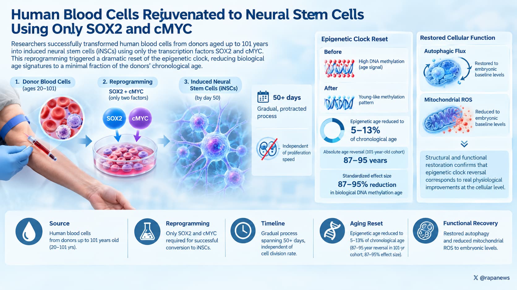
Researchers successfully transformed human blood cells from donors aged up to 101 years into induced neural stem cells (iNSCs) using solely the transcription factors SOX2 and cMYC. This specific reprogramming protocol triggered a severe reset of the cells’ epigenetic clocks, reducing biological age signatures to a minimal fraction of the donors’ chronological age. This rejuvenation mechanism is a gradual, protracted process spanning over 50 days and occurs independently of cellular proliferation speed.

The oldest person to donate cells to this experiment was 101 years old. Their blood carried, written in chemical marks across the DNA, a near-perfect record of that century. By the time those cells had been converted into established neural stem cell lines, the same chemical record read as under five years.

That much was known in outline. The same group at the University of Bonn reported in 2018 that converting human blood directly into neural stem cells erased most signs of age. The new paper asks a harder question: how fast does it happen, and what is driving it?

The answer to the first part is slower than anyone expected. Conventional reprogramming into induced pluripotent stem cells drives the methylation clock to an embryonic reading in about twenty days. Here the cells needed roughly fifty days to shed the majority of their age marks, and individual donor lines were still sitting at a fifth of their original age after seven weeks. The team sampled the same cultures repeatedly across that window and watched the number fall along a smooth decay curve rather than dropping off a cliff.

The answer to the second part is where the interest lies. The standard explanation for epigenetic rejuvenation is dilution. Cells divide, the enzymes that copy methylation marks onto new DNA strands fall behind, and age marks wash out of the population over successive divisions. If that were the whole story, slowing division should slow the reset.

The team tested this twice. They treated converting cells with glycerol, and separately with thymidine, two agents that work by different means and both of which cut the number of accumulated cell divisions by roughly half over five weeks. The methylation age kept falling at close to the same rate. Then they went further and forced the cells out of the cycle altogether, switching on a gene called NGN2 that converts neural stem cells into neurons, which do not divide. The clock kept running down.

If that holds, epigenetic age is not being passively washed out. Something is actively erasing it, and that something keeps working in cells that have stopped dividing. The point matters because the tissues people most want to rejuvenate, brain and heart muscle among them, are built largely from cells that no longer divide.

A second finding runs alongside the first. Cellular identity also consolidates far more slowly than it appears to. Within twelve days the converting cells look like neural stem cells, stain for the expected markers, and grow in the expected shape. Yet their gene expression shifted by almost as many genes between day fourteen and low passage as it had during the original exit from blood identity. The visible markers of a cell’s job arrive weeks before the job is finished, which is a caution for every protocol that validates reprogrammed cells by marker staining alone.

Actionable Insights

The application of SOX2 and cMYC is sufficient to initiate a deep erasure of epigenetic age without requiring full dedifferentiation to an embryonic state. Targeted, partial reprogramming strategies utilizing these factors could decouple functional cellular rejuvenation from cellular replication. For developers designing clinical longevity protocols, this strongly suggests that transient transcription factor expression holds more practical promise for translation than full Yamanaka factor application due to mitigated oncogenic risk.

The magnitude of the intervention is substantial. Epigenetic age reduction reached 5% to 13% of the donor’s chronological age, yielding an absolute age reversal of 87 to 95 years in the 101-year-old cohort. This translates to a standardized effect size of an 87% to 95% reduction in biological DNA methylation age. The intervention also restored autophagic flux and reduced mitochondrial reactive oxygen species to embryonic baseline levels. This structural restoration confirms the epigenetic clock reversal corresponds to concrete, functional physiological improvements at the cellular level.

Context & Source Evaluation

Novelty

Novelty: What This Adds

  1. The de-aging process is slow and continuous rather than switch-like. Prior work on iPSC reprogramming established a complete reset in roughly 20 days, and the field has generally carried that timescale over to other reprogramming paradigms. This maps roughly 50 days in a human blood-to-neural system using repeated sampling of the same cultures, which no previous study had done. [Confidence: High]
  2. De-aging proceeds without proliferation. Two chemically distinct methods of slowing division, plus forced permanent cell cycle exit into neurons, all failed to arrest the clock. If real, this directly contradicts the mitotic dilution model, which has been the default explanation for why reprogramming rejuvenates and why post-mitotic direct conversion does not. [Confidence: Medium, constrained by N=3 and by the fact that the claim rests on accepting a null]
  3. Cell fate consolidation is protracted and dissociable from the markers routinely used to certify it. Day 14 cultures that pass standard immunostaining are transcriptionally almost as far from a finished neural stem cell as they were from blood. This is an immediately usable result for anyone manufacturing these cells and it was not previously documented. [Confidence: High]
  4. The donor age range now extends to 101. The group’s earlier work covered 31 to 62 years. Near-complete epigenetic reset in centenarian cells had not been shown, and there was no prior basis for assuming the process would work identically at the extreme of human lifespan. [Confidence: High]
  5. Transcriptomic and epigenetic age reset on different schedules. The age-associated expression signature collapsed from 1,871 genes to 20 within 14 days while methylation age was still falling for weeks afterward. That dissociation suggests the transcriptome is responding directly to the overexpressed factors while the methylome is following a slower, separate process. [Confidence: Medium]
  6. A cross-paradigm hint pointing at SOX2. Running the Horvath clocks on published methylation data from a different group’s fibroblast-to-neural-plate-border conversion showed the same de-aging effect. SOX2 is the only transcription factor shared between the two protocols, which makes it the obvious candidate for the common driver. [Confidence: Low, because the supporting analysis is marked data not shown]

What this does not add: any evidence that an aging human can be rejuvenated, any evidence that the de-aged cells are functionally younger rather than simply different, and any mechanism.

Biomarker Data: Effect Size Extraction

First, what an effect size is and why it matters here. A p-value tells you only whether a difference is likely to be real. It says nothing about whether the difference is big enough to care about. An effect size tells you how large the difference is relative to the natural spread of the measurements. The common version, Cohen’s d, works like this: 0.2 is a small difference, 0.5 is moderate, 0.8 is large, and anything above about 2 means the two groups barely overlap at all. A drug trial reporting d of 0.5 would be considered a solid success.

This paper reports almost no effect sizes and no confidence intervals. Where a standardized figure is given below, it has been estimated from the published figures rather than read from reported statistics, and is flagged accordingly.

The primary outcome: epigenetic age reduction

Measure Value
Methylation age of iNSCs at low passage about 13 percent of donor chronological age
Methylation age of iNSCs at high passage, transgene-free under 5 percent
Relative reduction, low passage about 87 percent
Relative reduction, high passage over 95 percent
Absolute reduction, 101-year-old donor roughly 84 clock-years down to about 13, then under 5
Time to reset the majority of age marks about 50 days
Time to reach 20 percent of chronological age about 7 weeks
Benchmark: full iPSC reprogramming complete reset in about 20 days

Estimated standardized effect size for the de-aging itself: in donors over 60, source blood cells read between roughly 67 and 84 clock-years while their derived neural stem cells read between roughly 8 and 15. The two distributions do not overlap anywhere. A conservative estimate places Cohen’s d above 6, which is an order of magnitude beyond what any pharmacological intervention produces. The direction and rough magnitude are not in doubt. The precise value is, because it is reconstructed from a published scatter plot rather than from reported means and standard deviations. [Confidence: High on magnitude band, Low on the exact value]

The mechanistic claim: proliferation inhibition

This is the result the paper is actually built on, and it is the weakest numerically.

Comparison Outcome p
Population doublings, untreated vs glycerol significantly reduced 0.002
Population doublings, untreated vs thymidine significantly reduced 0.001
Population doublings, glycerol vs thymidine no difference 0.842
Relative methylation age, untreated vs glycerol reported as not significant 0.056
Relative methylation age, untreated vs thymidine not significant 0.977
Relative methylation age, glycerol vs thymidine significant 0.044

Proliferation was genuinely suppressed. Reading off the published figure, untreated cultures accumulated roughly 14 to 16 population doublings by day 49 while treated cultures reached roughly 5 to 8, a reduction on the order of 50 to 60 percent. Despite that, methylation age fell at a comparable rate in all three arms.

The honest reading of those three p-values is more equivocal than the paper’s framing. A value of 0.056 is not an absence of effect, it is a result sitting directly on the threshold. And glycerol-treated cultures were significantly epigenetically older than thymidine-treated ones at 0.044, a positive finding the authors note and do not reconcile.

The power problem is decisive. With three donors per group and alpha at 0.05, this design could reliably detect only effects of roughly d equal to 3.4 or larger. An effect of d equal to 1.5, which in most of biology would be called very large, would have been caught perhaps one time in five. The defensible statement is that no difference was detected, not that no difference exists. [Confidence: High]

The forced differentiation experiment carries the same ceiling: relative methylation age of NGN2-differentiated cultures versus proliferating iNSCs at day 42, Welch two-sample t-test, p equal to 0.635, N equal to 3.

Transcriptomic age convergence

This is the cleanest effect size in the paper and it does not depend on reading a figure.

Stage Genes differentially expressed between young and old donors
Day 0, source blood cells 1,871
Day 14 of conversion 20
Low passage iNSC 12

That is a 98.9 percent collapse in the transcriptional age signature within 14 days, and 99.4 percent by low passage. Methylation age was still falling well after this point, which means the two readouts of cellular age are not tracking the same process. [Confidence: Medium, because the authors themselves flag that the much larger fate-shift signal may be masking residual age-associated signal]

Fate acquisition, which is the paper’s other headline

Contrast Genes differentially expressed
Blood cells to day 14 iNSC 6,270
Day 14 to low passage iNSC 5,978
Low passage to transgene-free iNSC 2,267

The practical translation: the amount of transcriptional change that happens after the cells already look like finished neural stem cells is 95 percent as large as the change that made them look that way. Marker staining at day 12 is not evidence of a finished cell.