70% Lifespan Extension: Immune-Derived "Telomere Rivers"—A Transferable Youth Signal?

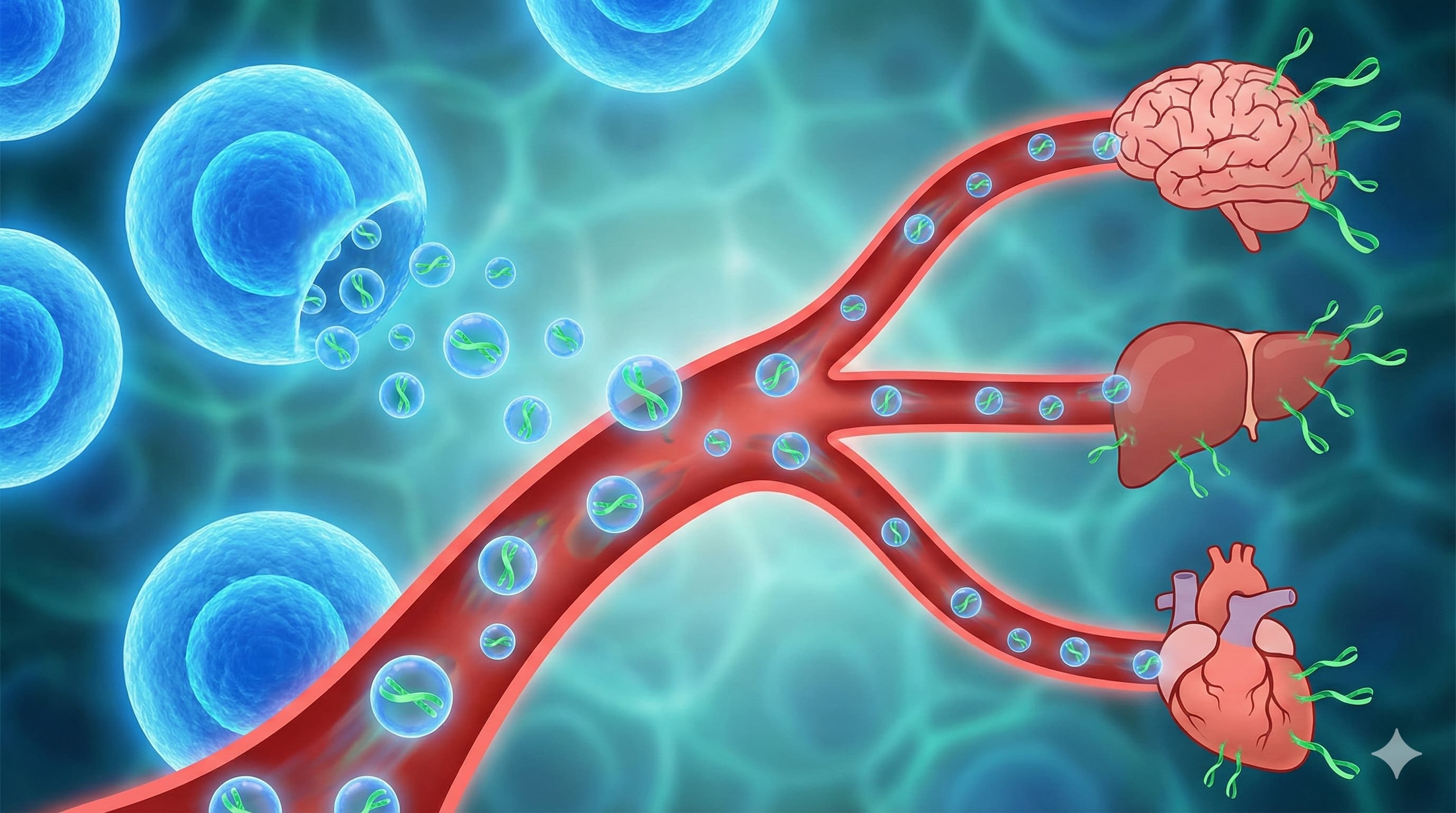
A research team led by Prof. Alessio Lanna (CEO of Sentcell) has released a provocative manuscript describing a new “fluid” organ of the immune system: “Telomere Rivers.” Building on their previous discovery that antigen-presenting cells (APCs) donate telomeres to T cells, this study claims that specific CD4+ T cells subsequently release these telomeres into the bloodstream as extracellular vesicles. These “Rivers” reportedly travel systemically, acting as a “quorum-sensing” youth signal that elongates telomeres in distant tissues (brain, liver, heart) and reverses senescence markers.

The most explosive claim is the lifespan data: 20-month-old mice treated with these telomeric vesicles allegedly survived to a median of ~47 months, with some reaching nearly 5 years (~60 months). If replicated, this would vastly outperform current gold standards like Rapamycin (which typically offers ~15–25% extension). The mechanism hinges on a metabolic switch: the process requires Fatty Acid Oxidation (FAO) and the exclusion of the glycolytic enzyme GAPDH from the vesicles. The authors suggest that “Artificial Rivers”—bioengineered vesicles lacking GAPDH—can replicate this rejuvenation, effectively creating a transplantable “program of youth” that functions independently of the donor’s T cells.

Source:

Related reading: New World record lifespan achieved in mice by Rejuvenating T Cells - #2 by EnrQay


Biohacker Analysis: Technical Breakdown

Study Design Specifications

  • Type: Pre-clinical In vivo (Murine) & In vitro (Human/Mouse cells).
  • Subjects: C57BL/6J mice.
    • Recipients: Aged 20-month-old males (equivalent to ~60-year-old humans).
    • Donors: Young (3-month) or “rejuvenated” old T cells.
    • N-numbers:
      • Lifespan Cohorts: n=10 for River transplant; n=10 for Artificial Rivers; n=10 for Control vesicles; n=8 for DOS-rejuvenated T cells; n=8 for Old T cells; n=8 for Untreated Old Serum.
      • Baseline Control: n=26 (Transfer-free old animals).
      • Tissue Analysis: n=5 per group.

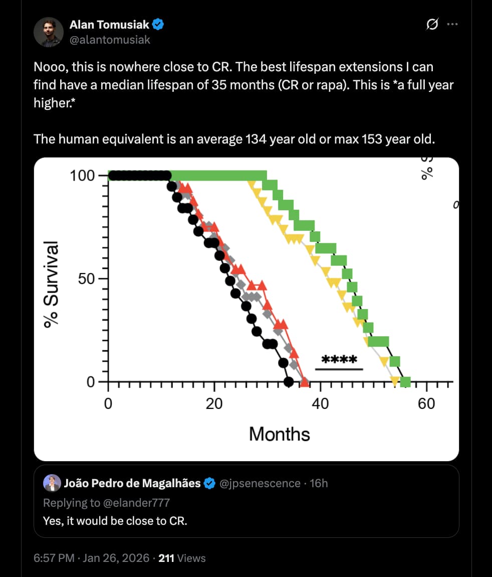
Lifespan Analysis & The “Control Problem”

  • Control Group Performance: The control mice in this study (treated with saline or inactive vesicles) exhibited a median lifespan consistent with standard laboratory conditions, dying between 26–28 months of age.
  • Benchmarking against Pabis et al. (2023):
    • Context: The preprint The impact of short-lived controls on the interpretation of lifespan experiments (2023)argues that many “successful” longevity interventions only appear effective because the control animals die prematurely (Median <900 days/30 months) due to stress or poor husbandry.
    • Verdict: By Pabis’s strict “900-Day Rule,” the controls in the Lanna study are indeed “short-lived” (falling short of the ~30-month gold standard).
    • The Anomaly: However, the magnitude of the effect in the treatment group renders the “weak control” argument moot. The treated mice did not just recover to the 900-day baseline; they shattered it, living to ~1,400–1,800 days. While short-lived controls typically inflate relative (%) gains, they cannot explain the absolute survival duration observed here, which exceeds the species’ known biological ceiling.

Lifespan Data

  • Median Lifespan Extension:
    • Absolute: ~17 months extension beyond controls.
    • Relative: ~65% increase (Treatment Median ~43–45 months vs. Control Median ~26–28 months).
  • Maximum Lifespan:
    • Absolute: Several subjects survived to ~60 months (5 years).
    • Relative: ~70–90% increase over historical maximums (typically ~34–36 months for C57BL/6J).
    • Comparative Significance: For context, Rapamycin (the current gold standard) typically delivers a 15–25% median extension. This intervention claims an effect size roughly 300% greater than Rapamycin.

Mechanistic Deep Dive

  • The “River” Payload: The vesicles are not just bags of telomeres; they are enriched with stemness factors (Wnt5a, Notch1, Runx2) and depleted of GAPDH.
  • Metabolic Gating: The formation of these vesicles is gated by CPT1A (the rate-limiting enzyme of fatty acid oxidation). Senescent T cells fail to produce Rivers because they are stuck in glycolysis/ceramide synthesis.
  • GAPDH as the “Aging Brake”: The study posits GAPDH as a competitive inhibitor of stemness factors within vesicles. Silencing GAPDH in APCs created “Artificial Rivers” that rejuvenated tissues even without T cells.
  • Target Tissues: Rejuvenation was observed in the Brain, Liver, Kidney, Heart, and Lung, suggesting the vesicles cross the blood-brain barrier.

Novelty

  • Extracellular Telomeres: Shifts the paradigm from telomeres being purely intracellular “clocks” to intercellular “signaling particles.”
  • Transplantable Youth: Demonstrates that the product of the immune interaction (the vesicle) is sufficient for rejuvenation, bypassing the need for successful T-cell engraftment.

Critical Limitations & Red Flags

  • The “Too Good to Be True” Problem: A ~70%+ increase in median lifespan from late-life intervention is virtually unheard of in mammal literature.
  • Conflict of Interest: The lead author is the CEO of Sentcell, the biotech company holding the IP for the “DOS” compound and Artificial Rivers.
  • Tumorigenesis Risk: Delivery of active Wnt/Notch stemness factors + telomeres to aged tissues is a textbook recipe for cancer. The paper claims extended healthspan, but rigorous cancer assays are missing.
  • Dosing Obscurity: The treatment used ~5,000 particles. This is an incredibly low quantity for systemic EVs, raising questions about the signal amplification mechanism.

6 Likes

Claims Investigation

Claim 1: CD4+ T cells release extracellular telomere vesicles (“Rivers”).

Claim 2: “River” therapy extends median mouse lifespan by ~17 months.

  • Evidence Level: D (Single Animal Study)
  • Status: Unverified & Outlier. This claim exceeds the effects of Rapamycin, Acarbose, and 17-a-Estradiol combined.
  • Translational Gap: Human lifespan is not limited by telomere length to the same degree as mice (who express telomerase in somatic tissues more than humans).
  • Source: CD4+ T cells confer transplantable rejuvenation via Rivers of telomeres (2025)

Claim 3: Fenofibrate (PPARa agonist) restores the mechanism in plants/mammals.

Claim 4: GAPDH inhibits the packaging of stemness factors.

  • Evidence Level: D (Mechanistic)
  • Status: Novel finding. GAPDH is traditionally a glycolytic enzyme; its “moonlighting” role in vesicle gating is a new biological claim.
  • Source: Source unverified in live search outside this preprint.

3 Likes

1 Like

Andrew is looking at the wrong plot. The mice above in the control group are aged naturally (seems like they should have started with 20 month old mice in the placebo group), the test group they have been aged to 20 months as they only apply the vesicles at 20 months… Maybe they expected the mice to live to at least 20 months and they didn’t. The other experiment uses 20 month old mice in both experiments.

1 Like

Nice to know we’re all gonna live an extra 50 years.

1 Like

3 Likes

I think he’s using linear interpolation on the plots, which would hide the actual number of animals - though I would assume it’s not deliberate, but hopefully bad with plotting software.

Let’s hope its replicable.

4 Likes

I have run checks on three LLMs (subscription accounts) and the public shares are below. In the end a big claim requires solid evidence.

https://claude.ai/share/05796a9d-2cd6-4048-81be-658ea588f1ff

3 Likes

More on the SenTcell discussion:

I. Executive Summary

Dr. Alessio Lanna’s presentation outlines a novel and radical biological paradigm regarding immune aging, asserting that T-cell longevity is dictated not solely by telomerase, but by the intercellular transfer of telomeres from antigen-presenting cells (APCs). According to this model, upon forming an immunological synapse, APCs package and donate cleaved telomeric DNA via extracellular vesicles to CD4+ T-cells. This transfer allegedly extends T-cell telomeres by roughly 3,000 base pairs—a massive structural reset compared to the minor additions provided by telomerase.

The core of Lanna’s recent thesis introduces “telomere rivers.” He posits that after acquiring these telomeres, CD4+ T-cells undergoing active fatty acid oxidation (FAO) utilize a portion of the genetic material to reverse their own senescence. The T-cells then actively filter out glycolytic enzymes (specifically GAPDH), enrich the remaining telomeric vesicles with stemness factors (such as Wnt), and release them systemically into the bloodstream. In murine models, adoptive transfer of these “rivers” reportedly generated a systemic rejuvenation cascade, extending maximum lifespan to nearly 60 months without inducing oncogenesis—an unprecedented claim in mammalian longevity research.

To translate this therapeutically, Lanna’s group has developed DOS (Disruptor of sMAC), a proprietary small molecule intended to degrade the sestrin-MAPK complex. This complex aggressively drives immune senescence by inhibiting FAO. DOS purportedly restores FAO, re-enables telomere transfer, and licenses the immune system to undergo TCR (T-cell receptor) revision, granting broad-spectrum immunity against novel pathogens without specific vaccination.

While the fundamental biology of APC-to-T-cell telomere transfer is documented in peer-reviewed literature, the systemic lifespan claims regarding “rivers” and DOS remain confined to pre-prints and pre-clinical murine models, representing an immense translational gap. Furthermore, Lanna’s aggressive dismissal of mTOR inhibitors (like rapamycin) as incapable of immune rejuvenation contradicts existing clinical trial data, highlighting a semantic dispute between “delaying aging” and “active structural rejuvenation.”


II. Insight Bullets

  • Non-Canonical Telomere Extension: T-cells acquire telomeres from APCs via extracellular vesicles at the immunological synapse, circumventing total reliance on telomerase.
  • Magnitude of Extension: Vesicular transfer extends T-cell telomeres by approximately 3,000 base pairs, roughly 30 times the capacity of a standard telomerase cycle.
  • Metabolic Gating: Telomere transfer strictly requires the recipient T-cell to be actively engaging in Fatty Acid Oxidation (FAO) via the CPT1A enzyme.
  • Senescence Pathology: Senescent T-cells lose FAO capacity, leading to ceramide accumulation that prevents proper T-cell receptor (TCR) clustering at the synapse.
  • “River” Generation: CD4+ T-cells consume ~50% of donated telomeres; the remainder is repackaged into larger extracellular vesicles and released systemically as “rivers.”
  • Molecular Filtration: T-cells actively deplete these systemic rivers of GAPDH (a glycolytic enzyme) while enriching them with stemness markers.
  • Extreme Lifespan Claims: Adoptive transfer of these rivers extended the lifespan of 20-month-old mice to 4.5–5 years (~60 months) with no observed increase in cancer rates.
  • Artificial Engineering: “Artificial rivers” (GAPDH-depleted APC vesicles created ex vivo) reportedly bypass the need for T-cell processing and exert an even stronger systemic rejuvenation effect.
  • The sMAC Bottleneck: The sestrin-MAPK activation complex (sMAC) is identified as the primary driver of T-cell metabolic failure and senescence during aging.
  • Pharmacological Intervention: The investigational drug DOS degrades sMAC, restoring FAO and re-enabling natural telomere transfer.
  • Antigen-Independent Readiness: DOS administration in mice reportedly triggered peripheral TCR revision, generating cross-reactive T-cells capable of clearing novel lethal infections (like influenza) without prior vaccination.
  • CD4+ Exclusivity: CD8+ T-cells do not generate telomere rivers; the systemic longevity effect is strictly dependent on the CD4+ helper compartment.
  • Accelerated Clinical Senescence: Lanna notes a dramatic post-2019 acceleration in human immunosenescence, with 20-year-olds presenting immune profiles previously typical of 70-year-olds.
  • Contrarian Stance on mTOR: Lanna argues that rapamycin and other mTOR inhibitors only slow aging but fail to rejuvenate immunity, claiming senescent T-cells lack the active mTOR signaling required for these drugs to work.

III. Adversarial Claims & Evidence Table

Claim from Video Speaker’s Evidence Scientific Reality (Current Data) Evidence Grade (A-E) Verdict
APC-to-T-cell telomere transfer elongates telomeres independently of telomerase. Human in vitro assays, murine models. Verified in pre-clinical settings. APCs use TZAP to cleave telomeres and transfer them via RAD51-positive vesicles. Lanna et al., 2022, Nature Cell Biology. Level D (Translational Gap) Plausible
Circulating “telomere rivers” extend murine lifespan to ~60 months. Murine adoptive transfer survival curves. Documented in a recent pre-print. The magnitude of lifespan extension is extreme and currently lacks independent replication. Lanna et al., 2025, bioRxiv. Level D (Translational Gap) Speculative
Disrupting sMAC (via DOS) restores FAO and triggers systemic immune rejuvenation. Murine models, pending Phase 1 trials. The role of sestrins in T-cell senescence is well-established Lanna et al., 2017, Nature Immunology. Clinical systemic rejuvenation via pharmacological disruption is untested in humans. Level D (Translational Gap) Speculative
mTOR inhibitors (Rapamycin) do not rejuvenate the immune system. Assertion that senescent cells lack mTOR targets. Adversarial. Human RCTs show mTOR inhibition significantly decreases infection rates and improves vaccine responses in the elderly. Mannick et al., 2014, Sci Transl Med. Level B Unsupported

IV. Technical Mechanism Breakdown

1. Synaptic Telomere Cleavage and Packaging:
Upon specific antigen recognition, the APC downregulates the shelterin complex (which normally protects chromosome ends). It simultaneously upregulates TZAP (telomeric zinc finger-associated protein), which trims the unshielded telomeric DNA. This DNA is packaged into extracellular vesicles alongside RAD51, a recombination protein necessary for subsequent integration into the recipient genome.

2. The Fatty Acid Oxidation (FAO) Gateway:
For a T-cell to receive this telomeric material, it must establish a highly ordered, stable immunological synapse. This requires the T-cell to be actively engaged in FAO, rate-limited by the enzyme CPT1A. FAO drives the production of phosphatidylethanolamine (PE), a lipid essential for clustering CD3/TCR complexes at the center of the synapse (the “bullseye”).

3. Senescence and the sMAC Bottleneck:
In aged or senescent T-cells, metabolic flexibility is lost. Stress-sensing proteins called sestrins bind to MAP kinases to form the sestrin-MAPK activation complex (sMAC). This complex halts FAO, forcing the cell into glycolysis. As a byproduct, ceramides accumulate, activating ceramidase, which ultimately disrupts PE synthesis. Without PE, the synapse fails to form properly, and telomere transfer is aborted.

4. Asymmetric Division and River Biogenesis:
Once a competent T-cell acquires telomeres, the RAD51 machinery integrates them, rescuing ultra-short chromosomal ends. The T-cell then undergoes an asymmetric division. The resulting stem-like daughter cell (which retains high CPT1A expression) repackages the surplus APC telomeres. Crucially, the cell strips the glycolytic enzyme GAPDH from these vesicles, replacing it with stemness factors (like Wnt), before exocytosing them into the plasma as “rivers” to exert systemic, paracrine rejuvenation effects on distal tissues.

4 Likes

The unresolved questions remain unresolved. vide twitter.

It’s an incredible discovery if it’s actually true. Hopefully it can be replicated.

3 Likes

New, from Mike Lustgarten:

Telomere Rivers: Rejuvenation And Lifespan Extension

This analysis synthesizes the claims presented in the provided transcript regarding a highly controversial, pre-clinical longevity study involving extracellular telomere transfer, evaluated against current medical and biological literature.

I. Executive Summary

The provided transcript features a defense of a highly disruptive, pre-clinical longevity breakthrough involving immune-mediated systemic rejuvenation. The core thesis asserts that CD4+ T cells, upon interacting with antigen-presenting cells (APCs), receive telomeres via extracellular vesicles. These T cells subsequently release these telomere-loaded vesicles—dubbed “Rivers”—into systemic circulation. The speaker claims that administering these Rivers into 20-month-old aged mice triggers an unprecedented lifespan extension, allowing the animals to survive up to five years. This represents an alleged median lifespan extension of approximately 17 months, an effect size the author explicitly claims is 20 to 30 times greater than established geroprotectors like mTOR inhibitors (e.g., rapamycin) or traditional telomerase activation therapies.

A significant portion of the video is dedicated to defending the study’s statistical methodology against peer critiques, specifically regarding a 30% background mortality rate in the control cohort prior to the intervention. The speaker insists that the divergence in survival curves post-intervention (at exactly 20 months) remains robust and valid even when early background mortality is excluded from the analysis.

Biologically, the mechanism relies on a strict metabolic prerequisite: fatty acid oxidation at the T cell–APC synapse. This metabolic state triggers ceramide activation and CD3 receptor clustering, which prompts a calcium flux in the APC. The APC then expels Rad51-loaded telomere vesicles. Upon absorption, the T cell undergoes a “stem-like switch” and a rapid asymmetric division, deliberately excluding the enzyme GAPDH from the vesicles. The released Rivers then allegedly circulate throughout the body to rejuvenate peripheral tissues by increasing systemic stem-like signaling. Despite the staggering magnitude of these claims, the data remains strictly confined to murine models. Independent replication is entirely absent, and the extreme statistical outliers reported in the lifespan data demand rigorous clinical skepticism. At present, this represents a profound biological hypothesis, but not a validated medical intervention.

II. Insight Bullets

  • The “River” Mechanism: CD4+ T cells acquire telomeres from Antigen-Presenting Cells (APCs) and release them into systemic circulation as extracellular vesicles known as “Rivers.”
  • Unprecedented Lifespan Extension: The experimental intervention allegedly extends median mouse lifespan by roughly 17 months, enabling subjects to reach nearly 5 years of age.
  • Efficacy vs. Standard Paradigms: The researcher claims the lifespan gain achieved via River therapy is 20 to 30 times greater than established protocols like mTOR inhibition or telomerase activation.
  • Intervention Timing: The experimental divergence in survival curves occurred precisely at the 20-month mark, the exact point of therapeutic intervention for the aged cohort.
  • Addressing Mortality Criticisms: The author directly counters peer critique regarding a 30% baseline mortality in the control group, maintaining that the survival gap remains statistically robust independent of early background deaths.
  • Delivery Modalities: The rejuvenation effect was achieved via three distinct laboratory methods: direct injection of Rivers, adoptive transfer of donor cells, and the depletion of artificial rivers.
  • Metabolic Trigger: Telomere transfer is strictly dependent on the T-cell engaging in fatty acid oxidation (FAO) at the immune synapse.
  • Ceramide & Receptor Clustering: The structural formation of the synapse requires ceramide activation and the clustering of CD3 receptors on the T-cell boundary.
  • Calcium-Driven Expulsion: The physical release of telomeres from the APC is facilitated by a targeted intracellular calcium flux.
  • Rad51 Cargo Inclusion: The transferred telomere vesicles are loaded with Rad51, a critical protein essential for DNA repair and homologous recombination.
  • Stem-Like Cellular Switch: Receiving these vesicles triggers a rapid “stem-like switch” in T-cells, extending their functional lifespan and preventing normal immune senescence.
  • Asymmetric Division: Following the telomere transfer, T-cells undergo an asymmetric division, preserving the telomere payload in one lineage while the other is depleted.
  • GAPDH Exclusion: The daughter cells utilize a distinct metabolic tone, specifically linked to the targeted exclusion of GAPDH from the telomere vesicles.
  • Systemic Tissue Rejuvenation: Unlike localized immune actions, the Rivers circulate systemically, dramatically increasing pan-tissue stem-like signaling and overall organismal fitness.

Red Flag Zone (Safety Data & Gaps)

  • Extreme Efficacy Claims (Safety Data Absent): The assertion that this therapy yields 20–30x the lifespan extension of mTOR inhibition is based entirely on a single, unverified pre-print murine study: Lanna et al., 2025.
  • Severe Translational Gap: Transferring extracellular vesicles (Rivers) or donor T-cells for systemic rejuvenation has not been tested for safety, dosing, autoimmune risk, or oncogenic risk in human subjects. Attempting to replicate this via unverified biologic injections carries severe, potentially lethal risks.
  • Methodological Dispute: As acknowledged directly in the transcript, peer reviewers have flagged abnormal background mortality (~30%) in the control groups. Independent replication by third-party longevity laboratories is strictly required before the underlying survival data can be considered scientifically sound.
3 Likes

I dont think this deals with the baseline mortality or the other issues.

1 Like

He mentioned in this video (if I understand correctly), that further administration did not extend lifespan further. This implies that some other mechanism kills the mice, maybe it’s mitochondria or maybe you need epigenetic reprogramming at that point. I thought the epigenetic reprogramming only extended mouse life by about 20% so maybe that is limited by these other things.

Andrew Steele is spot on here, and your logic is completely off the mark.

Think about it: if the experimental group only gets injected at 20 months of age, those mice are already survivors who successfully made it to that 20-month milestone. Taking a cohort of “survivor mice”—with a guaranteed 100% survival rate at 20 months—and putting them on the exact same starting line to compare lifespans against a “naturally aging” control group tracked from birth (which inevitably includes mice that died prematurely) is a massive statistical violation.

2 Likes

There’s a second plot which looks like it’s correct at 20 months. With siCtrl and SiGAPDH and that is what I meant by (wrong plot). The second one I don’t think was posted on twitter - but it looks ok if they started at 20 month. The one with DOS and Old is definitely wrong unless they started at 10-15ish months or earlier.

Alessio promised a second paper on HIV clearance using the same molecule DOS. Here that paper is, tested on human blood infected with HIV https://www.biorxiv.org/content/biorxiv/early/2026/01/05/2026.01.04.697570.full.pdf

I wasn’t personally inclined to spend the time to review the paper on HIV, but I put it into chatgpt and asked for errors:

I found many internal red flags. I would not treat this paper as reliable without independent replication and raw data. The biggest problems are not just “weak evidence”; several claims are internally inconsistent with the methods and figures.

Major errors / inconsistencies

1. The paper repeatedly says “integrated HIV DNA”, but the main assay is not integration-specific.
In the methods, the core qPCR uses LTR primers and ΔΔCt. LTR qPCR can detect HIV/LTR-containing DNA, but by itself it does not prove integration. Integration-specific evidence would require Alu–HIV PCR, integration-site sequencing, or equivalent. Yet the text and Fig. 1 repeatedly interpret ordinary LTR qPCR as “integrated HIV DNA” and “proviral eradication.”

2. The plotted units look impossible: “absolute copies ×10⁻⁹”.
Fig. 1 plots “Integrated HIV DNA (Absolute copies)” with values around 10⁻⁹ copies. Absolute copy number cannot be 0.000000003 copies in a biological sample. That strongly suggests either a relative ΔΔCt value was mislabeled as absolute copies, or the quantification is fundamentally mishandled. This is a serious quantitative error.

3. The model extrapolates from a tiny sample to “less than one intact provirus per individual”.
The methods say only 2 × 10⁵ CD4⁺ T cells were interrogated per sample, then use a claimed assay limit “LOD 10⁻¹²; Ct ≥ 40” to infer M < 1 expected intact provirus per individual. That is not justified. A negative qPCR in 200,000 cells cannot establish absence across the whole memory CD4 pool, let alone across anatomical reservoirs. Ct ≥40 is a threshold, not a whole-body cure bound.

4. The claimed mechanism depends on HIV integrase still being bound to latent integrated provirus.
The proposed model is that RAG displaces HIV integrase from BRD9/chromatin and thereby causes excision/degradation of integrated HIV. But in established latent reservoirs, HIV integrase is not expected to remain sitting on every integrated provirus as a persistent target. This makes the mechanism especially problematic for patient-derived latent HIV reservoirs. The paper’s own model says cure occurs by displacing integrase from infected DNA, but that is not a convincing explanation for old integrated provirus.

5. GFP⁺ “infected” cells and “cured” cells are conceptually muddled.
For scRNA-seq, they sort GFP⁺ cells from lentiviral vector infection and compare cells described as “cured” or not. But if the lentiviral GFP cassette has been removed or silenced sufficiently to represent “cure,” the basis for still sorting GFP⁺ “cured” cells is unclear. GFP protein persistence after only 4 hours could confound the sorting, and scRNA-seq does not itself prove that the individual sequenced cells had lost integrated provirus.

6. Lentiviral-vector infection is repeatedly treated as equivalent to HIV reservoir cure.
The first experimental system uses primary CD4⁺ T cells infected with lentiviral vectors encoding GFP, not necessarily replication-competent HIV latency. That can be a useful model, but the paper jumps from vector clearance to “HIV cure.” The in vivo “sanctuary” model also uses CD45.1 mouse CD4 cells “HIV infected with empty backbone lentiviral vectors,” which is not the same as natural HIV infection in human tissue reservoirs.

7. The mouse experiments are overclaimed.
The abstract says humanised mice had adoptive transfer or in vivo reprogramming of murine T cells with months-long clearance. The methods describe NSG mice receiving patient CD4⁺ T cells plus APCs, then DOS/ART; and a CD45.1/CD45.2 donor-recipient system using lentiviral vectors. That is not a robust human HIV reservoir model. Calling brain and gut recovered transferred cells “virological sanctuaries” is particularly weak.

8. The RAG claim is not supported by the kind of evidence needed.
They claim RAG1/RAG2 “eradicates integrated HIV DNA,” but the decisive experiment appears to incubate extracted DNA with recombinant RAG1/RAG2 and then qPCR for HIV DNA. Loss of a PCR signal could mean cleavage or damage at/near the amplicon, not precise proviral excision. There is no clear demonstration of excision junctions, integration-site loss by long-read sequencing, or genome-wide off-target safety.

9. “Safe and transient” is overinterpreted.
The paper says transient RAG/integrase expression, reversible γH2AX, and no Annexin V increase “confirmed” safety. That is far too strong. RAG activity can cause rare but serious rearrangements; absence of short-term Annexin V positivity and resolution of γH2AX does not prove genomic safety. One would need karyotyping, long-read/WGS, translocation assays, clonal expansion studies, and longer follow-up.

10. Reference numbering and citation support are inconsistent.
In the main text, it says viral outgrowth assays, IPDA and Alu-PCR confirm clearance, citing refs 24–26. But ref. 24 is Simonetti/IPDA-type benchmarking, ref. 25 is Alu-PCR, and ref. 26 is an old HIV integration mechanism paper, not IPDA validation. More seriously, ref. 27 is cited for RAG silencing in mature T cells, but ref. 27 is Siliciano on latent reservoir stability, not RAG biology.

11. The conflict-of-interest statement is awkwardly inconsistent.
The paper says the study was funded by Sentcell and that the funder had no role in study design or decision to publish, but also says A.L. is Sentcell’s CEO, shareholder, sole inventor of the DOS pharmaceutics/methods, discovered the cure, directed the study, analysed the data and wrote the paper. That does not invalidate the data, but it makes independent validation and raw data access essential.

Bottom line

This paper has severe internal problems. The most damaging are the misuse/mislabeling of qPCR as integrated proviral DNA, the impossible “absolute copies ×10⁻⁹” values, the unsupported whole-body cure modelling, and the mechanistic dependence on persistent HIV integrase at latent proviruses.

I would classify it as extraordinary claims with inadequate and internally inconsistent evidence, not as a credible demonstration of HIV cure.

1 Like