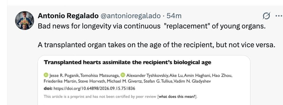
https://www.biorxiv.org/content/10.64898/2026.09.15.751836v1
I think the sharing of mitochondria around the body is part of this, but also other factors have effects (such as IL-10)
chatGPT(6AstraMaxPaid):
The paper provides substantial evidence that recipient age influences ageing-related molecular patterns in transplanted mouse hearts. Its evidence for rejuvenation of human hearts is preliminary, and the claim that hearts acquire the recipient’s biological age is stronger than the results establish.
The paper is Transplanted hearts assimilate the recipient’s biological age, by Poganik and colleagues, posted on 17 September 2026. The supplied version is a preprint that has not undergone peer review.
Summary. The researchers ask whether a transplanted heart retains its original ageing characteristics or changes in response to its new host.
They combine controlled mouse experiments, DNA methylation measurements from human heart biopsies, and clinical assessments of transplant recipients.
| Study component | Design and scale | Main finding |
|---|---|---|
| Mouse transplantation | Hearts exchanged between genetically matched mice aged approximately 3, 12, or 18-20 months; usually 4-6 animals per group; assessment 4-6 months later | Young hearts in older recipients developed older molecular signatures, while old hearts in younger recipients developed younger signatures |
| Human molecular analysis | Biopsies from 11 recipients: five receiving older hearts and six receiving younger hearts | Two of three methylation clocks showed significant differences between the two transplant groups |
| Human functional analysis | Between 151 and 317 recipients per measurement, assessed one year after transplantation | Older recipient age predicted lower exercise capacity after adjustment for donor age and recipient sex |
Several details strengthen the mouse findings:
- The result extends beyond clock outputs. Methylation changes across approximately 260,000 measured sites correlated with normal ageing changes in young hearts transplanted into old mice, and opposed normal ageing changes in old hearts transplanted into young mice. The respective correlations were approximately 0.42 and -0.53.
- Gene expression changed in broadly corresponding directions. Mitochondrial translation, oxidative phosphorylation and respiratory electron transport pathways increased in old hearts placed into young recipients and decreased in young hearts placed into old recipients.
- Transplantation itself produced additional effects. Grafted hearts generally showed inflammatory and ageing-associated changes, including in age-matched transplants.
- Effects on the recipient were much weaker. There were no consistent methylation-age changes in the recipient’s native heart, liver or blood.
The authors interpret these findings as evidence that the systemic environment governs tissue biological age. They suggest that younger recipients might rejuvenate older donor hearts, potentially expanding the usable donor pool.
Novelty. The strongest contribution is the combination of a controlled heart transplantation experiment, several molecular readouts and preliminary human evidence.
The broader idea that the host environment influences ageing is already established through heterochronic parabiosis and related experiments. Human evidence also predates this paper: a 2024 study found that epigenetic ageing of donor-derived blood cells was influenced by the recipient’s age following haematopoietic stem cell transplantation. (PMC)
What this paper adds is:
- Evidence from an intact transplanted heart. This extends the question beyond circulating cells and shared-circulation models.
- Changes in both directions. The mouse experiments show ageing-associated shifts in young grafts and opposing shifts in old grafts.
- Comparison of graft and recipient tissues. The finding that graft changes were much stronger than reciprocal host changes helps distinguish transplantation from parabiosis.
- A clinically accessible setting for studying tissue plasticity. Routine transplant biopsies could support larger longitudinal studies.
The mitochondrial expression changes are interesting mechanistic clues. They do not, however, identify a new causal rejuvenation mechanism.
Critique. The experimental design has real strengths: genetically matched mouse transplants reduce rejection-related confounding, age-matched transplants help separate host-age effects from surgery, and agreement between methylation and RNA measurements strengthens the central observation.
The following limitations substantially affect its interpretation.
1. Molecular age shifts do not establish comprehensive rejuvenation.
The clocks predominantly estimate age from methylation patterns. A younger prediction establishes a change in those patterns; it does not establish reversal of fibrosis, vascular damage, mutations or impaired cellular function.
Similarly, increased expression of oxidative phosphorylation genes does not demonstrate improved mitochondrial respiration or ATP production.
The mouse evidence therefore supports partial molecular remodelling towards a younger profile. Complete restoration of a younger heart’s condition remains untested.
2. Bulk tissue measurements cannot establish which cells changed.
Heart samples contain cardiomyocytes, fibroblasts, endothelial cells and immune cells. Changes in their proportions can alter both methylation and gene expression profiles.
Recipient-derived cells are particularly relevant. A human cardiac allograft study showed that recipient-derived macrophages populate transplanted hearts and that donor-derived resident macrophages decline over time. (PMC)
Consequently, a graft sample could acquire a more recipient-like molecular profile partly because it contains recipient-derived cells. That possibility does not explain away the findings, but it needs to be quantified.
Analyses of isolated donor-derived cardiomyocyte nuclei, alongside other identified cell populations, would provide much stronger evidence that the original heart cells changed age.
3. The human analysis has a substantial age-confounding problem.
The 11 patients were selected for contrasting donor-recipient age differences. Young recipients received older hearts, while older recipients received much younger hearts. Donor age and recipient age therefore vary together in opposite directions.
The principal outcome, AgeDev, is predicted methylation age minus donor chronological age. If a clock tends to underestimate older tissue and overestimate younger tissue, this design can generate an apparent recipient-age association even without a host-induced age change.
This is a plausible statistical alternative requiring explicit testing, rather than proof that the result is artefactual.
A stronger analysis would include pre-transplant measurements, repeated biopsies, appropriate heart-tissue calibration and enough overlapping donor-recipient age combinations to distinguish their effects.
Moreover, the human correlations use recipient chronological age, not an independently measured systemic biological age.
4. The human rejuvenation result depends considerably on the clock.
Figure 4 is less uniform than the paper’s wording suggests:
- With the original Horvath clock, most old-to-young grafts have age deviations close to zero, with one clearly negative result.
- Universal Clock 2 does not show a statistically significant group difference.
- Universal Clock 3 gives the clearest old-to-young rejuvenation pattern.
The group-comparison p values are 0.015, 0.088 and 0.002, respectively.
A significant difference between opposite transplant groups also does not establish that the old-to-young group underwent rejuvenation from its own starting point. Without baseline measurements, that remains an inference.
5. The functional analysis does not independently validate heart rejuvenation.
This is a major weakness in the clinical argument.
Exercise capacity and maximal oxygen consumption depend on the recipient’s skeletal muscles, circulation, lungs, conditioning and other characteristics. An older person can have lower exercise capacity with an otherwise well-functioning young donor heart.
A randomised exercise study in heart transplant recipients demonstrated improved oxygen consumption and muscle function without a significant improvement in measured exercise left ventricular systolic function. This illustrates why exercise capacity cannot be treated as a heart-specific ageing measurement. (PubMed)
Adjusting for donor age, sex and BMI does not resolve this problem. The present paper also does not demonstrate that individual graft methylation changes predict individual functional improvements.
6. The mouse model and timing limit the conclusions.
The transplanted mouse heart sits in the neck, while the native heart continues to support circulation. Its workload and physiological context differ substantially from those of a replacement human heart.
Age-matched controls help, but cannot exclude interactions between recipient age and the graft’s unusual loading conditions.
Furthermore, measurements 4-6 months after transplantation do not reveal how quickly the changes occurred. Describing the process as rapid requires earlier measurements. Small groups also mean that an absence of consistent recipient-tissue effects cannot establish an absence of systemic effects.
7. The proposed damage-exchange mechanism is speculative.
The discussion argues that exchange or dilution of accumulated damage explains the results and supports damage accumulation as the essence of ageing.
The experiments do not directly test that explanation. Altered inflammatory signalling, hormones, nutrient availability, cell composition or stress responses could also contribute.
The mitochondrial expression results identify an association. They do not measure mitochondrial damage removal, citrate export, acetyl-CoA availability, histone acetylation or splicing fidelity. Those mechanisms remain open questions.
8. Generalisation to other organs and allocation policy is premature.
Relevant evidence from another organ differs: a study of 28 lung transplant recipients found that airway epithelial methylation age tracked donor age, with no association with recipient age at one year. Differences in tissue, cell selection and experimental methods prevent a direct contradiction, but this argues against assuming universal recipient-age assimilation. (PMC)
For allocation policy, the necessary outcomes are durable graft function, vascular disease, graft survival and recipient survival. This paper does not establish that molecular age shifts overcome the structural risks associated with older donor hearts.
The most informative follow-up would combine paired pre-transplant and serial post-transplant samples, identification of donor-derived cell types, direct mitochondrial and cardiac functional measurements, and long-term clinical outcomes.
The supplied PDF references Methods and supplementary tables that are not included. Consequently, some details of clock calibration, sample selection, quality control and statistical analysis could not be assessed fully.