Starting longevity in childhood: what do/would you do with your kids?

Why High Stress Events and Environments during Childhood Result in Frequent Stresses for the rest of a Person’s Life

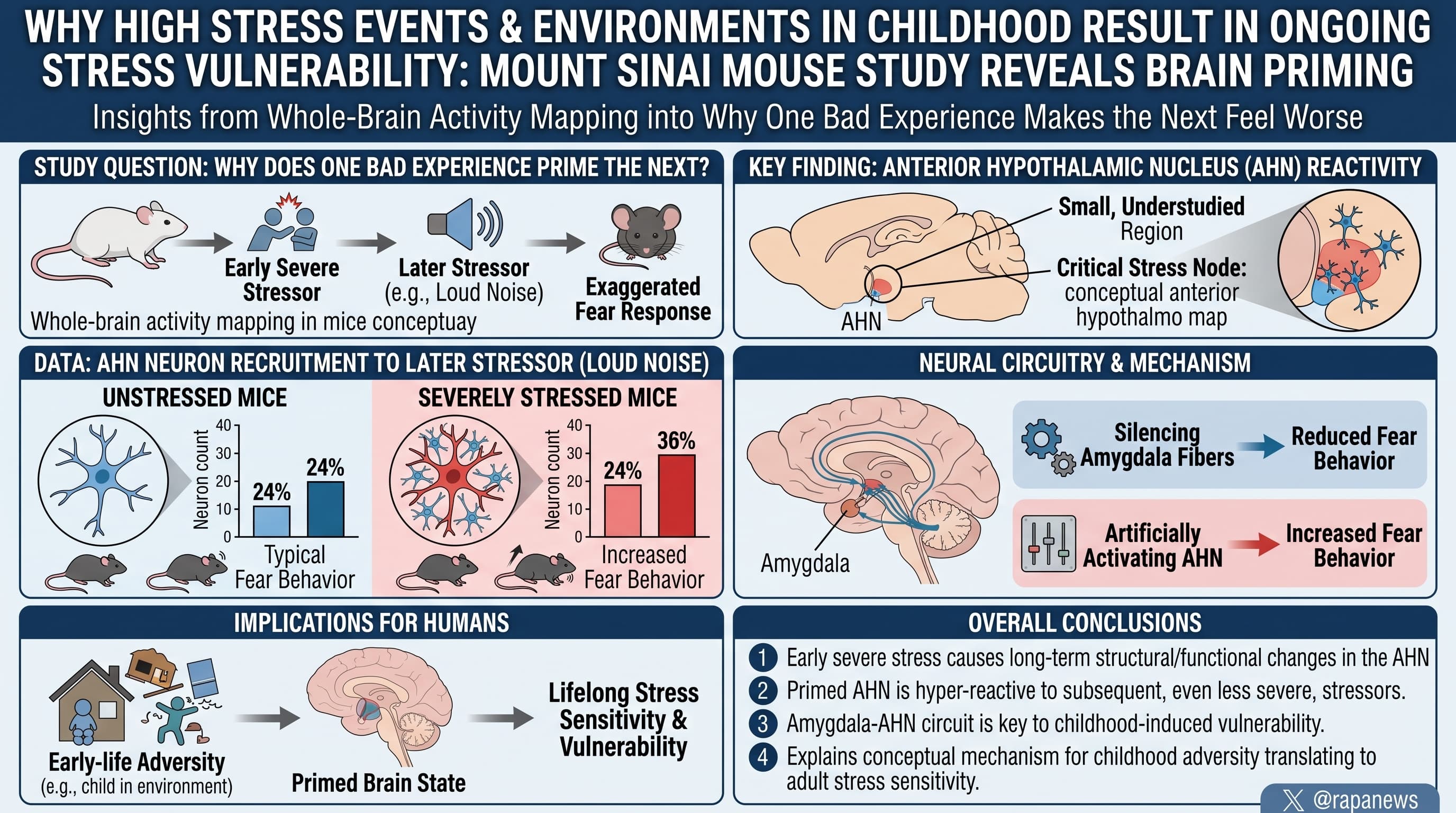
Mount Sinai researchers used whole-brain activity mapping in mice to ask why one bad experience makes the next one feel worse. They found that a small, understudied region, the anterior hypothalamic nucleus (AHN), becomes more reactive after a severe stressor. Previously stressed mice recruited about 36 percent of AHN neurons in response to a later loud noise, versus 24 percent in unstressed mice. Silencing these neurons, or the amygdala fibres feeding them, reduced fear behaviour, and artificially activating them increased it. The effects on behaviour are large by statistical convention, but they rest on one behavioural measure, in young mice, with no human data and no drug target.

People who have lived through severe stress tend to react more strongly to later stressors, even mild ones. This “stress sensitization” raises the risk of PTSD and depression. Where it is stored in the brain has been unclear, because most research has looked in the same few places: amygdala, prefrontal cortex, hippocampus.

A team led by Zachary Pennington and Denise Cai at the Icahn School of Medicine at Mount Sinai took a different route. They gave mice ten footshocks, waited ten days, then played a loud burst of noise. Shocked mice froze about twice as long after the noise as mice that had never been shocked. The researchers then cleared the animals’ brains to make them transparent and counted active cells across 452 regions.

The cortex of previously stressed mice was quieter. Two deep regions were more active. One was the AHN, a hypothalamic nucleus better known for temperature control, sleep and aggression. Its activity was also more tightly correlated with a cluster of threat-related regions, including most of the amygdala.

To see what AHN cells respond to, the team mounted miniature microscopes on the heads of mice and watched hundreds of inhibitory neurons fire. The cells responded to shocks and loud sounds, more strongly to stronger ones, and went quiet when a thirsty mouse drank water or sugar solution. That pattern suggests the cells track how bad something is, not merely how attention-grabbing it is.

The key finding concerns what prior stress changed. Individual responsive neurons did not fire harder. Instead, more neurons joined in. The share of cells responding to the noise rose from roughly a quarter to more than a third.

The team then tested cause and effect. Switching AHN neurons off with light during the footshocks cut freezing during the shocks and in later memory tests. Switching them off later, when the mice were returned to the shock chamber or heard the noise, also cut freezing, and the behaviour returned within moments of the light going off. Chemically activating the cells made mice freeze about ten times longer after the noise. Finally, silencing amygdala nerve endings inside the AHN during the noise reduced freezing.

Manipulating the AHN did nothing to a standard anxiety test, and activating it without a stressor did not cause freezing. The AHN therefore looks like an amplifier for threats that are actually present.

Some of the paper’s wording outruns its data. The abstract says silencing amygdala inputs “abolished” sensitized responses. The figure shows a reduction of about nine percentage points, with no unstressed group in that experiment to show what a non-sensitized response would be. The work was done in young mice, with freezing as the only behavioural readout, and no stress hormone measurements.

Actionable Insights

  1. Prior severe stress roughly doubled the fear response to a later, unrelated mild stressor: freezing rose from about 22 to about 47 percent of the time. In statistical terms that is a “large” effect (Cohen’s d near 1.0, meaning the average stressed mouse reacted more than about 84 percent of unstressed mice).
  2. The brain change behind it was recruitment, not intensity: 36 versus 24 percent of cells responding, a 49 percent relative increase and 12 points in absolute terms.
  3. The effect was reversible in real time when the circuit was switched off. That hints sensitization is an active, ongoing process and not permanent damage.
  4. The circuit did not drive general anxiety. This supports the idea that heightened reactivity to stressors and background anxiety are separable problems
  5. The implication that limiting severe stress exposure limits later over-reactivity

Context/Source

  • Open Access Paper: An amygdala to anterior hypothalamic circuit gates stress sensitivity
  • Institutions: Nash Family Department of Neuroscience and Department of Pharmacology, Icahn School of Medicine at Mount Sinai, New York; Medical University of South Carolina
  • Country: USA
  • Journal: Nature. published online 30 September 2026.
  • Impact evaluation: The impact score of this journal is 56.1, evaluated against a typical high-end range of 0 to 60+ for top general science, therefore this is an Elite impact journal.

Related Reading:

1 Like

So @relaxedmeatball and others here with kids, how old is old enough for anti-lipid medications? At what age might we want to get our kids on statins or other medications to lower LDL-C / APOB?

See: Nearly 1 in 10 young adults could have fatty, narrow arteries

To my knowledge, guidelines now say 6 years old for those at risk. I’m going through this right now:

I have heterozygous familial hypercholesterolemia (HeFH), so one copy of the allele for high LDL-C, giving me a total LDL-C of 200-220mg/dl. My mother also has it, and her family has a terrible history of early MIs.

I also have high Lp(a) - 60-85mg/dl. Lucky me!

My wife and I tested our first kid: aged 6 her LDL-C was 150, age 7 it was 147, age 8 it was 154, and age 9 her LDL-C jumped to 193mg/dl. The old guidelines said 150mg/dl was the cutoff for children, so she was always around that and so we held off for a while. New guidelines basically recommend treating to adult targets - i.e. 100mg/dl, and her latest test came back at 193mg/dl, so she has started statins (5mg Rosu per day)

Her Lp(a) also came back as 80mg/dl (only tested once, but obviously high). So she got both risk factors from me.

Second kid, tested at age 6, LDL-C 102mg/dl, Lp(a) 64.5mg/dl. So he has normal LDL-C but did they the high Lp(a) from me.

So both of them have elevated risk, with the 9 year old having the highest risk. We’re now doing the trial runs now to see what dosage gives what reduction. If she’s anything like me, statins alone will do nothing. Guidelines also now recommend that kids can also have Ezetimibe. So I think that’s where we will end up; most likely 5mg Rosu + 10mg Ezetimibe per day.

For those at extreme risk (Homozygous FH, LDL-C can be 400mg/dl or higher), I think you can start immediately. Those kids can have other problems like tendonopathy, cholesterol xanthomas and deposits in joints. And they have risk of heart attacks beginning from their teenage years. That’s something a specialist should handle though.

2 Likes

Good feedback. This got me thinking more broadly about what the key blood tests we should be doing on our kids for optimal health and longevity, with a long-term perspective in mind. Here is the prompt, and the result I got from Claude Opus 5.5 below:

Prompt:

System Role: Act as a preventative medicine physician practicing “Medicine 3.0,” leveraging the longevity and healthspan optimization frameworks of Dr. Peter Attia. Your objective is the proactive identification and mitigation of metabolic, cardiovascular, and hormonal disease risk decades before clinical manifestation.

Task: Formulate a prioritized, tiered list of blood tests and biomarkers for 16- and 17-year-old males and females. The primary goal is the early identification of harmful physiological trends to minimize lifetime disease burden.

Output Requirements:

  1. Tiered Prioritization: Categorize the tests into distinct tiers:
    • Tier 1: Essential Baseline & Cardiovascular (e.g., ApoB, Lp(a))
    • Tier 2: Metabolic & Glycemic Control (e.g., Fasting Insulin, Uric Acid, ALT)
    • Tier 3: Sex-Specific Hormonal & Nutritional (e.g., Iron panels, Gonadal hormones)
  2. Sex-Specific Variations: Differentiate the testing priorities and reference ranges for 16/17-year-old males versus females (e.g., the necessity of comprehensive iron/ferritin tracking in menstruating females vs. establishing optimal hormonal baselines in males).
  3. Optimal vs. Normal Ranges: For each biomarker, define the “Medicine 3.0” optimal target range for an adolescent and explicitly contrast it with standard commercial laboratory reference ranges (which represent population averages of a progressively metabolically unhealthy society).
  4. Clinical Rationale & Cumulative Risk: Justify the early measurement of these markers. Specifically address the concept of “area under the curve” (cumulative lifetime exposure) for ApoB and the early pathogenesis of insulin resistance.
  5. Scholarly Debate & Knowledge Gaps: Acknowledge the current limitations and controversies in applying aggressive adult longevity protocols to pediatric populations. Address the lack of longitudinal data on early pharmacological interventions in normolipidemic youth, discuss the debate regarding aggressive lipid-lowering and adolescent neural development, and define what empirical data is currently missing to form a clinical consensus.
  6. Formatting: Deliver the response in strict Markdown format. Use concise, academic prose. Embed URLs to primary source material, pediatric lipid guidelines, or Dr. Attia’s published literature where applicable.

Response from Claude Opus 5.5 below:

Adolescent Biomarker Screening Through a “Medicine 3.0” Lens: A Tiered Protocol for 16–17-Year-Olds

Scope note. The “optimal” targets below are mostly extrapolations. They come from adult Mendelian randomization data, from population percentiles in metabolically healthy youth, and from mechanistic reasoning. Few have been validated against hard outcomes in adolescents, and each table says so. Pediatric guideline cutoffs are cited wherever they exist. Testing a minor also requires age-appropriate consent and shared decision-making that includes the adolescent.


Framing: Why Measure at 16–17?

Late adolescence is a good window for baseline testing for three reasons:

  1. Puberty is mostly finished (typically Tanner 5). The physiological insulin resistance of puberty, which peaks around Tanner 3, has largely resolved, and lipids have recovered from their mid-pubertal dip. Values measured now are close to the person’s adult phenotype.
  2. It is the second universal lipid-screening window (ages 17–21) in the NHLBI Expert Panel guidelines (2011).
  3. Habits are still forming. Lifestyle interventions started here act on the longest possible remaining exposure period.

Tier 1: Essential Baseline & Cardiovascular

Rationale. Atherosclerosis is a disease of cumulative exposure to apoB-containing lipoproteins. It begins in childhood. Autopsy data from PDAY and the Bogalusa Heart Study show fatty streaks and raised lesions in adolescents, and these scale with non-HDL-C and other risk factors.

Biomarker Standard lab / pediatric reference Medicine 3.0 adolescent target Evidence for target
ApoB Adult lab flag usually ~>120–130 mg/dL. NHLBI pediatric: acceptable <90, borderline 90–109, high ≥110 <70 mg/dL (roughly the lower quartile of healthy youth) Extrapolated from MR and adult trial data; no pediatric outcome validation
LDL-C NHLBI: acceptable <110, borderline 110–129, high ≥130. Adult lab: <100 <80–90 mg/dL. LDL ≥160 mg/dL (≥130 with family history) should trigger an FH evaluation FH thresholds are guideline-based (EAS 2015); the optimum is extrapolated
Non-HDL-C NHLBI: acceptable <120, borderline 120–144, high ≥145 <100 mg/dL Extrapolated. Usable non-fasting, which is practical for screening
Triglycerides (ages 10–19) NHLBI: acceptable <90, borderline 90–129, high ≥130 <70 mg/dL. TG/HDL-C ratio <1.5 Ratio is a proxy for insulin resistance; cutoffs vary by ancestry
HDL-C NHLBI: acceptable >45; low <40 >45 (descriptive only, not a target) Raising HDL pharmacologically has no causal benefit
Lp(a) (measure once, report in nmol/L) Labs often flag >30 mg/dL or >75 nmol/L <75 nmol/L (low risk). >125 nmol/L means elevated lifetime risk (EAS 2022) Genetically determined. It rises modestly through childhood, so an adolescent value approximates the adult level
CBC + CMP Standard Within reference range Needed to interpret Tiers 2–3 (iron, liver, renal)
Blood pressure (not a blood test) AAP 2017: ≥13 y, normal <120/<80 <115/75 Essential companion to lipids; cumulative SBP also behaves like an area-under-the-curve exposure

Clinical use of Lp(a). Lp(a) cannot be modified today with approved therapies. Its value is that it sets how aggressively lifetime ApoB should be controlled, and it identifies parents and siblings for cascade screening. A high adolescent Lp(a) is one of the strongest arguments for an early, deliberate lifestyle focus on ApoB.

Area Under the Curve: The ApoB Argument

  • Cumulative exposure model. Atherosclerotic burden scales roughly with concentration × time (often written as “LDL-years”). Untreated homozygous FH reaches the event threshold in childhood. Heterozygous FH reaches it around age 35–45. Typical Western LDL levels reach it in the 50s–60s.
  • Mendelian randomization. Genetic variants that lower LDL for a whole lifetime produce about 2–3× the coronary risk reduction per mmol/L seen in 5-year statin trials (EAS consensus, Ference et al. 2017). Black carriers of PCSK9 loss-of-function variants had about 28% lower LDL-C and about 88% fewer CHD events (Cohen et al., NEJM 2006).
  • Illustrative arithmetic. Holding LDL-C at 80 instead of 110 mg/dL from age 17 to 50 avoids about 1,000 mg/dL-years of exposure, the same burden as several extra decades at a modestly lower level. This is the core logic behind the Attia framework’s preference for early, lifelong suppression of ApoB (Outlive, 2023).
  • Prospective validation. The i3C consortium linked childhood risk factors, including non-HDL-C and triglycerides, to adult cardiovascular events (Jacobs et al., NEJM 2022).

Tier 2: Metabolic & Glycemic Control

Rationale. Hyperinsulinemia and ectopic fat in the liver and muscle precede hyperglycemia by years. HbA1c and fasting glucose are late markers: by the time they become abnormal, a substantial share of β-cell function may already be lost. Youth-onset type 2 diabetes progresses faster than the adult form. TODAY found high rates of complications within about 15 years of diagnosis.

Biomarker Standard lab reference Medicine 3.0 adolescent target Evidence for target
Fasting insulin ~2–25 µIU/mL (assay-dependent) <8 µIU/mL, ideally ~3–6 Distribution-based. Insulin assays are poorly standardized, so trend results within the same lab
Fasting glucose 70–99 mg/dL 70–90 mg/dL Extrapolated from adult cohort data
HbA1c <5.7% (prediabetes 5.7–6.4%) <5.3% Iron deficiency falsely raises A1c, so interpret alongside Tier 3
HOMA-IR (calculated) No standardized pediatric cutoff <1.5–2.0. Values >2.5–3.0 in post-pubertal adolescents suggest insulin resistance Cutoffs vary by population and assay
ALT Many labs use an upper limit of 35–55 U/L Males <25; females <20 U/L. SAFETY upper limits of normal are 26 (M) and 22 (F) NASPGHAN 2017: ALT persistently >2× the SAFETY ULN warrants MASLD evaluation
AST, GGT AST <40; GGT <50–60 GGT <20 U/L Heavy training raises AST and ALT, so avoid testing within 48 h of hard exercise
Uric acid M ~3.5–7.2; F ~2.5–6.0 mg/dL M <5.5; F <4.5 mg/dL Associations with BP, fructose intake, and MASLD; causal role debated
2-h OGTT with insulin (selective) Glucose <140 mg/dL at 2 h Insulin returning toward baseline by 2 h For obesity, family history of T2D, PCOS, or acanthosis; consistent with AAP 2023 obesity CPG

Tier 3: Sex-Specific Hormonal & Nutritional

Females (menstruating)

The main issue in this group is iron deficiency without anemia. It affects roughly 40% of US females aged 12–21 (Weyand et al., JAMA 2023), and a hemoglobin-only screen misses it.

Biomarker Standard lab reference Adolescent target Notes
Ferritin Lower limit often 10–15 ng/mL (WHO deficiency cutoff <15) >50 ng/mL; treat if <30 Ferritin rises with inflammation, so pair with CRP. Recheck every 6–12 months if menses are heavy or the patient is an endurance athlete
TSAT / serum iron / TIBC TSAT 15–50% TSAT 20–45%
Hemoglobin / MCV / RDW Hb ≥12 g/dL Within range A rising RDW is an early sign of iron restriction
TSH, free T4 TSH ~0.5–4.5 mIU/L TSH ~1–2.5 Relevant to menstrual irregularity and fatigue
Hormones (if indicated): total testosterone, SHBG, LH, FSH, estradiol, prolactin Lab-specific Diagnostic use, not “optimization” PCOS in adolescents requires both irregular cycles (>1–2 years post-menarche) and hyperandrogenism. The 2023 international PCOS guideline advises against ultrasound or AMH for diagnosis within 8 years of menarche. Athletes with amenorrhea need evaluation for RED-S / functional hypothalamic amenorrhea

Males

Biomarker Standard lab reference Adolescent target Notes
Total testosterone (fasting, before 10 a.m.) Tanner 5 range about 300–1,000+ ng/dL Baseline documentation, not a target The main value is a lifelong comparison point and detection of obesity-related hypogonadism
SHBG, calculated free T, LH, FSH, estradiol Lab-specific Coherent axis Suppressed LH/FSH with high testosterone or high hematocrit suggests exogenous androgen use, which is a real and underappreciated problem in this age group
Hematocrit ~39–50% <50%
Ferritin / TSAT Ferritin ~30–300 Ferritin 50–150; TSAT <45% TSAT >45% warrants HFE testing. Endurance athletes can become depleted

Both sexes (selective)

  • 25-OH vitamin D. The Endocrine Society 2024 guideline recommends empiric supplementation for ages 1–18 and against routine testing in healthy people. This is a direct conflict with longevity-clinic practice.
  • B12 and folate for vegetarian or vegan adolescents.
  • Omega-3 index (target ≥8%). Popular in the Attia framework, but outcome data in youth are lacking.
  • hs-CRP. Low yield in adolescents and easily confounded by intercurrent infection. Use it mainly to interpret ferritin.

Scholarly Debate & Knowledge Gaps

1. Screening itself is contested

  • NHLBI/AAP recommend universal lipid screening at ages 9–11 and 17–21.
  • The USPSTF (2023) gives an “I” statement (insufficient evidence) for lipid screening in asymptomatic youth (JAMA 2023). It cites the lack of trials linking pediatric screening to adult outcomes.
  • Real-world uptake of universal screening is low. FH, the clearest beneficiary (prevalence about 1 in 250), stays largely undiagnosed until adulthood.

2. Pharmacotherapy evidence is confined to FH

  • The strongest data come from children with heterozygous FH. In 20-year follow-up, statin therapy started in childhood was associated with fewer cardiovascular events than in affected parents, and no growth or pubertal harms appeared (Luirink et al., NEJM 2019). Evolocumab was effective in pediatric HeFH in HAUSER-RCT (Santos et al., NEJM 2020).
  • There are no randomized or long-term cohort data on lipid-lowering drugs in normolipidemic adolescents. Extending Medicine 3.0’s “lower for longer” logic to drug treatment in a 16-year-old with LDL 110 mg/dL is hypothesis, not evidence. For non-FH youth, the defensible current position is measurement plus lifestyle.

3. Lipid lowering and neural development

  • Concern. Myelination and synaptic pruning continue into the mid-20s, and myelin is cholesterol-rich.
  • Counterpoints. Brain cholesterol is synthesized locally, and circulating LDL does not cross the blood–brain barrier. PCSK9 antibodies do not enter the CNS. Hydrophilic statins (rosuvastatin, pravastatin) penetrate less than lipophilic ones. Lifelong low LDL from PCSK9 loss-of-function variants shows no neurocognitive phenotype.
  • Caveats. Pediatric FH trials were not powered for subtle cognitive or psychiatric outcomes. Severe hypobetalipoproteinemia does cause neurological disease, though mainly through fat-soluble vitamin malabsorption, which is a different mechanism. For adolescent females, statins are still generally stopped before conception, which complicates long-horizon therapy.

4. Harms of “optimization” culture in adolescents

  • Over-interpreting borderline results can label healthy teens, drive anxiety, and push toward restrictive eating. Adolescents have the highest incidence of eating disorders, so framing matters.
  • Hormonal “optimization” language is especially risky for adolescent males, given their access to androgens and SARMs. Hormone testing at this age should be diagnostic and protective, not aspirational.

5. What empirical data are missing for a consensus

  1. Outcome-anchored pediatric thresholds for ApoB, fasting insulin, ALT, and Lp(a), as opposed to percentile-derived ones.
  2. Long-term cohorts connecting adolescent ApoB and insulin trajectories to imaging (CAC, CCTA plaque in the 30s–40s) and to events.
  3. Pragmatic trials testing whether disclosing biomarkers to adolescents changes long-term behavior and outcomes, including psychological effects.
  4. Assay standardization for insulin and Lp(a) (nmol/L, isoform-insensitive methods).
  5. Safety data on CNS and endocrine outcomes for any lipid-lowering class started before age 25 in people without FH.
  6. Cost-effectiveness modeling of universal versus risk-based panels, beyond FH detection.

Summary Priority List

Priority Males (16–17) Females (16–17)
Tier 1 ApoB, lipid panel, Lp(a) once, CBC, CMP, BP Same
Tier 2 Fasting insulin, glucose, HbA1c, HOMA-IR, ALT/GGT, uric acid; OGTT if risk factors Same; interpret HbA1c alongside iron status
Tier 3 AM total T, SHBG, LH/FSH (baseline and androgen-misuse screen); ferritin/TSAT Ferritin, TSAT, CBC indices (high priority); TSH; androgens/LH/FSH only if PCOS or amenorrhea is suspected

I agree with most of that in principle. However, I think we also need to consider that a 16-17 year old should be having their own life, and subjecting them to a battery of medical tests is something you probably want to be careful with. There’s a balancing act IMO between recognising and dealing with problems early, and making them have some sort of pathogenic health anxiety issues. As we know, eating disorders, body dysmorphia etc are prevalent. I can imagine negative consequences if you start emphasising optimisation to teenagers who are just trying to find their independence, and already have enough to deal with from the Manosphere and crazy influencers telling them they need to look a certain way.

For me personally, I would still prefer to be symptom-led for a lot of those. For example, if your 16-17 year is active, fit, eats appropriately etc, there’s no real reason to be worrying about their HBA1C, fasting insulin etc. There’s almost no chance they would be pre-diabetic. And, if they are unfortunately obese, or skinny-fat, inactive and drink 8 cokes a day, you have a very obvious problem that doesn’t really require a blood test before you take action.

The case of FH is different, since it is lifelong, completely out of the persons control, and not modifiable by lifestyle. So medicating it is really the only sensible option.

For kids, I would be really prioritising sleep, establishing good dietary patterns, establishing good habits of activity and exercise. (Other people drive their kids the 5 mins to school. We walk every day, climb the hill to our house most days. I take stairs if it’s less than 3 floors. Things like that.)

From what I’ve heard, anemia and bone density are two critical issues - but again, they’re quite reasonably addressed through very simple lifestyle practices. So if I have an otherwise healthy 16 year old who has stayed doing her gymnastics, drinking milk etc, I’m not really thinking she needs a DEXA scan.

Hope that makes sense.

1 Like