SPECT scan from amenclinics?

Expensive, but could be a way to diagnose some conditions, and AGI timelines might be short enough to make this matter… (The most important thing s can do is do all we can to make human brains suck less - even eliezer yudkowsky strongly believes this)

Eros MarcelloEros Marcello • 1stVerified • 1stMultidisciplinary Product Designer, Human-AI Interaction | Deep Tech | prev: Meta, Multidisciplinary Product Designer, Human-AI Interaction | Deep Tech | prev: Meta, 1yr • Edited • 1 year ago • Edited • Visible to anyone on or off LinkedIn

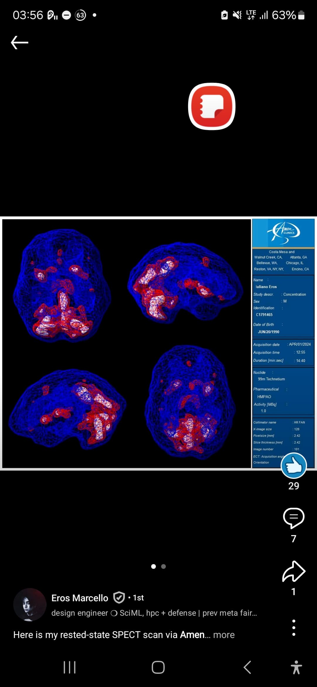
Here is my rested-state SPECT scan via Amen Clinics San Francisco Bay Area along with a key result of my Full Body Intelligence test via Viome

In my SPECT scan, notice the hyperactivity in the Basil Ganglia and activation of my Thalmus (all highly dependent on adequate bioavailability of GABA and serotonin for nominal regulation).

With these tests and scans, along with aggressive self-education to fashion a modicum of domain expertise, I have come to establish the understanding that I have systemic GABA, serotonin and dopamine deficiencies which has contributed / caused an array of seeming mental health related ailments. I’ve now been able to calibrate and subsequently leverage precision supplementation + neuromedicine to remedy this.

All it took was $7,000.00+, several months to actually schedule and undergo these procedures, weeks to wait for the raw data to be sent in a CD-ROM, an external optical drive (Apple actually just discontinued theirs), teaching myself how to read medical imaging scans, fine-tuning Llama 3 for neuroscientific applications to aid in domain-specific knowledge curation, dodging the plethora of powerful psychotropic medications “experts” were urging me to ingest, and years of basically being like “WTF is wrong?”

If you are in any sort of mental health crisis, you’re either institutionalized or God forbid in some cases, decided to unalive yourself, well before any of the above can even be commenced.

This is the problem I’m trying to solve. Our brains are literally how we perceive reality. Getting access to its activity and functioning on a granular level is paramount, just like how we now have democratized purview over the rest of our vitals (heart rate, blood oxygen, etc.)

Consumer neurotech, specifically BCIs, is quickly becoming a saturated market. To not make the neural data gleaned from these devices actionable (and in conjunction with brain imaging) is lunacy to me.
1 Like

Here was the image he posted

This may be much cheaper in Japan…

Japanese centers commonly use e-ZIS or 3D-SSP, which compare regional perfusion statistically against a normative database. Tokyo Hospital explicitly describes doing this quantitative analysis. �

Worked for 2m 29s

Yes. I dug for places that actually perform cerebral-perfusion SPECT and have a plausible pathway for a foreign/self-pay patient, rather than just hospitals mentioning SPECT somewhere on a radiology page. There are some surprisingly workable options.

Best candidates I found

Place Why it’s interesting Access / price

:india: Ganesh Diagnostic & Imaging Centre Probably the lowest-friction option. They literally sell “Brain Perfusion (SPECT)” through their website. Currently listed at ₹30,000. Requires a physician prescription and ID. Report advertised within 24 h.
:jp: Tokyo Metropolitan Ohtsuka Hospital Excellent transparent pricing. Offers both 99mTc-ECD and 123I-IMP cerebral perfusion SPECT. Full self-pay published price: ¥69,000 ECD / ¥66,000 IMP. Generally referral + appointment based; without a referral there’s an additional ¥7,000 first-visit charge.
:jp: National Hospital Organization Tokyo Hospital My favorite if the goal is analyzable data. Uses ECD or IMP and explicitly performs 3D-SSP + eZIS quantitative analysis instead of merely eyeballing rainbow slices. No simple self-pay price published that I found. More hospital-like access, so clinician order is likely.
:kr: Samsung Medical Center Especially interesting because its own SPECT documentation explicitly includes psychiatric disorders among brain-perfusion SPECT indications. Very large nuclear-medicine program. International center takes overseas/out-of-pocket patients and can arrange tests after physician consultation.
:kr: Asan Medical Center Very standardized resting protocol: 99mTc-ECD, 20-min uptake, ~30-min scan in a quiet dark environment. Excellent international-patient machinery. International center coordinates exams, billing and explicitly issues imaging CDs. Ask for a cash quote.
:india: Hero DMC Heart Institute This is particularly relevant if you want to reproduce the Amen scan as closely as possible. They explicitly offer 99mTc-HMPAO OR ECD brain-perfusion SPECT. No public price found. Contact/physician-order route rather than online checkout.
:india: Fortis Memorial Research Institute More hospital-grade alternative in India. Their nuclear medicine department explicitly lists brain SPECT for perfusion, dementia and epilepsy, and handles international patients directly. They will provide international patients a written cost estimate before travel.
:us: North Texas Medical Center A surprisingly straightforward US cash option. $1,660 currently through MDsave. Explicitly requires a physician order.

Ganesh Brain Perfusion SPECT booking page
Ohtsuka Hospital published SPECT prices
Tokyo Hospital brain-perfusion SPECT and eZIS information
Samsung international-patient appointments
Asan international healthcare center

There’s one important wrinkle with reproducing your Amen scan

Your screenshot says 99mTc-HMPAO.

Japan overwhelmingly seems to favor 99mTc-ECD and 123I-IMP. Asan’s standard resting protocol is ECD. These all measure regional cerebral perfusion, but HMPAO and ECD aren’t interchangeable if your goal is a genuine longitudinal A→B comparison. Their regional distributions differ somewhat because their intracellular trapping mechanisms aren’t identical.

So there are really two different goals:

If you just want a high-quality cerebral perfusion measurement:
I’d lean toward Tokyo Hospital, because eZIS/3D-SSP is far more scientifically useful to me than an Amen-style thresholded 3-D “surface.”

If you want “Amen scan #2” and want to compare it to the image you posted:
I’d prioritize a facility willing to use 99mTc-HMPAO under a standardized resting protocol. Hero DMC is the clearest non-Amen facility I’ve found explicitly advertising HMPAO cerebral perfusion. A US nuclear-medicine department may also have HMPAO available, but you need to verify it before scheduling.

What I’d ask before paying anyone

Send the center these exact technical questions:

  1. “Do you perform resting cerebral perfusion SPECT using 99mTc-HMPAO/exametazime, or only 99mTc-ECD?”

  2. “Is tracer injection performed at rest in a quiet, dim room, and are eyes open or closed?”

  3. “Can I receive the complete reconstructed SPECT dataset in DICOM format, not just JPEG/PDF images?”

  4. “Can you also provide the raw projection data if your PACS permits it?”

  5. “Do you provide voxelwise normative analysis such as eZIS, 3D-SSP/Neurostat, SPM, or CortexID?”

  6. “What scanner, collimator, reconstruction algorithm, attenuation correction and smoothing parameters do you use?”

  7. “How should caffeine, nicotine and medications be handled before injection?” For prescribed medications, get their imaging physician’s instructions rather than stopping anything yourself.

That transforms the scan from colorful brain oracle :rainbow::brain: into something you can actually register to MRI, normalize, extract ROIs from, and compare later.

One especially interesting possibility is having the scan acquired cheaply in India/Japan/Korea and doing the quantitative analysis yourself afterward. If they give you the DICOM volume, you do not need to depend on their proprietary visualization. You could MRI-register it, calculate cortical/subcortical ROI perfusion ratios, make Z maps against an appropriate reference database, and compare repeat scans with much less thresholding theater.

For your specific objective, my ranking would be Tokyo Hospital for quantitative rigor → Hero DMC if matching HMPAO matters → Samsung/Asan for easiest sophisticated international-hospital experience → Ganesh Delhi for cheapest/easiest acquisition.

Even if you pay 7000 extra yen without referral, 73,000 yen is like $450…

Quirónsalud, Spain

Worked for 23s

Taken together, you’d be building a multilayer map of the same brain rather than five redundant scans. Each modality asks a different question:

Modality What it tells you What you could actually do with it

T1/T2 structural MRI Exact cortical anatomy, sulci, gray/white matter, volumes Neuronavigation, place TMS/tFUS in your anatomy instead of using scalp coordinates
Resting-state fMRI Which regions fluctuate together, i.e. functional networks Potentially choose an individualized cortical stimulation target based on connectivity to deeper symptom-related networks
Diffusion MRI / HARDI Estimated white-matter fiber orientations and pathways Ask whether candidate cortical targets are structurally connected to ACC, striatum, thalamus, etc.; HARDI handles crossing fibers better than simple DTI
ASL perfusion Quantitative resting cerebral blood flow, often in mL/100 g/min Identify relatively hypo/hyperperfused regions and repeat after an intervention without radiation or contrast
MRS Local metabolic/neurochemical environment Measure things such as Glu/Glx, NAA, Cho, Cr, myo-inositol; with edited MRS, GABA. Potentially compare target chemistry before/after treatment
DSC perfusion Contrast-bolus hemodynamics, especially relative blood volume/flow and transit characteristics Much more useful for tumors, stroke and vascular questions than for your sort of neuromodulation mapping; requires gadolinium

The really interesting thing is cross-modal convergence. Suppose rs-fMRI says cortical patch A is strongly connected to a deeper network you care about. HARDI says there is also a plausible white-matter route linking those territories. ASL says that same network has unusual resting perfusion. MRS says the target region has a different Glu/GABA/metabolic state. That is much more informative than any one pretty brain map.

For TMS, rs-fMRI is probably the highest-value exotic sequence. Connectivity-informed targeting, particularly selecting left prefrontal sites based on connectivity with regions such as the subgenual cingulate, is a serious research direction. But it is not yet settled clinical doctrine, and individualized target reproducibility remains an issue. A major TMS consensus review explicitly notes that a pivotal trial proving individualized functional-connectivity targeting superior to conventional targeting has not yet been done.

For tFUS, anatomy + network connectivity are valuable for deciding what node to target, but MRI alone doesn’t solve how the ultrasound reaches it. You’d still ideally want skull CT or a validated ZTE/UTE-derived skull model for acoustic simulation. That’s the missing sixth layer.

ASL versus DSC, specifically

For what you’re contemplating, I’d choose ASL almost every time.

ASL magnetically labels your own arterial blood water, so there is no injection and you get an approximately quantitative CBF map. It’s especially attractive for repeated before/after measurements.

DSC injects gadolinium and watches the bolus traverse the brain. It has stronger signal and is clinically excellent for questions like tumor vascularity or acute cerebrovascular hemodynamics, but its measurements are generally more relative/semiquantitative.

So if a Barcelona clinic merely says “perfusion MRI,” I would not pay extra until they answer:

Is this 3D pCASL/ASL with quantitative CBF maps, or gadolinium DSC?

For this project, pCASL is the interesting answer.

MRS has another important caveat

MRS does not measure neurotransmitter firing. A GABA measurement is not “how much inhibitory signaling is occurring right now,” and Glu is not equivalent to synaptic glutamatergic transmission. You’re measuring relatively large tissue metabolite pools inside a voxel.

At 3T, good short-TE spectroscopy can resolve several metabolites substantially better than 1.5T. GABA generally needs an edited acquisition such as MEGA-PRESS, which is technically demanding.

So MRS would be scientifically fascinating as phenotyping / pre-post intervention measurement, but I wouldn’t use one MRS value to decide “therefore target X” or “therefore take medication Y.”

And that’s the larger distinction I’d make:

Most actionable now:
T1/T2 → neuronavigation
rs-fMRI → candidate network target
CT/ZTE → tFUS acoustic targeting

Very useful supporting physiology:
HARDI → structural network architecture
ASL → perfusion phenotype
MRS → metabolic/neurochemical phenotype

Currently weak: using any of these scans to pick a particular psychiatric medication.

If Barcelona really will sell a single 3T session containing T1/T2 + 20–30 min rs-fMRI + multi-shell diffusion + pCASL + MRS, that is far more scientifically useful to me than spending comparable money on an Amen-style SPECT. It gives you five mutually co-registered views of the same neural terrain rather than one perfusion snapshot.