Should Biological Age be measured in mV (milliVolts)?

I have just posted this:

People who study NCD (Non Communicable Diseases) and particularly those that focus on the diseases of aging (which I think include many of the NCDs, but not all of them) have a concept of someone having a biological age as well as a chronological age. This guides things like how frail someone is, how well their cognition works, whether they are at the risk of cancer or diabetes or indeed how they look.

Human beings are quite good at judging each other’s general health by appearance. That is because the various NCDs tend to correlate with how well the body maintains the skin and when someone is frail it is quite easy to see this by how they move around.

Regular readers of my blog will know that I think this is because the genome stops functioning properly as people get older. In essence cells stop producing the right proteins. I provide full details of this hypothesis on this web page Further thoughts from the world of independent researchers.

Various approaches have been used to calculate a biological age. Some look at methylation marks on DNA. Others take various biomarkers and try to calculate an age from those. These are always done in years (or months).

However, if the biological age actually primarily depends upon the efficiency of the mitochondria and the membrane potential of mitochondria then perhaps the biological age should reflect that. The membrane potential of mitochondria is measured in millivolts. (normally in the low 100s) Although practically measuring the combination of the membrane potentials of all of the mitochondria in all of the cells is not practical the question is still raised as to whether there is a reason for looking at things this way. I think there is.

What is obviously the case although not necessarily thought about that much is that the average membrane potential of a human egg mitochondria when fertilised is not constant. It is this, however, that drives where development goes both as the egg becomes an embryo and then gets born as a baby. Also when the baby grows up. There is solid scientific research that demonstrates that this starting position is a key foundation as to how good someone’s health will be. Also it would sound a bit odd that a baby would be born at a varying biological age.

There are good reasons why it is worth people knowing the state of their mitochondria. There is plenty of evidence that the state of the mitochondria can be improved (and also made worse). Therefore for people who start out with a lower membrane potential it is more important to them to look for interventions that will help prevent cancer or diabetes in later years. In a sense this moves away from the concept of biological age as “age”, but more towards it being a health and development status.

At the same time the fact that people start out with different membrane potential averages shows that the links between membrane potential and development are not just linked to an absolute value. The variation in membrane potential links to hormones and the variation in hormones drives development. Hence although for example a lower membrane potential will tend to speed up development such that puberty happens earlier, it probably won’t link directly to a multiple of the membrane potential.

People will still want to work out where they are compared to the average and there will be a mechanism to associate an average membrane potential to an age. However, understanding what is happening with the biology is perhaps more useful to people who want to improve their health.
5 Likes

Related paper, Harvard and NIH: Mitochondrial membrane hyperpolarization modulates nuclear DNA methylation and gene expression through phospholipid remodeling 2025

Maintenance of the mitochondrial inner membrane potential (ΔΨm) is critical for many aspects of mitochondrial function. While ΔΨm loss and its consequences are well studied, little is known about the effects of mitochondrial hyperpolarization. In this study, we used cells deleted of ATP5IF1 (IF1), a natural inhibitor of the hydrolytic activity of the ATP synthase, as a genetic model of increased resting ΔΨm. We found that the nuclear DNA hypermethylates when the ΔΨm is chronically high, regulating the transcription of mitochondrial, carbohydrate and lipid genes. These effects can be reversed by decreasing the ΔΨm and recapitulated in wild-type (WT) cells exposed to environmental chemicals that cause hyperpolarization. Surprisingly, phospholipid changes, but not redox or metabolic alterations, linked the ΔΨm to the epigenome. Sorted hyperpolarized WT and ovarian cancer cells naturally depleted of IF1 also showed phospholipid remodeling, indicating this as an adaptation to mitochondrial hyperpolarization. These data provide a new framework for how mitochondria can impact epigenetics and cellular biology to influence health outcomes, including through chemical exposures and in disease states.
In summary, our results provide insights into the molecular and genomic adaptations associated with a chronic rise in resting ΔΨm. Our data adds to the growing body of evidence identifying a relationship between phospholipids and mitochondria while revealing that the ΔΨm can serve as a signal to communicate with the epigenome. Notably, this occurs in the absence of mitochondrial dysfunction. Overall, our data raise fundamental questions about the potential insidious effects of environmental exposures that affect the ΔΨm, including their potential to influence health outcomes. It also highlights the possibility of modulating the ΔΨm to treat diseases, such as ovarian cancer.

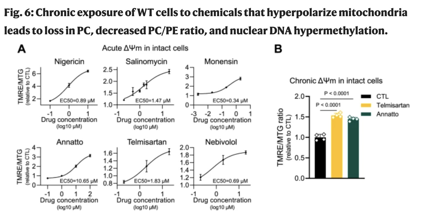
Exposure to telmisartan or annatto for 10 consecutive days led to significant increases in the resting ΔΨm of WT cells when compared to vehicle-treated controls (Fig. 6B). Like in the KO cells, this chronic rise in resting ΔΨm did not lead to mitochondrial dysfunction as judged by lack of defects on oxygen consumption or increased extracellular acidification (Fig. 6C, D). Analysis of phospholipids showed that the levels of PC were decreased in the treated cells while PE levels did not change, decreasing the cellular PC/PE ratio (Fig. 6G). More importantly, the nuclear DNA of treated cells was hypermethylated (Fig. 6H), collectively recapitulating the main phenotypes reported for the chronically hyperpolarized IF1-KO cells.

So higher ΔΨm is not better @John_Hemming?

This is hyperpolarisation, not just the normal range/steady state. What the methylation does is to hold back transcription so that the higher output of citrate does not cause difficulties. The normal range perhaps goes as high at 180mV and hyperpolarisation can be another 10-30mV above that.

Thanks. So could telmisartan be detrimental? :thinking:

I haven’t studied telmisartan. I asked chatGPT for an explanation and got:

  • Primary action — AT₁ receptor blockade (inverse agonist):
    Telmisartan selectively binds the angiotensin II type-1 (AT₁) GPCR on vascular smooth muscle, adrenal zona glomerulosa, kidney, heart, and CNS. By blocking (and partly inverse-agonizing) AT₁, it prevents the Gq/PLC → IP₃/DAG → Ca²⁺ signaling that normally drives vasoconstriction, aldosterone release, sympathetic facilitation, ADH release, and proximal tubular Na⁺ reabsorption.
    Physiologic results: arterial vasodilation, ↓ aldosterone → natriuresis (with a tendency to retain K⁺), less vasopressin effect, and dilation of the efferent renal arteriole (↓ intraglomerular pressure).

  • Ang II “rebalancing”:
    With AT₁ blocked, circulating angiotensin II may signal more through AT₂ receptors, which are generally vasodilatory and anti-proliferative, further aiding BP reduction and vascular remodeling.

  • No ACE inhibition:
    It does not inhibit ACE, so it doesn’t raise bradykinin—hence cough and bradykinin-mediated angioedema are less common than with ACE inhibitors (though angioedema can still rarely occur).

  • Metabolic “extra”: partial PPAR-γ modulation:
    Telmisartan uniquely (among ARBs) acts as a selective partial agonist of PPAR-γ, modestly improving insulin sensitivity and aspects of lipid/glucose metabolism in some patients. This effect is weaker than thiazolidinediones but can be clinically helpful.

  • Why once daily works:
    High lipophilicity and strong receptor affinity give a long functional duration (~24 h) with good tissue penetration, so a single daily dose provides sustained AT₁ blockade.


I don’t see from the above why it would be relevant.

It’s based on the above paper:

Next, we performed time-course experiments to establish doses to which WT cells could be chronically exposed without toxicity, thus allowing them to adapt to the rise in resting ΔΨm. The agents ultimately selected for further analyses were telmisartan, a medication to control blood pressure, and annatto—a food additive used in South American cuisine. These test articles provided the means to model a physiological setting in which the resting ΔΨm may be chronically increased through voluntary environmental exposure. WT cells were exposed for 10 days to either of these chemicals, which proved to be well-tolerated (i.e., no loss of cell viability; Supplementary Fig. 6B), at which point we analyzed the ΔΨm, PC, and PE content, as well as nuclear DNA methylation. Exposure to telmisartan or annatto for 10 consecutive days led to significant increases in the resting ΔΨm of WT cells when compared to vehicle-treated controls (Fig. 6B). Like in the KO cells, this chronic rise in resting ΔΨm did not lead to mitochondrial dysfunction as judged by lack of defects on oxygen consumption or increased extracellular acidification (Fig. 6C, D).

I have had an initial look at this paper, but not seen how they think telmisartan has effects that it does. That makes it difficult to make a stab at its merits.

1 Like