chatGPT(6Max):
The paper makes a potentially valuable observational contribution through its regional map of age-associated intronic RNA and its analysis of differences between people. Its findings support further investigation of RNA processing in ageing, while leaving the underlying mechanism unresolved.
This assessment concerns Regional and person-associated variation in age-related intronic RNA across human tissues dated 4 November 2026. I reviewed the manuscript and relevant literature; I have not independently rerun the deposited analyses.
The paper asks three distinct questions: whether ageing changes transcript abundance, the choice between completed splice junctions, or the amount of intron-containing RNA.
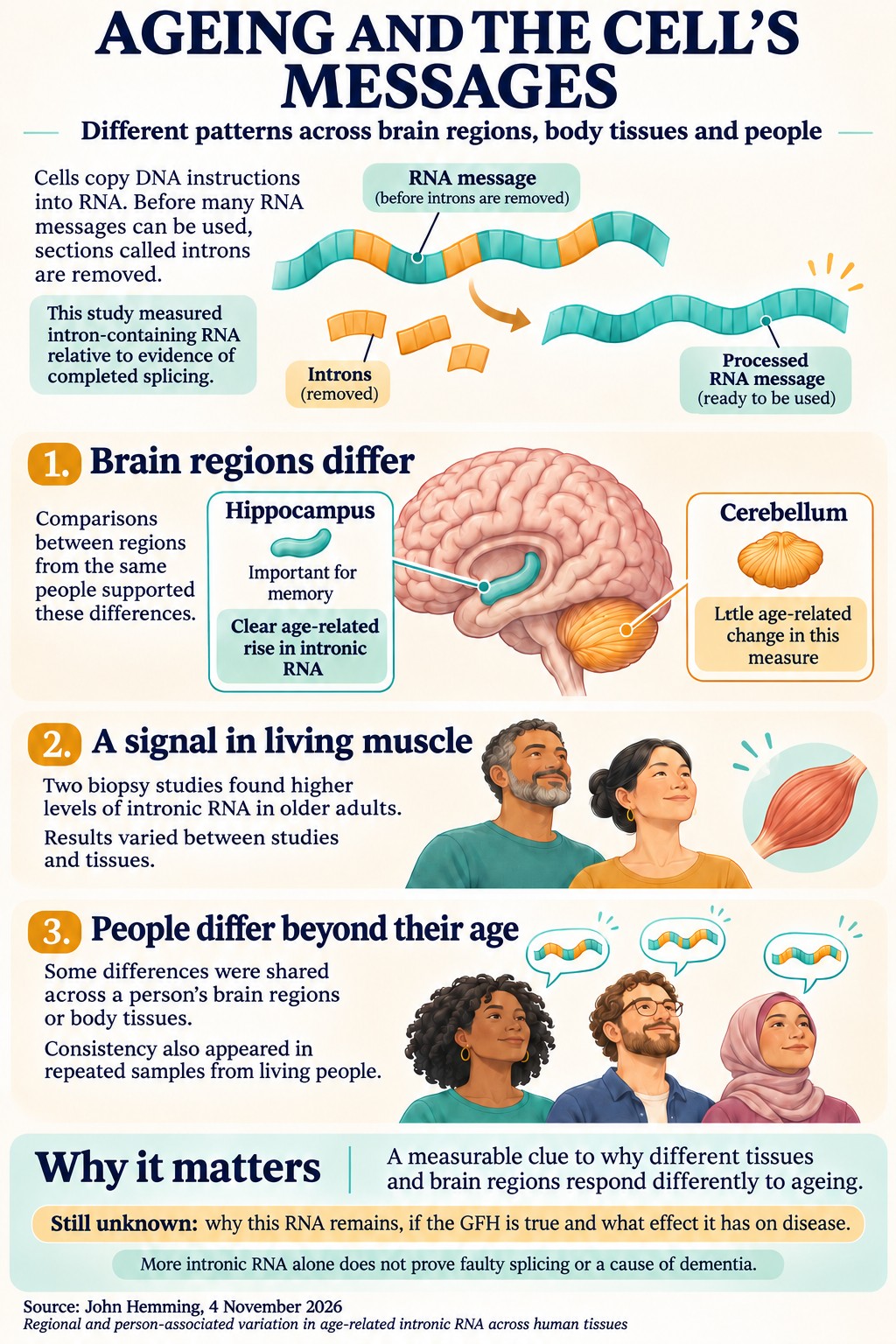
It analyses recount3-processed GTEx data across 39 non-CNS tissues and 13 CNS regions, with additional analyses of independent cohorts, disease datasets and experimental perturbations. Its clearest result is an increase in intronic RNA relative to junction reads in particular tissues and brain regions.
| Tissue or region | Estimated increase per decade | 95% confidence interval | Testing family |
|---|---|---|---|
| Hippocampus | 7.4% | 2.9% to 12.2% | Primary |
| Caudate | 7.2% | 1.9% to 12.8% | Primary |
| Tibial artery | 7.0% | 4.2% to 10.0% | Primary |
| Skeletal muscle | 4.2% | 2.3% to 6.1% | Primary |
| Kidney cortex | 12.8% | 4.3% to 21.9% | Secondary |
| Spleen | 4.5% | 1.8% to 7.2% | Secondary |
| Oesophageal mucosa | 4.2% | 1.9% to 6.5% | Secondary |
| Testis | 2.2% | 0.9% to 3.6% | Secondary |
All eight pass the stated false-discovery threshold within their respective families. These percentages describe relative changes in an RNA ratio, rather than percentage-point increases in defective transcripts.
Several findings strengthen the biological interest:
- Hippocampus, caudate, amygdala and anterior cingulate show increasing intronic RNA relative to the same donors’ cerebellum.
- The hippocampal signal extends across many introns and survives direct adjustment for gene-body coverage bias.
- Two independent living-muscle cohorts show higher intronic RNA in older adults.
- Approximately 18% of residual variation is associated with the donor within the brain or body compartment, including comparisons across extraction batches. Approximately 4% is shared between compartments.
- Person-associated consistency also appears in repeated living samples and independent datasets.
Detected changes in completed splice-junction choice are comparatively sparse, but the paper demonstrates that these tests have limited power. The mitochondrial and acetyl-CoA analyses produce weak or inconsistent support for the proposed mechanism.
The strongest novelty lies in the combination and resolution of these analyses.
Age-associated intron retention itself is established. Adusumalli and colleagues reported it in ageing fly, mouse and human brain. Wang and colleagues mapped age-associated splicing across human tissues, and Garcia-Ruiz and colleagues examined splicing inaccuracies using recount3 GTEx data. These precedents limit any broad claim to discovering age-related splicing deterioration. PubMed
The more defensible contributions are:
-
A detailed regional map supported by comparisons within donors. Comparing hippocampus and caudate with cerebellum from the same people provides stronger evidence of regional differences than comparing separate groups of donors.
-
Quantification of person-associated intronic RNA variation. The distinction between shared brain-body variation and variation specific to each compartment is particularly interesting. Repeated living samples support persistence over weeks or months.
-
Joint examination of transcript abundance, junction choice and intronic RNA. Analysing these together helps establish which observations support which biological interpretations.
-
Concrete demonstrations of analytical artefacts. The examples involving small age bins, death circumstances and depth-sensitive proportion tests are useful methodological contributions.
The developmental decline followed by a possible later-life rise has more limited novelty. Mazin and colleagues previously described lifespan splicing patterns in prefrontal cortex and cerebellum, including preferential inclusion of potentially disruptive gene segments in infants and elderly people. That directly relevant primary study should be cited and discussed. PMC
The regional findings could be important for understanding selective vulnerability in neurodegeneration. However, their precise implication is that this RNA phenotype varies between regions. They do not establish a single difference between CNS ageing and peripheral ageing.
The study has several substantial strengths.
It makes a serious effort to challenge its own interpretations: alternative denominators, covariate models, donor comparisons, coverage analyses, different intron weightings and external cohorts all contribute useful evidence. Reporting contradictory findings and unsuccessful perturbations improves its credibility.
The manuscript also distinguishes observation from mechanism reasonably carefully. Its conclusion that bulk RNA sequencing cannot establish the compartment, identity or fate of the intronic RNA is scientifically appropriate.
The main concerns are the following.
-
The measurements establish relative intronic signal more securely than delayed splicing.
Higher intronic RNA relative to junction reads could reflect increased precursor RNA, nuclear detention, altered nuclear recovery, changing cell composition, reduced mature RNA abundance or slower RNA clearance.
Consequently, the term “accumulation” should remain explicitly relative. Without absolute measurements or kinetic experiments, the analysis cannot establish an increased number of intronic molecules per cell or a reduced splicing rate.
The proposed fractionation and full-length transcript experiments address this central uncertainty well.
-
The simplified retention measure leaves unresolved technical questions.
The manuscript acknowledges that its method omits several IRFinder safeguards, including masking problematic intronic features and checking library suitability. These safeguards matter because overlapping transcription and poor mappability can distort intronic coverage. PMC
The coverage-bias analyses are valuable, but validation using a standard retention pipeline on a representative subset would materially strengthen the paper. GC content, mappability and updated annotation remain relevant sensitivities.
-
Effect size and breadth depend on the summary measure.
In hippocampus, the pooled retention index increases by approximately 7.1% per decade, while equal-weight and gene-balanced estimates are approximately 1.5%, with weaker statistical support.
These measures answer different questions: the pooled index describes an abundance-weighted signal; equal weighting describes the typical analysed intron. Their disagreement does not invalidate either result, but it limits claims of a uniform increase in processing failure.
Likewise, preferential involvement of longer introns remains uncertain because the association persists on the rank measure but disappears on the depth-adjusted log-ratio measure.
-
RNA-quality and batch sensitivities deserve greater prominence.
Restricting hippocampal samples to higher RNA integrity reduces the pooled estimate from approximately 7.1% to 3.7% per decade, with a wide confidence interval. Reduced sample size contributes to that uncertainty, so this does not demonstrate that the original result is artefactual.
More concerning is the caudate estimate: Table 4 shows a reduction from 7.2% under the primary model to 2.1% under the fuller model including expression batch. The paper should explain whether this reflects batch confounding, age-batch overlap, model instability or reduced precision.
Statements that the principal findings survive batch adjustment should be reconciled with this result.
-
Within-donor comparisons reduce confounding without eliminating every alternative.
They remove shared additive donor effects, but tissues can respond differently to the same agonal state, handling or degradation. The manuscript acknowledges this in Section 2.18, although some earlier wording is more absolute.
Cell-marker adjustment also provides incomplete control. Marker expression can change within cells, and a few markers cannot capture all relevant cell populations or nuclear-cytoplasmic differences.
-
External replication supports a restricted conclusion.
The two positive living-muscle cohorts are important evidence that the muscle association can occur without post-mortem collection. However, they do not independently validate the hippocampal pattern.
Only three of the initial 19 external cohorts show a significant rise after correction. Some null results agree with GTEx’s tissue-specific predictions, but the opposite result in one muscle cohort and the smaller surgical-kidney estimate deserve continued emphasis.
Independent replication of the matched hippocampus-cerebellum contrast would be particularly valuable.
-
Person-associated consistency has unresolved biological and technical sources.
Repeated samples and different-batch comparisons support a reproducible component. They do not identify whether it reflects genotype, stable cell composition, exposures, RNA recovery or mitochondrial state.
Nor does stability over months establish a lifelong starting level or an individual ageing rate. The paper appropriately recognises this; its strongest claim is reproducible person-associated variation.
-
Mechanistic and statistical claims need to remain proportionate.
None of the 28 perturbation contrasts passes the stated false-discovery threshold, and resemblance to the ageing pattern is weak. HDAC inhibition frequently produces changes opposite to the simple directional prediction. These results provide little affirmative evidence for the proposed acetyl-CoA mechanism, although heterogeneous interventions and limited power prevent a decisive rejection.
Similarly, few detected junction-choice changes cannot establish stability when benchmark power is low.
The exploratory regional comparisons should receive confidence intervals and clearly identified multiplicity treatment. A sensitivity analysis correcting across all 52 tissue-region tests would also help readers assess the two-family arrangement.
The manuscript would benefit from tighter presentation. It repeatedly recounts earlier versions, withdrawn arguments and successive analysis additions. Consolidating these into one account of the final methods and results would make its contribution easier to assess.
My publication assessment is favourable toward the descriptive contribution, with substantive revision needed around measurement validation, sensitivity and presentation. The strongest publication case rests on regional intronic-RNA differences and reproducible person-associated variation. Establishing their cellular source and functional consequences would determine how far these findings advance an explanation of ageing and neurodegenerative disease.