In a landmark study that challenges several prevailing theories of longevity science, researchers have constructed the first comprehensive “Metabolic Atlas” of mammalian aging, mapping 12 organs across five distinct life stages in both sexes. The findings, published in Cell Metabolism, offer a radical departure from the “systemic decline” model. Instead of a uniform metabolic collapse, the study reveals a mosaic of organ-specific aging clocks. The most shocking revelation? Contrary to the billion-dollar supplement industry’s central premise, NAD+ levels and the NAD+/NADH ratio remained largely unchanged across most tissues during healthy aging.
The research identifies the thymus as the “canary in the coal mine,” exhibiting the most dramatic metabolic rewiring (50% of metabolites altered) starting as early as adolescence. Conversely, the spleen’s metabolic profile largely stabilizes after early adulthood. The team also discovered a potent new aging biomarker: Hydroxyproline, a breakdown product of collagen, which consistently plummets across 11 of 12 organs, signaling a systemic failure in extracellular matrix turnover. Furthermore, they identified Carglumic acid not just as a waste product, but as a potential driver of aging synthesized by human cells, while reaffirming Alpha-Ketoglutarate (AKG) as a robust signature of youth. This study shifts the focus from “boosting” metabolism indiscriminately to targeting specific organ failures and waste-product accumulation.
Part 2: The Biohacker Analysis
Study Design Specifications
- Type: In vivo / Multi-organ metabolomic profiling.
- Subjects: C57BL/6NCrl mice (Note: C57BL/6N strain, not the standard C57BL/6J.
- Demographics: Male and Female.
- Organs: Heart, thymus, lungs, liver, pancreas, spleen, kidney, bladder, quadriceps, tongue, brain, plasma.
Lifespan Data
- Extension: Not a lifespan study. This is an observational atlas.
- Validation: The study cross-referenced findings with previous datasets, confirming Alpha-Ketoglutarate (AKG) as a youth-associated metabolite previously shown to extend lifespan (~12-15% in prior studies).
- Driver Identification: Identified Carglumic acid as a pro-aging metabolite; reducing it is a theoretical lifespan extension target, but untried in this paper.
Mechanistic Deep Dive
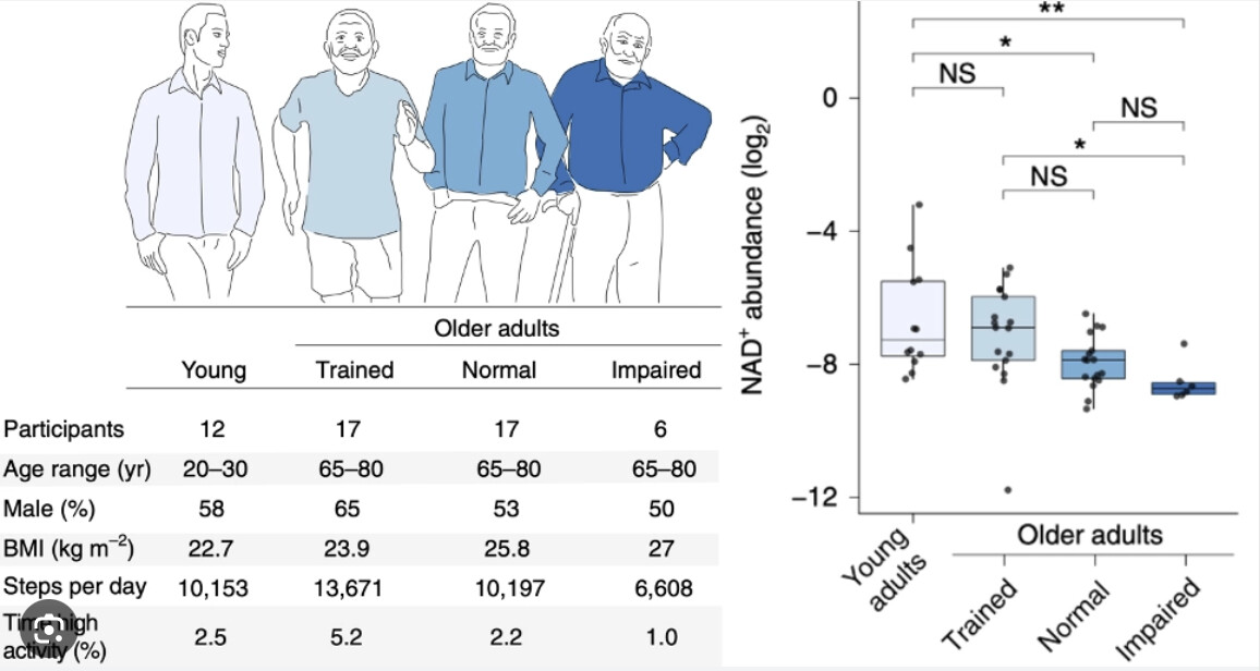
- The NAD+ Controversy: The study found no significant decline in NAD+ or the NAD+/NADH ratio in most tissues during healthy aging. This directly contradicts the rationale for indiscriminate precursor supplementation (NR/NMN) in healthy subjects, suggesting that localized flux, rather than total pool availability, may be the bottleneck—or that NAD+ decline is pathological, not intrinsic to aging.
- Collagen & Extracellular Matrix (ECM): Hydroxyproline levels dropped in 11/12 organs. This indicates a “stiffening” phenotype where collagen degradation slows down, leading to fibrosis and loss of tissue elasticity (the “stiff heart/stiff vessel” phenomenon).
-
Organ-Specific Hierarchy:
- Thymus: The primary site of metabolic aging. 50% of its metabolome shifts, mostly between 3 and 18 months. This confirms “immune aging” as a primary, early-onset driver.
- Sexual Dimorphism: Male livers showed increased hydroxyproline (fibrosis risk), whereas females followed the systemic decrease.
- Network Analysis: Essential fatty acids were identified as critical connectors in the aging metabolic network, suggesting lipid membrane composition is a central, under-appreciated aging node.
Critical Limitations
- Strain Specificity: The use of C57BL/6N mice is a double-edged sword. While it avoids the NNT mutation of the “J” substrain, it makes comparison with the vast majority of longevity literature (which uses J mice) difficult. The “stable NAD+” finding might be specific to this “healthier” mitochondrial background.
- Descriptive vs. Functional: The paper establishes correlations (Atlas). It proves AKG predicts youth but does not prove that blocking Carglumic acid reverses aging.
- Missing Data: No functional assays (e.g., grip strength, cognitive tests) were paired with the metabolic snapshots to correlate specific metabolite levels with functional decline.
Part 3: Actionable Intelligence
The Protocol (Translational Hypotheses)
-
Optimize Collagen Turnover (The Hydroxyproline Protocol):
- Intervention: Do not just “boost collagen” (peptides). You must enhance collagen turnover (degradation and resynthesis).
- Tools: Pulsed dosing of proteolytic enzymes (Serrapeptase/Nattokinase) away from food to target fibrotic tissue, combined with Glycine/Proline rich inputs (Bone broth/Hydrolyzed collagen) to fuel resynthesis.
-
Re-evaluate NAD+ Strategies:
- Pivot: If you are healthy and active, high-dose NR/NMN may be chemically redundant. Focus on NAMPT activation (exercise, circadian fasting) to maintain the flux rather than the pool. Save precursors for stress states (viral infection, sleep deprivation) or known pathology.
-
AKG Stacking:
- Dosage: Continue/Start Calcium-AKG (Ca-AKG) at ~1g/day (standard human extrapolated dose). This paper reinforces AKG as a fundamental “youth signal” metabolite.
Biomarkers (N=1 Experiments)
- Serum Hydroxyproline: Track this to assess collagen turnover. A drop suggests fibrotic stagnation.
- Cystatin C & eGFR: To proxy kidney health, as the kidney and bladder showed distinct metabolic aging signatures.
- Essential Fatty Acid Panel: specifically Omega-3 index and AA/EPA ratio, as lipid handling was a key network node.
Feasibility & ROI
- Cost: Low to Moderate. AKG is cheap. Collagen modulation is cheap.
- ROI: High. Moving away from expensive NAD+ boosters (if healthy) saves ~$100/month. Targeting fibrosis (collagen stiffness) addresses a root cause of hypertension and fragility with low-cost interventions.
Population Applicability
- Sex-Specific: High. The study emphasizes that male and female livers age differently. Men need to be more aggressive about liver fibrosis prevention (alcohol modulation, fructose reduction) than women based on the hydroxyproline data.
Context:
- Institution: University of Southern California (USC) & University of Michigan, USA.
- Journal: Cell Metabolism
- Impact Factor: ~30.9 (2024)
- Source: Cell Metabolism Abstract | BioRxiv Preprint