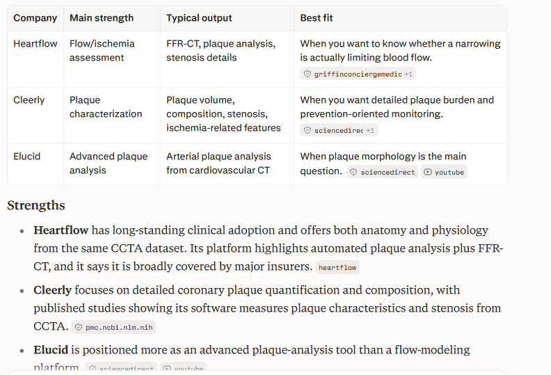
Here’s a way to check how much atherosclerosis plaque you have and the risk from that, and medical therapies under evaluation based on stage.
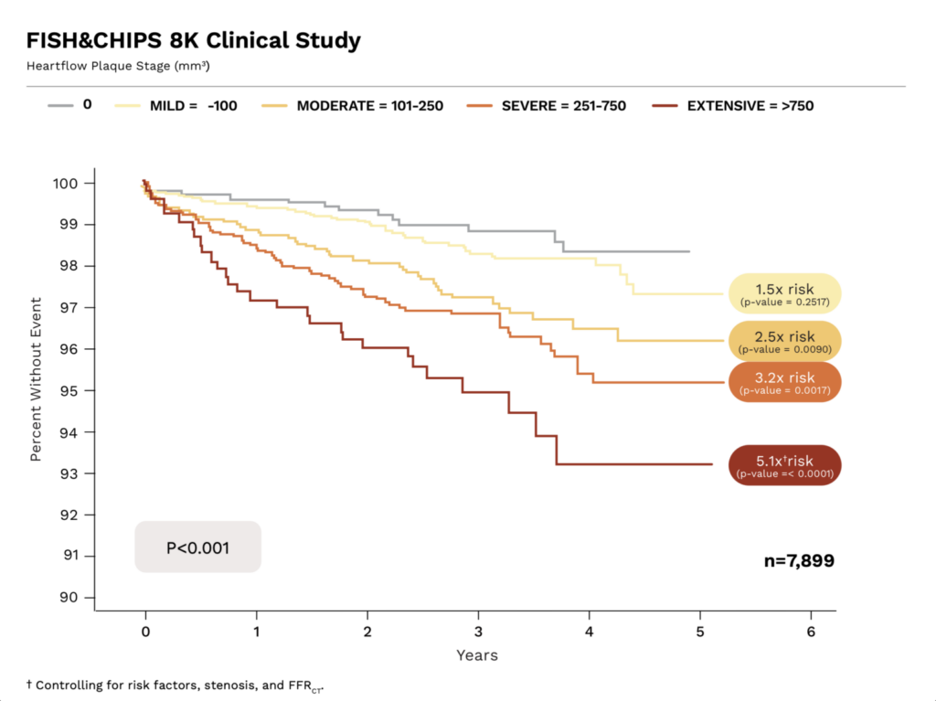
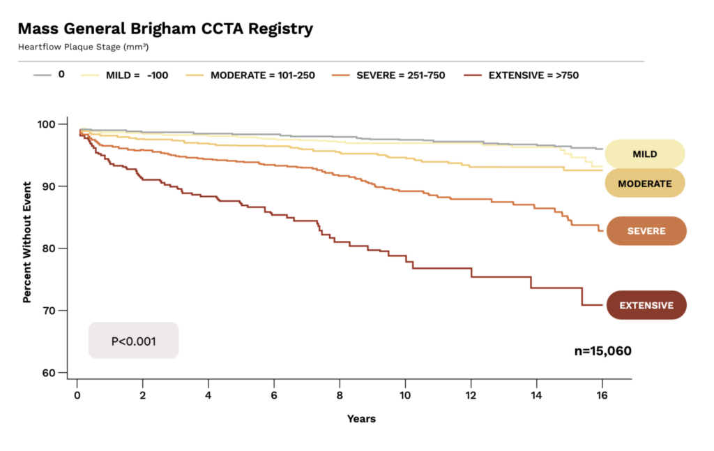
Plaque Stage and event risk
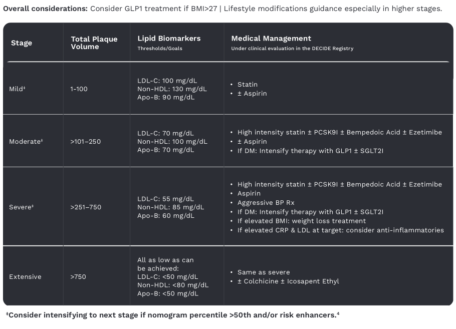
Medical management, statins are king
Video from Simon Hill

Video AI Summary
Advancements in Coronary Artery Disease Diagnosis and AI-Driven Plaque Analysis
Overview of Coronary Disease Diagnosis and HeartFlow Technology [00:00:00 - 00:10:35]
Dr. Campbell Rogers, an interventional cardiologist with extensive experience at Brigham and Women’s Hospital and Harvard, discusses revolutionary advances in the diagnosis and management of coronary artery disease (CAD). Traditionally, diagnosing CAD required invasive procedures performed in catheterization labs (“Kath labs”) such as angiograms followed by treatments like balloon angioplasty or stenting. However, only about one-third of patients undergoing invasive angiography actually benefit from intervention; two-thirds undergo invasive procedures without requiring treatment.
HeartFlow technology, developed from Stanford University research, improves patient care by using non-invasive coronary CT angiograms (CTA) combined with artificial intelligence and computational fluid dynamics to analyze coronary blood flow and plaque burden. This approach calculates fractional flow reserve (FFR) from CT images, helping decide whether invasive procedures are necessary or if medical therapy suffices ([00:06:38 - 00:09:38]).
Evolution from Blood Flow Assessment to Detailed Plaque Quantification [00:10:35 - 00:19:59]
While original HeartFlow products focused on blood flow limitation, the company expanded to advanced AI coronary plaque analysis (AI CPA). This allows detailed quantification of plaque volume and composition (calcified, non-calcified, and low attenuation plaque), providing a data-driven assessment of disease burden beyond traditional categorical reports of mild, moderate, or severe disease seen in standard CT angiograms.
Dr. Rogers compares this advancement to the introduction of X-rays over a century ago—where physical exams gave way to definitive imaging, enabling precise diagnosis. Similarly, AI CPA removes guesswork from CAD risk assessment, offering patient-specific, quantitative measurements of plaque, which were never previously possible ([00:14:48 - 00:20:59]).
Clinical Utility, Risk Stratification, and Plaque Composition [00:20:59 - 00:35:40]
Understanding plaque type is important: non-calcified and especially low attenuation plaque correlates with higher risk for heart attacks and adverse cardiovascular events, although the field is still learning and evolving. The presence and amount of plaque should be viewed relative to normative data for age and sex — for example, a 40-year-old with plaque volume in the 90th percentile has higher risk than peers.
HeartFlow’s plaque measurements have been prospectively validated against intravascular ultrasound (IVUS), the invasive gold standard, demonstrating strong correlation and reliability in quantifying plaque burden on a lesion-by-lesion basis ([00:21:24 - 00:25:16], [00:24:43 - 00:27:34]).
Additionally, CAC (coronary artery calcium) scoring, a widely used but simpler test, identifies calcium in coronaries but does not detect non-calcified plaque. A calcium score of zero implies low short-term risk but does not exclude coronary disease, as soft plaque may still be present and identifiable only on CTA with AI analysis ([00:30:25 - 00:32:25]).
Patient Selection, Indications for CT Angiography and AI Plaque Analysis [00:32:22 - 00:38:33]
Current guidelines recommend coronary CTA with FFR analysis primarily for patients who have symptoms suggestive of CAD (e.g., chest pain, exertional dyspnea). CTA is now preferred over older tests like stress nuclear imaging.
There is growing consideration for using CTA and AI analysis in:
- Patients with ambiguous calcium scores (especially 1 to 300 range), where treatment decisions can be uncertain.
- High-risk asymptomatic individuals (e.g., pilots, first responders).
- Patients desiring a more precise understanding of their coronary health, although insurance coverage for asymptomatic screening is limited ([00:32:22 - 00:38:33]).
Process and Technical Considerations for AI Analysis of CT Angiograms [00:38:33 - 00:40:55]
HeartFlow receives coronary CTA images, first assesses image quality (around 97% of scans are adequate), followed by automated AI analysis with quality checks by trained analysts. Consistency in using the same CT scanner and imaging protocol is crucial in serial imaging studies to assess plaque progression or regression accurately.
Case Study: Simon’s Baseline and Follow-Up HeartFlow Analysis [00:44:43 - 00:53:40]
Simon shares his personal HeartFlow analysis, revealing:
- Baseline (at age 38) plaque volume of 116 mm³, placing him in the 89th percentile for his age group, indicating high plaque burden compared to peers.
- Despite this burden, fractional flow reserve was normal, indicating no blood flow limitation.
- Plaque distribution included primarily non-calcified plaque, with evidence of positive arterial remodeling—plaque growing outward without obstructing blood flow ([00:44:45 - 00:49:31]).
On the follow-up scan 16 months later:
- Total plaque volume decreased to 60 mm³, dropping to the 80th percentile despite aging.
- Plaque in the left main artery disappeared, and non-calcified plaque in the left anterior descending reduced significantly, indicating plaque regression ([00:50:53 - 00:53:40]).
Interpretation of Results and Insights on Plaque Changes [00:53:40 - 01:02:19]
The discussion highlights that minor variability can exist between different plaque quantification tools, although HeartFlow is uniquely validated prospectively against IVUS. Dr. Rogers emphasizes appropriate interpretation of serial changes, especially over shorter intervals and low plaque burden scenarios where noise can influence measurements.
Simon’s experience shows how detailed coronary imaging and AI analysis can guide clinical decisions, such as initiation and intensification of lipid-lowering therapy despite “normal” blood biomarkers (e.g., modest ApoB levels) but with documented plaque presence and regression seen on imaging ([00:53:40 - 01:02:19]).
Evidence for Using AI Plaque Analysis in Treatment Monitoring and Outcomes [01:02:19 - 01:16:06]
Emerging data demonstrate that AI plaque analysis can track treatment response accurately. For example, a randomized trial in men undergoing androgen deprivation therapy for prostate cancer used HeartFlow analysis to reveal differences in coronary plaque progression between drugs.
Statin therapy often correlates with increased calcium scores, reflecting stabilization and calcification of soft plaque, which is considered a positive healing sign. While low attenuation plaque indicates higher risk, overall non-calcified plaque burden remains the most reliable risk marker.
Early clinical evidence links use of these AI tools to significant lowering of LDL and ApoB levels, correlating with reduced cardiovascular events. Ongoing trials, like the NIH-funded PREEMPT study and others, aim to establish whether plaque regression detected by AI plaque analysis translates into fewer heart attacks and deaths ([01:09:01 - 01:16:06]).
Practical Recommendations and Future Directions [01:16:06 - 01:18:33]
Dr. Rogers recommends clinicians consider coronary CTA and AI plaque analysis for:
- Symptomatic patients (class I, guideline-supported).
- Patients with positive coronary calcium scores in the intermediate range where treatment decisions are unclear.
- High-risk asymptomatic individuals, especially those interested in proactive cardiovascular health monitoring (though insurance coverage remains a challenge).
The technology is transforming cardiology by enabling individualized, precision-based risk assessment and targeted treatment far earlier and more accurately than ever before. Clinical studies continue to build the evidence base, paving the way for widespread guideline incorporation and insurance reimbursement.
Key Takeaways:
Topic Summary Traditional Diagnosis Relied on invasive angiograms and procedures with risk and cost, only 1/3 had obstructive disease needing intervention HeartFlow Technology Non-invasive CT angiography plus AI for blood flow (FFR) and detailed coronary plaque assessment Plaque Analysis Quantifies volume and composition (calcified vs non-calcified vs low attenuation), improves risk prediction Clinical Impact Guides need for invasive procedures, helps tailor lipid lowering therapy, tracks plaque regression Validation Strong prospective validation against IVUS gold standard Patient Selection Ideal for symptomatic patients, ambiguous calcium scores, high-risk asymptomatic populations Serial Imaging Requires standardized scanning technique; detects meaningful plaque changes over ~1-2 years or more Emerging Evidence Shows AI plaque analysis improves LDL management and correlates with cardiovascular outcomes Limitations & Risks Radiation exposure (though low), intravenous contrast risks (minimal for most), cost and access issues This dialogue signals a major shift in cardiology toward personalized, image-guided coronary disease management with promising implications for preventing heart attacks and deaths through early detection and targeted therapy.