“Mammalian tissue regeneration largely depends on the capacity of adult stem cells to differentiate. Stem cell differentiation is generally viewed as unidirectional and follows the hierarchical model originally established through the study of haematopoietic stem cells9,10,11. This theory proposes that stem cells (undifferentiated state) have two distinct fates: one to sustain themselves through self-renewal and the second to produce transit-amplifying (TA) progeny (intermediate differentiated state) that ultimately give rise to functional differentiated cells during tissue regeneration12,13,14. In this model, the life-long durability of self-renewing tissues is typically sustained by a functionally and molecularly heterogeneous pool of stem and progenitor cells.
The organization of the McSC system, responsible for hair pigmentation, is thought to parallel that of hair follicle stem cells (HFSCs)5,6,7,8. McSCs are located in the bulge and hair germ (HG) area in telogen-phase hair follicles (HFs)4,5, where they are surrounded by HF epithelial stem cells (bulge cells)14 and progenitor cells (HG cells)15,16 that constitute to the McSC niche. At the onset of the anagen growth phase, McSCs regenerate differentiated melanocytes that migrate downwards into the hair bulb, where they produce pigment for the hair. Similar to HG epithelial cells, HG McSCs activate WNT signalling and undergo differentiation at the onset of regeneration7. Furthermore, McSCs in the bulge cycle more slowly than those in the HG during HF regeneration6. On the basis of these studies, McSCs in the bulge are postulated to represent long-term stem cells6. However, their distinct functions and self-renewal capacities have yet to be characterized. Despite the close relationship between HFSCs and McSCs, there are disparities in their durability over time: McSCs become exhausted earlier than HFSCs in most animals and humans, which results in hair greying during ageing1,2,3. The high prevalence of hair greying suggests that there may be specific disadvantages in the long-term maintenance of McSCs.“
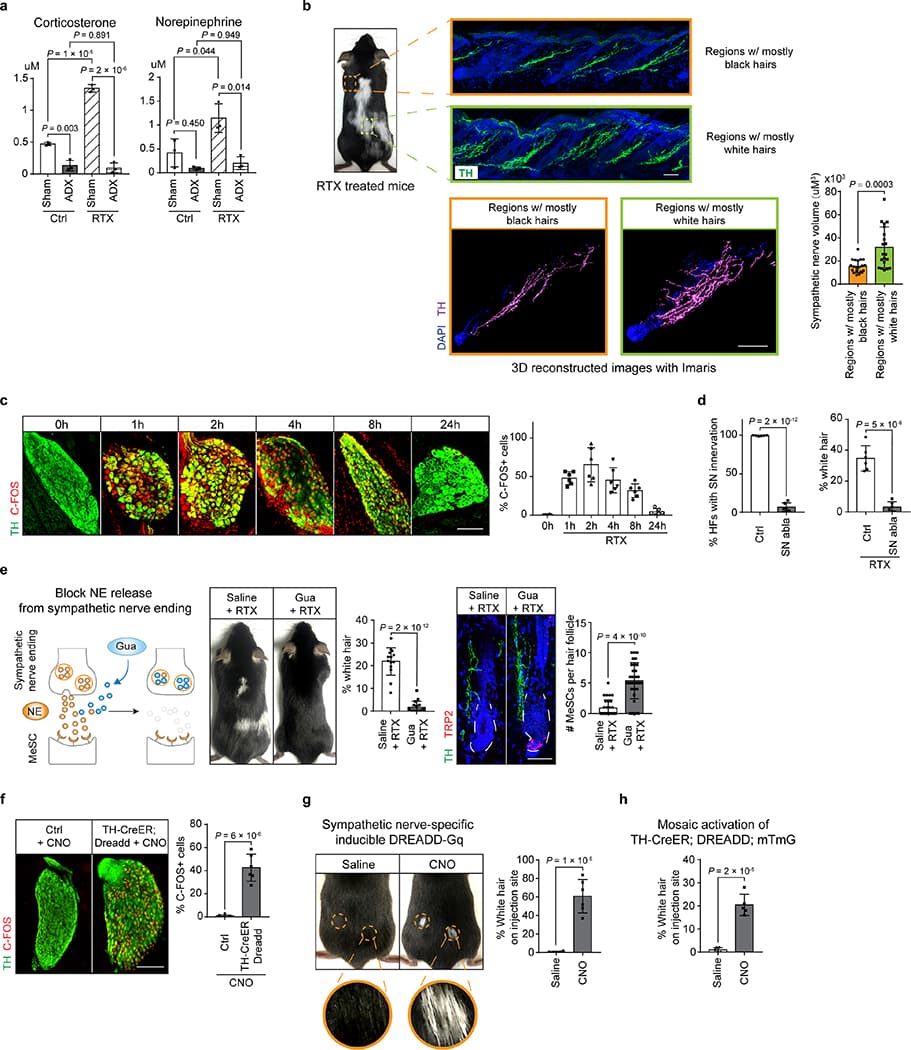
A 2020 Nature paper seemed to suggest that acute stress can permanently deplete melanocyte stem cells — not through adrenal hormones, but via norepinephrine released directly from sympathetic nerves.
That norepinephrine causes the stem cells to over-proliferate and exit the niche, which means no more pigment in future hair cycles…
Separately, copper deficiency has also been linked to greying.
Tyrosinase — the key enzyme for melanin production — is copper-dependent, so low copper can impair pigment formation even if the melanocyte stem cells are still present. And since high zinc intake can interfere with copper absorption, it would then follow that excessive zinc could contribute to greying by tipping that balance.
Anecdotally, I subjectively observed more greying when I was supplementing zinc 50mg nearly every day and experienced improvement when I reduced that to 2-3 days a week.
That said, it’s worth noting that many if not most people are actually high in copper and low in zinc.
I have been aware of the neccessity to balance zinc and copper to prevent hair graying for awhile. After experimenting with dosages I now take 25mg zinc (from 202,5 mg zinc gluconate) for 2 days and then 1 day copper 2mg (in the evening before sleep on an empty stomach). This gets both levels at the high end of the reference range for me.
Total copper was 110 μg/dL on my latest test, reference range is 63.7-140.12 μg/dL. When I took zinc 25mg every other day alternating with copper 2mg every other day, it was 138 μg/dL and free copper was above the reference range.
A 2020 Nature paper seemed to suggest that acute stress can permanently deplete melanocyte stem cells — not through adrenal hormones, but via norepinephrine released directly from sympathetic nerves.
That norepinephrine causes the stem cells to over-proliferate and exit the niche, which means no more pigment in future hair cycles…
Given the Adrb2-dependence, topical propranolol might be a solution to this.
On another note, topical propranolol is also a potential hair loss treatment. There was recently a small Chinese RCT in FASEB reporting efficacy against hair loss.
I’ve looked into this a bit since I started taking buproprion/Wellbutrin, which has hair loss as a common side effect. Given that stress can also cause hair loss, I was wondering if (nor)epinephrine might lead to HFSC depletion via a similar mechanism, but it seems there are some differences (excerpt from Cell Types Promoting Goosebumps Form a Niche to Regulate Hair Follicle Stem Cells):
The sympathetic nerve is known to influence melanocyte stem cells (MeSCs), a distinct stem cell population also located around the bulge that regenerates the pigment to color the hair (Zhang et al., 2020). Hyperactivation of sympathetic neurons, as occurs in severe stress, depletes MeSCs, forming the basis for stress-induced hair graying. There are several interesting differences regarding how the sympathetic nerve regulates MeSCs versus HFSCs. First, HFSCs are more sensitive to low levels of sympathetic nerve activity than MeSCs. HFSCs respond to both basal and modest elevation of sympathetic tone (such as in cold), a characteristic likely facilitated by the synapse-like connections between sympathetic nerve terminals and HFSCs. By contrast, MeSCs are only depleted upon sympathetic nerve hyper-activation. MeSCs are outside of the synaptic transmission range (∼1–2 μm for sympathetic nerve) and are likely influenced by norepinephrine mostly through diffusion, which is only effective at high concentrations. Moreover, whereas HFSCs are positively regulated by the sympathetic nerve to produce tissues, MeSCs are negatively affected. It is likely that the sympathetic nerve innervates the hair follicle to regulate HFSCs, whereas depletion of MeSCs is an undesired side effect when the nerve activity is abnormally high. Future studies are needed to explore how the sympathetic nerve drives different outcomes for distinct stem cells on the basis of differences in the amplitude and duration of nerve activation.