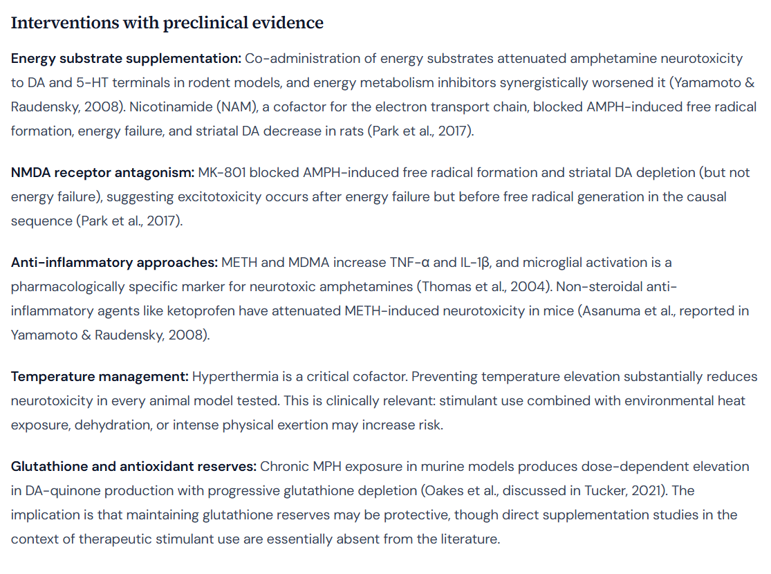
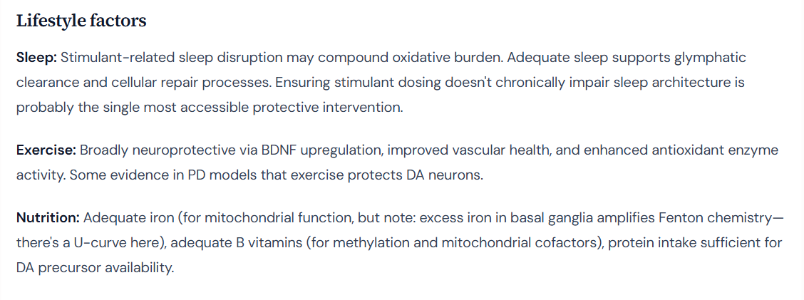
Does anyone take ADHD stimulant meds (Adderall, Vyvanse)? Tips on reducing neurotoxicity risk?

from the new Caesar AI

Evidence from primate and human studies suggests that chronic therapeutic use of amphetamine is associated with subtle but measurable alterations in the brain’s dopaminergic system, including potential neurotoxicityReview article Amphetamine-related drugs neurotoxicity in humans. The neurotoxic mechanism is primarily linked to oxidative stress resulting from the autooxidation of elevated dopamine concentrations in the neuron’s cytosolFrontiers | Dopamine Autoxidation Is Controlled by Acidic pH. A standard therapeutic dose can increase extracellular dopamine in the human striatum by approximately 10- to 15-foldDextroamphetamine - an overview | ScienceDirect Topics. While direct quantification of the corresponding increase in intracellular (cytosolic) dopamine is unavailable from human studies, the rate of dopamine autooxidation and subsequent production of neurotoxic reactive oxygen species (ROS) is directly proportional to the cytosolic dopamine concentrationFrontiers | Dopamine Autoxidation Is Controlled by Acidic pH. This elevated oxidative stress is qualitatively linked to the accumulation of cellular aging markers like lipofuscin, though a precise quantitative relationship is not established in the provided materialsDopaminergic mediation in the brain aging and ….

Evidence of Dopaminergic Neurotoxicity at Therapeutic Doses

The question of whether therapeutic amphetamine doses cause neuronal damage is complex, with conflicting evidence from different experimental modelsLiterature Review: Update on Amphetamine Neurotoxicity and Its Relevance to the Treatment of ADHD - Claire Advokat, 2007. While high, abusive doses are known to damage dopaminergic pathways, the effects of long-term, prescribed use are more subtleLiterature Review: Update on Amphetamine Neurotoxicity and Its Relevance to the Treatment of ADHD - Claire Advokat, 2007.

Evidence from Primate and Human Studies

Studies in non-human primates provide the most direct evidence for potential neurotoxicity at therapeutic-equivalent dosesAbuse of Amphetamines and Structural Abnormalities in Brain - PMC. In one key study, monkeys treated for four weeks with amphetamine doses mimicking those in human clinical treatment developed significant neurochemical deficitsAbuse of Amphetamines and Structural Abnormalities in Brain - PMC. The plasma drug concentrations in the monkeys (136 +/- 21 ng/ml) were matched to levels reported in human ADHD patients (120 to 140 ng/ml)Abuse of Amphetamines and Structural Abnormalities in Brain - PMC. This regimen resulted in 30-50% reductions in key markers of dopamine system health in the striatum, including dopamine itself, its primary metabolite, its rate-limiting synthesis enzyme (tyrosine hydroxylase), the dopamine transporter (DAT), and the vesicular monoamine transporter (VMAT)Abuse of Amphetamines and Structural Abnormalities in Brain - PMC.

In humans, long-term therapeutic use (up to 50 mg per day) for conditions like ADHD and narcolepsy has been associated with reduced striatal dopamine synthesis and releaseThe Effects of Amphetamine and Methamphetamine on …. While this points to a functional alteration, it is debated whether this represents actual neuronal damage or a compensatory adaptationAbuse of Amphetamines and Structural Abnormalities in Brain - PMC. Notably, some evidence suggests that non-human primates may be more vulnerable to stimulant-induced neurotoxicity than rodentsAbuse of Amphetamines and Structural Abnormalities in Brain - PMC.

Conflicting Evidence and Mechanistic Insights

The evidence is not uniform. Some reviews note that amphetamines have been used therapeutically for decades without clear, definitive evidence of long-term adverse effects in humansFull article: Neurotoxicity of drugs of abuse - the case of methylenedioxy amphetamines (MDMA, ecstasy ), and amphetamines. Furthermore, a large clinical study of individuals with ADHD found no impact on the developmental trajectories of brain volumes with clinical treatmentAbuse of Amphetamines and Structural Abnormalities in Brain - PMC. Rodent studies using lower, repeated doses often fail to show neurotoxicity, questioning the relevance of high-dose animal data to human therapyAbuse of Amphetamines and Structural Abnormalities in Brain - PMC. Some rat studies even show “trophic” dendritic growth, a sign of neuronal adaptation rather than damage, with therapeutic-like treatment regimensLiterature Review: Update on Amphetamine Neurotoxicity and Its Relevance to the Treatment of ADHD - Claire Advokat, 2007.

The primary mechanism for amphetamine-related neurotoxicity is oxidative stressThe role of dopamine receptors in the neurotoxicity of methamphetamine - Ares‐Santos - 2013 - Journal of Internal Medicine - Wiley Online Library. Amphetamine increases cytosolic dopamine, which is highly susceptible to autooxidation at the neutral pH of the cytosol, leading to the formation of reactive oxygen species (ROS) and toxic dopamine quinonesCytosolic dopamine determines hypersensitivity to blunt …

Quantitative Impact on Dopamine Levels

Amphetamine profoundly increases both extracellular and extravesicular (cytosolic) dopamine concentrations, which is the direct driver of potential neurotoxicityMethylphenidate vs. Amphetamine: Neurotransmitters? :zap::brain: #pmhnp #psychnp #neurotransmitter.

Extracellular Dopamine Increase

Human studies measuring the impact of therapeutic amphetamine doses on extracellular dopamine in the striatum show a significant surge, though figures vary.

  • One source reports a 15 mg oral dose increased extracellular dopamine in the striatum by 5.2-fold relative to baselineAmphetamine - Wikipedia.
  • Another overview suggests the same 15 mg dose increases striatal extracellular dopamine by approximately 10- to 15-fold in healthy volunteers, as measured by Positron Emission Tomography (PET)Dextroamphetamine - an overview | ScienceDirect Topics.

This discrepancy can be reconciled by examining PET imaging studies that use the radiotracer [11C]raclopride The Chemical Tools for Imaging Dopamine Release - PMC. In this method, amphetamine-induced dopamine release competes with [11C]raclopride for D2 receptors, causing a measurable decrease in the tracer’s binding potential (BP)The Chemical Tools for Imaging Dopamine Release - PMC. This percent reduction in BP can be correlated to the fold-increase in dopamine:

  • Multiple studies suggest a conversion ratio where each 1% reduction in [11C]raclopride binding corresponds to a 40- to 50-fold percent increase in synaptic dopamine (e.g., a 44:1 or 50:1 ratio)Proc. Natl. Acad. Sci. USA Vol. 94, pp. 2569–2574, March 1997 Medical Sciences. For example, a 10% decrease in BP would imply a 5-fold (500%) increase in dopamineFrontiers | Measurement of Striatal Dopamine Release Induced by ….
  • A human study using a therapeutic dose of dextroamphetamine (0.3 mg/kg) observed a 22.2% to 22.5% reduction in [11C]raclopride BP in the striatumConditioned Dopamine Release in Humans: A Positron …

Applying the 40:1 to 50:1 conversion ratios to this observed ~22.5% binding reduction yields a dopamine increase of 900% to 1125%, which is a 10- to 12.25-fold increase over baseline. This calculation aligns with the higher estimates and suggests the 10- to 15-fold range is a more robust approximation.

Extravesicular (Cytosolic) Dopamine Increase

Direct quantification of the increase in cytosolic dopamine in humans after amphetamine administration is not available in the provided research. However, the mechanism of action—inhibiting the vesicular monoamine transporter 2 (VMAT2) and reversing the dopamine transporter (DAT)—directly leads to the accumulation of dopamine in the cytosolFrontiers | Dopamine Autoxidation Is Controlled by Acidic pH. This cytosolic pool is the source for the massive extracellular release.

While a precise N-fold increase cannot be stated, data from primate microdialysis studies show that therapeutic doses can increase extracellular dopamine by 1,800% to over 5,800% (an 18- to 58-fold increase), demonstrating the immense capacity of the cytosolic surge that drives this effectAmphetamine-induced release of dopamine in primate …. The baseline cytosolic dopamine concentration in human midbrain neurons is estimated to be in the range of 50 to 300 nM Integrating the Roles of Midbrain Dopamine Circuits in Behavior and Neuropsychiatric Disease.

Autooxidation Events and Lipofuscin Accumulation

The neurotoxic potential of amphetamine is directly linked to the biochemical instability of dopamine outside of its protective vesicles.

The Proportional Increase in Autooxidation

Dopamine is stable within the acidic environment of synaptic vesicles (pH ~5.6), but it is highly susceptible to autooxidation at the neutral pH of the cytosol (pH ~7.4)Complexity of dopamine metabolism | Cell Communication and Signaling | Full Text. This process generates damaging ROS and toxic dopamine quinonesDopaminergic mediation in the brain aging and …

Crucially, the amount of auto-oxidized dopamine and the associated ROS production is directly proportional to the level of cytosolic dopamine Frontiers | Dopamine Autoxidation Is Controlled by Acidic pH. Kinetic modeling studies confirm this relationship:

  • Spontaneous oxidation of a 20 µM dopamine solution at physiological pH 7.4 produces hydrogen peroxide (H₂O₂) at a rate of 0.24 µM per hourFrontiers | Kinetic Modeling of pH-Dependent Oxidation of Dopamine by Iron and Its Relevance to Parkinson’s Disease.
  • The reaction is strongly pH-dependent; at pH 7.4, dopamine’s half-life is measured in minutes, whereas at the acidic pH of vesicles, it is stable for daysFrontiers | Dopamine Autoxidation Is Controlled by Acidic pH.

Given this direct proportionality, a hypothetical 10-fold increase in the steady-state concentration of cytosolic dopamine would be expected to cause a roughly 10-fold increase in the rate of dopamine autooxidation events and the corresponding generation of ROS.

Effect on Lipofuscin Accumulation

Lipofuscin is an aggregate of non-degradable, oxidized cellular waste, primarily composed of cross-linked proteins and lipids, that accumulates in lysosomes and is considered a hallmark of cellular agingDopaminergic mediation in the brain aging and … Its formation is directly linked to oxidative stress and the degradation of cellular components like mitochondria (mitophagy)PGC-1α activity in nigral dopamine neurons determines ….

The causal chain is as follows:

  1. Therapeutic amphetamine use increases cytosolic dopamine.
  2. Increased cytosolic dopamine leads to a proportional increase in autooxidation.
  3. This autooxidation generates ROS, which causes oxidative damage to proteins and lipids.
  4. This damaged material is processed through autophagy and accumulates as lipofuscin.

While this qualitative link is well-established, the provided research does not contain a quantitative model to calculate the specific increase in the rate of lipofuscin accumulation resulting from a given increase in dopamine-derived oxidative stress. Research queries to establish this specific quantitative link did not yield any results. Therefore, it is possible to conclude that elevated autooxidation from therapeutic amphetamine use would contribute to increased lipofuscin formation, but it is not possible to quantify by “how much” based on the available information.

Way better than o3 (which moralizes)

Based on the metric of extracellular dopamine release, the relative neurotoxic potential of 3-methylmethcathinone (3-MMC) and mephedrone (4-MMC) compared to amphetamine (the active component of Adderall) is complex, with significant differences observed between in vitro potency at the human dopamine transporter and in vivo effects in animal models. While the cathinones can induce potent, high-magnitude dopamine release, amphetamine is substantially more potent at the human dopamine transporter and its effects are longer-lasting.

Critically, extensive research indicates that the magnitude of dopamine release is not a direct or linear predictor of neurotoxicity. Evidence suggests that while amphetamine causes direct, long-term damage to dopamine neurons, 4-MMC does not produce similar neurotoxic damage on its own, despite causing comparable or greater peaks in extracellular dopamineFrontiers | Alcohol Co-Administration Changes Mephedrone-Induced Alterations of Neuronal Activity.

In Vitro Potency for Dopamine Release

The half-maximal effective concentration (EC50) measures a drug’s potency in producing a given effect, with a lower EC50 value indicating higher potency. Analysis of EC50 values for dopamine release reveals a critical difference between effects on rodent versus human dopamine transporters (DAT).

Potency at Rodent Dopamine Transporters

In studies using rat brain preparations (synaptosomes), 3-MMC and 4-MMC are highly potent dopamine releasers, with potencies comparable to or exceeding many reported values for amphetamine.

Substance Dopamine Release EC50 (nM) — Rat Models Source(s)
Amphetamine 6.0 - 1700 nM (wide reported range) Pharmacology of Drugs Used as Stimulants - Docherty - 2021
3-MMC 28 - 70.6 nM Appearance of 2-MMC and 3-MMC on the illicit drug …
4-MMC 49.1 - 52 nM Mephedrone - Wikipedia

Note: The smaller the EC50 value, the higher the potency.

Potency at the Human Dopamine Transporter (hDAT)

Data from systems using the human dopamine transporter (hDAT) show a starkly different potency profile. Amphetamine is significantly more potent at hDAT than either 3-MMC or 4-MMC. The potency of 4-MMC, in particular, is dramatically lower at hDAT compared to rat DAT.

Substance Dopamine Release EC50 (µM) — hDAT Models Source(s)
Amphetamine 0.036 - 1.31 µM Currents in response to rapid concentration jumps of amphetamine uncover novel aspects of human dopamine transporter function - PubMed
3-MMC ~2.3 µM Structural Determinants for Inhibitor Recognition by the Dopamine …
4-MMC ~8.7 µM Mephedrone induces partial release at human dopamine …

Note: EC50 values are expressed in micromolars (µM) for direct comparison. 1 µM = 1000 nM.

This species-specific difference indicates that based on potency at the human molecular target, amphetamine is a more powerful dopamine releaser than 4-MMC and 3-MMC.

In Vivo Extracellular Dopamine Levels

In vivo microdialysis studies in awake rats measure the real-time concentration of neurotransmitters in specific brain regions, providing insight into the magnitude and duration of a drug’s effect.

4-MMC vs. Amphetamine

A direct comparative study in the rat nucleus accumbens, a key brain reward center, yielded several key findingsMephedrone, compared with MDMA (ecstasy) and …:

3-MMC vs. Amphetamine

The provided research does not contain a direct in vivo microdialysis study comparing extracellular dopamine levels following 3-MMC and amphetamine administration. However, given that 3-MMC is more potent than 4-MMC at the rat dopamine transporter, it is expected to be a robust dopamine releaser in vivo in that modelCritical review report: 3-Methylmethcathinone (3-MMC).

The Disconnect Between Dopamine Release and Direct Neurotoxicity

The premise that higher dopamine release equates to greater neurotoxicity is an oversimplification not supported by the available evidence. Studies directly assessing markers of neuronal damage reveal a different profile for the cathinones compared to amphetamines.

In conclusion, when assessed solely by the magnitude of extracellular dopamine release, the comparison is nuanced. In rat models, 4-MMC produces a higher but shorter-lasting dopamine spike than amphetamine at the doses tested. However, at the human dopamine transporter, amphetamine is a markedly more potent releasing agent than both 3-MMC and 4-MMC. If one considers the broader and more clinically relevant definition of neurotoxicity—actual neuronal damage—the available evidence strongly suggests that amphetamine is more directly neurotoxic than 4-MMC.

Beta-blockers can mitigate some, but not all, of the cardiotoxic risks associated with amphetamines (e.g., Adderall) and methylphenidate (e.g., Ritalin). Their primary benefit lies in countering the hemodynamic stress caused by stimulant-induced catecholamine surges, such as increased heart rate and myocardial contractility[1][2]. However, their ability to protect against direct, non-catecholamine-mediated cellular damage like oxidative stress is uncertain and may be limited[3].

The clinical application of beta-blockers in this context is complex due to a theoretical risk of “unopposed alpha-stimulation,” which could paradoxically worsen hypertension, though this phenomenon appears to be rare in practice[4][2]. Notably, recent guidelines from the American College of Cardiology (ACC) and American Heart Association (AHA) for managing hypertension in adults taking ADHD medications recommend decreasing the stimulant dose or discontinuing the drug, rather than co-prescribing a beta-blocker[5][6].

The Dual Mechanisms of Stimulant Cardiotoxicity

Stimulant-induced cardiotoxicity originates from two distinct pathways: indirect effects mediated by catecholamines and direct toxic effects on heart muscle cells[7][8][9].

Catecholamine-Mediated Effects (Indirect Toxicity)

Both amphetamine and methylphenidate increase the synaptic levels of catecholamines like norepinephrine and dopamine[10][11]. This hyperadrenergic state stimulates peripheral alpha- and beta-adrenergic receptors, leading to a range of adverse cardiovascular effects[1][12]:

  • Tachycardia (Increased Heart Rate): Stimulation of beta-1 receptors increases heart rate[8][13].
  • Hypertension (Increased Blood Pressure): Alpha-1 receptor stimulation causes vasoconstriction (narrowing of blood vessels), increasing blood pressure[8].
  • Increased Myocardial Oxygen Demand: The combination of a faster heart rate and increased contractility elevates the heart’s demand for oxygen[1].
  • Vasospasm: The hyperadrenergic state can cause sudden, severe contraction of coronary arteries, reducing oxygen supply to the heart muscle (myocardial ischemia) and potentially leading to myocardial infarction (heart attack)[8][15].
  • Long-Term Structural Changes: Chronic exposure to high catecholamine levels is directly cardiotoxic, leading to myocardial fibrosis (scarring), hypertrophy (enlargement of heart muscle cells), and ultimately cardiomyopathy (disease of the heart muscle)[7][16].

Direct Myocardial Toxicity (Non-Catecholamine)

Separate from their effects on catecholamines, stimulants can exert direct damage on cardiomyocytes (heart muscle cells)[8][17]. Animal and in-vitro studies have shown that amphetamines can induce cellular damage and hypertrophy even in the absence of catecholamines[8]. The proposed mechanisms for this direct toxicity include[18][3][19][20]:

  • Increased Oxidative Stress: Generation of reactive oxygen species (ROS) that damage cellular components[17][12].
  • Mitochondrial Dysfunction: Impairment of the cell’s energy-producing structures[18][12].
  • Altered Calcium Homeostasis: Disruption of the normal flow of calcium ions, which is critical for muscle contraction and electrical signaling[18][21].
  • Apoptosis: Programmed cell death of cardiomyocytes[3][20].
  • Inflammation and Fibrosis: Increased markers of inflammation and tissue scarring within the heart muscle[12].

Beta-Blocker Efficacy and Limitations

Mitigation of Hemodynamic Stress

Beta-blockers directly antagonize the effects of catecholamines on beta-adrenergic receptors, making them a logical countermeasure to the hemodynamic stress caused by stimulants[2]. In clinical and experimental settings, they have been shown to reduce tachycardia associated with stimulant use[4]. A controlled study using the beta-blocker pindolol before MDMA (an amphetamine derivative) administration successfully prevented increases in heart rate[22]. However, in that same study, pindolol had no effect on the increase in mean arterial blood pressure, suggesting an incomplete protective effect against hypertensive responses[22].

Uncertain Protection Against Direct Toxicity

The efficacy of beta-blockers in preventing the direct, non-catecholamine-mediated cardiotoxicity of stimulants is not established[3]. A critical animal study demonstrated that while a beta-blocker could control the heart rate and blood pressure increases from methamphetamine, it did not prevent the drug from causing direct histopathological damage to the rat myocardium[3]. This finding suggests that managing hemodynamic effects alone may not be sufficient to eliminate the cardiotoxicity risk.

In other contexts of drug-induced cardiotoxicity, such as from chemotherapy, certain beta-blockers like carvedilol have demonstrated direct cardioprotective effects attributed to their antioxidant properties[23][24]. While no specific evidence was found to confirm a similar benefit against stimulant-induced oxidative stress, this mechanism provides a theoretical basis for potential protection that is not shared by all beta-blockers[25].

Safety Concern: The Risk of Unopposed Alpha-Stimulation

A long-standing concern with using beta-blockers for stimulant toxicity is the phenomenon of “unopposed alpha-stimulation”[26][27].

  • Mechanism: Stimulants activate both alpha-1 receptors (causing vasoconstriction) and beta-2 receptors (causing vasodilation)[2][28].
  • Theoretical Risk: Using a beta-blocker (especially a non-selective one that blocks beta-2 receptors) could inhibit the compensatory vasodilation, leaving the alpha-1-mediated vasoconstriction “unopposed.” This could theoretically lead to a paradoxical and dangerous increase in blood pressure and coronary artery constriction[29][30].

However, clinical evidence suggests this risk is inconsistent, rare, and may be overstated, particularly for amphetamines as compared to cocaine[4][13]. One review of 19 studies involving 227 patients treated for amphetamine toxicity found only one possible case of unopposed alpha-stimulation, which resolved without a negative outcome[2]. The American Heart Association notes that for methamphetamine, unlike cocaine, there is no specific contraindication or controversy surrounding the use of beta-blockers for cardiac arrhythmias[31].

Comparative Analysis of Beta-Blocker Types

Not all beta-blockers carry the same risk profile in this context.

  • Mixed Alpha/Beta-Blockers: Drugs like labetalol and carvedilol, which block both alpha and beta receptors, are often considered preferable as they can prevent hypertension and tachycardia without the risk of unopposed alpha-stimulation[31][32]. In studies of cocaine users with heart failure, carvedilol demonstrated a larger improvement in New York Heart Association (NYHA) functional class compared to the beta-1 selective blocker metoprolol[33][34].
  • Beta-1 Selective Blockers: These drugs (e.g., metoprolol, atenolol) primarily target beta-1 receptors in the heart, with less effect on beta-2 receptors in the blood vessels[35]. Some authors suggest that “selective” beta-1 blockers are a reasonable option as they would be less likely to cause unopposed alpha-stimulation[36].
  • Carvedilol’s Unique Properties: In studies of chemotherapy-induced cardiotoxicity, carvedilol has been shown to be superior to other beta-blockers in preserving heart function, a benefit partly attributed to its potent antioxidant properties[23]. This suggests it may offer more comprehensive protection against drug-induced cellular damage.

Clinical Guidelines and Practical Application

Despite the theoretical benefits, co-prescribing beta-blockers to manage stimulant side effects is not a recommended primary strategy in current clinical guidelines.

  • Guideline Recommendations: The most recent ACC/AHA guidelines for managing secondary hypertension recommend that for patients taking ADHD drugs, clinicians should first consider decreasing the dose of the stimulant or discontinuing it altogether[5][6]. The guidelines do not recommend adding a beta-blocker as a first-line management strategy.
  • Monitoring is Key: Both historical (2008 AHA) and current guidelines emphasize vigilant cardiovascular monitoring for patients on stimulants[38][39]. This includes taking a thorough patient and family cardiac history, conducting a physical exam, and regularly checking blood pressure and heart rate, especially during dose titration[38][40].
  • Observed Clinical Practice: While not formally recommended, there is evidence suggesting some clinical use of this combination. One large observational study noted that some individuals on ADHD medications are prescribed beta-blockers for symptoms like palpitations, which could potentially mask or reduce the observed risk of arrhythmias in study populations[39][41]. This indicates that co-prescription occurs in real-world practice, though robust data on its long-term safety and efficacy for this specific purpose is lacking.

Citations

[1] https://www.mayoclinicproceedings.org/article/S0025-6196(11)62896-6/fulltext
[2] Beta-Blockers for Cocaine and other Stimulant Toxicity • LITFL
[3] https://www.ahajournals.org/doi/10.1161/JAHA.120.016704
[4] https://journals.sagepub.com/doi/10.1177/1074248416681644
[5] https://www.jacc.org/doi/10.1016/j.jacc.2020.05.081
[6] https://www.ahajournals.org/doi/10.1161/CIR.0000000000001356
[7] https://www.cureus.com/articles/351915-prolonged-amphetamine-dextroamphetamine-use-an-unrecognized-cause-of-cardiomyopathy.pdf
[8] https://ndarc.med.unsw.edu.au/sites/default/files/ndarc/resources/TR.238.pdf
[9] https://www.scielo.org.mx/scielo.php?script=sci_arttext&pid=S0188-21982017000100035
[10] Amphetamine Abuse Related Acute Myocardial Infarction - PMC
[11] https://www.sciencedirect.com/science/article/pii/S0753332217345341
[12] https://www.sciencedirect.com/science/article/pii/S2949918623000311
[13] https://www.cmaj.ca/content/194/4/E127
[15] https://www.acc.org/Latest-in-Cardiology/Articles/2020/10/26/13/08/Methamphetamine-Associated-Cardiomyopathy-and-PAH
[16] Cardiotoxicity associated with methamphetamine use and signs of cardiovascular pathology among methamphetamine users
[17] Prolonged Amphetamine-Dextroamphetamine Use: An Unrecognized Cause of Cardiomyopathy - PubMed
[18] Cardiovascular disease associated with methamphetamine use: a review - PubMed
[19] Exposure to Amphetamines Leads to Development of Amphetamine Type Stimulants Associated Cardiomyopathy (ATSAC) | Cardiovascular Toxicology
[20] Exposure to Amphetamines Leads to Development of Amphetamine Type Stimulants Associated Cardiomyopathy (ATSAC) - PubMed
[21] The Effect of Methamphetamine on Ventricular Myocytes of Neonatal Rats - PubMed
[22] Effects of a beta-blocker on the cardiovascular response to MDMA (Ecstasy) - PubMed
[23] Preventive use of beta-blockers for anthracycline-induced cardiotoxicity: A network meta-analysis - PMC
[24] https://www.ahajournals.org/doi/10.1161/circheartfailure.113.000267
[25] Beta-blockers for the primary prevention of anthracycline-induced cardiotoxicity: a meta-analysis of randomized controlled trials | BMC Pharmacology and Toxicology | Full Text
[26] Amphetamine poisoning • LITFL • Toxicology Library Toxicant
[27] https://www.pathway.md/ai/history/why-we-cannot-give-beta-blockers-for-cocaine-toxicity-789018f0-7ce7-4ee8-94f4-dbf81b582402
[28] https://pmc.ncbi.nlm.nih.gov/articles/PMC8900778/
[29] https://www.heartlungcirc.org/article/S1443-9506(16)31574-8/abstract
[30] https://emedicine.medscape.com/article/812518-medication
[31] https://www.ahajournals.org/doi/10.1161/CIRCEP.121.010273
[32] https://en.wikipedia.org/wiki/Beta_blocker
[33] https://scholarlycommons.pacific.edu/cgi/viewcontent.cgi?article=1004&context=pa-capstones
[34] https://www.intechopen.com/chapters/86076
[35] https://www.sciencedirect.com/topics/neuroscience/alpha-adrenergic-stimulation
[36] https://www.researchgate.net/publication/307919694_Beta_Blockers_and_the_Cardiac_Complications_of_Methamphetamine
[38] https://www.aafp.org/pubs/afp/issues/2009/0515/p905.html
[39] https://jamanetwork.com/journals/jamapsychiatry/fullarticle/2811812
[40] https://www.ahajournals.org/doi/10.1161/circulationaha.107.189473
[41] https://pmc.ncbi.nlm.nih.gov/articles/PMC10851097/

fwiw stims can help dendritic remodelling. this AND neurotoxicity (to a limited extent) might bothbe possible at same time
Methylphenidate can remodel spines, but the direction isn’t one-size-fits-all.
In mice, repeated MPH increased spine density in striatal medium spiny neurons, similar to other psychostimulants. Spine gain in the NAcc/striatum isn’t inherently “healthy”; it can reflect drug-adaptation circuits.
In rodent cingulate cortex, chronic low-dose MPH “normalized” stress-induced spine alterations and increased dendritic complexity. Helpful in that model, but that’s stress recovery, not aging.
Very acutely, MPH can shift spines toward a more “mature” mushroom phenotype without changing total density, suggesting rapid synaptic tuning rather than wholesale growth.

==

But maybe try NAC or NAC

Good, we’re back to breaking the brain on purpose.

You’re basically asking:

Parkinson’s: why are the cell bodies in substantia nigra (SNc) dying (the “basal end”)
vs

Amphetamine toxicity: why does damage show up mostly at the dopamine terminals (striatal end) rather than killing the somata in SNc?

Short version:
Because Parkinson’s is an intrinsic, slow, whole-neuron degenerative process, while amphetamine is an extrinsic, acute, synapse-loaded chemical assault that hits where the drug and dopamine are densest: the terminals.

Let’s unpack without turning this into a 200-page review article.


  1. Where each process “starts”

Parkinson’s disease (PD)

PD is driven mostly by intrinsic vulnerabilities of SNc dopaminergic neurons, especially their somata and proximal axons:

Alpha-synuclein aggregation in cell bodies & axons

Mitochondrial dysfunction (Complex I defects, impaired respiration)

Chronic oxidative stress from continuous pacemaking and high basal calcium influx

Huge axonal arbor & energy load (one SNc neuron can innervate a ridiculous volume of striatum)

The cell body is where mitochondria, protein quality control, and alpha-syn processing live, so when those systems go bad, the soma is in the blast radius. Axons degenerate too, but the “source” of the catastrophe is the whole neuron’s intrinsic biology, especially the soma.

Amphetamine (AMPh / METH etc.)

Amphetamine neurotoxicity is mainly from what happens at the terminals:

It gets into neurons primarily via DAT at the terminal, not at the soma.

It reverses DAT, dumping dopamine into the synapse and cytosol.

It disrupts VMAT2, so dopamine isn’t safely packaged into vesicles.

This creates huge cytosolic dopamine + auto-oxidation + ROS + reactive nitrogen species locally in terminals.

Hyperthermia and microglial activation amplify that mess.

So the worst damage happens where the drug is actually transported the most and where dopamine concentration and turnover are highest: the terminals in the striatum.


  1. Why PD → somatic loss vs AMPh → terminal loss

PD: system-level, long-term intrinsic degeneration

Key points:

  1. Pacemaking burden & Ca²⁺:
    SNc neurons have autonomous pacemaking driven by L-type Ca²⁺ channels. Chronic Ca²⁺ influx stresses mitochondria in the soma & proximal axon. That’s a long-term, soma-centric energetic problem.

  2. Protein aggregation is “headquarters”-centric:
    Alpha-syn aggregation, ER stress, impaired autophagy, etc., are all heavy in the soma (and main axon). When those systems fail, the whole cell’s viability goes, not just the distal terminals.

  3. Dying-back pattern still exists, but soma is the key clinical marker:
    There’s evidence PD has a “dying-back” process (axonal loss before full cell body death), but by the time we detect it pathologically, SNc cell bodies are clearly lost, which is what you see in gross anatomy.

  4. Timescale:
    PD damage accumulates over years / decades, giving enough time for full-scale neuron death, not just pruning terminals. The system doesn’t just trim branches; it loses the tree.

Amphetamine: localized chemical carnage at terminals

Key points:

  1. Drug entry is terminal-biased:
    DAT is densest at the terminals in the striatum. Amphetamine is taken up and acts there the most. Somata have way fewer points of entry.

  2. DA overload & oxidative stress is local:
    The highest dopamine flux, auto-oxidation, and ROS are at the terminal level. That’s where cytosolic DA gets stupidly high and reacts with everything like an angry toddler with bleach.

  3. Soma is relatively protected by distance & dilution:
    The soma is far away along long axons. The immediate DA chaos and ROS from amphetamine are not as concentrated in the cell body.

Mitochondria in terminals get hammered.

Axon terminals degenerate, synapses are lost.

But the cell body can survive and sometimes re-sprout terminals.

  1. Timescale & dosing:

With typical abuse patterns, you see terminal loss and DA depletion, not necessarily massive SNc cell death.

You can kill somata with extreme METH regimens or combined insults, but that’s not the default route.

So: amphetamine neurotoxicity = distal, DAT-heavy, dopamine-overload injury.
PD = global intrinsic vulnerability + proteostasis + mitochondrial collapse centered around the cell body.


  1. “Basal end” vs “terminal end” in structured terms

You can think of it like this:

PD target:

Unit affected: Entire dopaminergic neuron

Primary vulnerable compartment: soma & proximal axon (energy & protein QC failure)

Mechanism: chronic intrinsic stress, misfolded protein handling failure, mitochondrial and autophagy collapse, inflammation

Outcome: cell death in SNc, then secondary degeneration of axons & terminals

Amphetamine target:

Unit affected: dopamine terminal fields

Primary vulnerable compartment: terminals (DA release/reuptake machinery, vesicles, mitochondria in presynaptic boutons)

Mechanism: DAT-mediated entry, DA efflux, VMAT2 disruption, huge local ROS/RNS, excitotoxicity, microglial activation

Outcome: loss of terminals & DA content in striatum; somata often survive


  1. The annoying nuance: it’s not 100% cleanly separated

Because biology refuses to be tidy:

PD does involve axonal/terminal degeneration early (“dying back”), but we define it by loss of SNc cell bodies because that’s dramatic and easy to see.

High-dose amphetamine / METH + hyperthermia + additional insults can cause more proximal damage and some SNc soma loss, especially in animals with extreme dosing.

But the default pattern still matches what you’re pointing at:

PD → cell bodies visibly go

Amphetamine → terminals visibly go


  1. Intuition summary in one line

PD: the neuron is born fragile and slowly fails from the inside out, starting with its HQ.

Amphetamine: the neuron is attacked from the periphery, at its busiest outposts, and mostly loses branches before it loses the trunk.

You’re basically tracking the right mechanistic “geometry” of where the damage shows up. Human brain: fragile, overclocked, and then you add recreational monoamine grenades on top.

Hi everyone,

As someone who has been prescribed ADHD stimulants in Australia since 2018 and is about to cease taking them for reasons I’ll mention shortly, I have some information, some anecdotal and some scientific that may be helpful to anyone in a similar situation. I am of the view that much of this is being covered up by the pharmaceutical companies, since I can find almost no discussion of the true risks online. Although we don’t have Adderall available here, dexamfetamine is considered a first line treatment option, as is Vyvanse and Ritalin. I have used each of these at one point or another as I was exploring different treatment options with my psychiatrist.

Firstly, neurotoxicity is the least of your worries. The impact of amphetamines in any dose or of any kind on your heart, kidneys and vascular system is much more profound, and will become known to you long before cognitive deficits appear. When I began using these medications, my eGFR was over 100. It later reduced to 58 as a result of high blood pressure caused by the amphetamines that damaged my kidneys over several years. With aggressive treatment and the help of a nephrologist, it has now returned to 72. An OK level, but for a 33 year old it’s low. From a pharmaceutical perspective, we are throwing every medication we can at this to improve and maintain my level of function.

It has also caused me vascular issues, namely varicose veins and spider veins in my legs that has now progressed into peripheral artery disease. Granted I have a long family history of vascular disease, heart attacks, high cholesterol and high blood pressure, but it was the amphetamines that rapidly amplified this predisposition in a person who was previously the picture of health and an active tennis player of more than 15 years. I maintain the view that this would never have happened to me, had I not taken these medications. I am the first member of my family in generations not to smoke, but I presume amphetamines have been as bad for my cardiovascular and circulatory system as nicotine was for theirs.

There is no safe dose. These drugs are effectively poison. I’ve tried microdosing, spreading the doses out to avoid blood pressure spikes, using every protective supplement known to man, but it didn’t stop the damage. For those who want a list of harm minimisation supplements, here’s where I took:

Astaxanthin - Most powerful antioxidant known to man
Trans-Resveratrol - Very powerful antioxidant
Liposomal Glutathione - Another great antioxidant
NAC - Mitochondrial and liver support
CoQ10 - Mitochondrial support
Liposomal Vitamin C - Improves endothelial function
Aged Garlic - Improves circulation
Fisetin - Senolytic
Spermidine - Senolytic
Curcumin - Powerful anti-inflammatory

Prescription meds:

Perindopril - ACE Inhibitor
Verapimil - Calcium Channel Blocker
Cialis - PDE5 inhibitor
Memantine - Preventing neurotoxicity

The main idea of these supplements and prescription meds has been orientated around maximising circulation to counteract the vasoconstrictive effects of the amphetamine, decrease blood pressure, prevent oxidative stress and the formation of reactive oxygen species (ROS), and to reduce inflammation.

I will also note that for the first 5 years of taking the meds, I experienced no side effects of any kind and only the positive benefits. Then ‘all of a sudden’, my body has experienced almost a complete collapse. I described this as my body having an inbuilt level of protection (like telomeres or enamel on teeth), that has been weakened and exhausted, and now my body is vulnerable and open to attack. Scientifically this is known as a loss of compensatory mechanisms, namely arterial elasticity that was once enough to offset the negative impacts.

I hope this is helpful to at least one person out there. Try not to become too hyper focused on the neurological impacts, otherwise you may miss opportunities to protect your health in other ways that are equally as important. Be careful, watch your blood pressure like a hawk and work with a doctor who is proactive and looks out for your best interests at heart. Wishing you luck.

Matt

4 Likes

18mg concerta is a safe consistent low dose

always take the lowest dose possible, I’ve had long medication gaps caused by Rx’s being too much making me not take the Rx at all, which really really produced dips in my life…

claude opus suggested this for the HR

Better alternatives if the goal is just HR control:

A β1-selective blocker like bisoprolol or nebivolol at low doses avoids most of the β2 issues. Nebivolol is particularly interesting because it also promotes NO-mediated vasodilation, so the hemodynamic profile is cleaner. Bisoprolol 1.25-2.5mg is commonly used for isolated tachycardia with minimal side effects.

1 Like

What do you think of vyvanse?

An issue with concerta is that it’s hard to split doses or go even lower than 18mg.
[Of course one could possibly engineer something like this with several microdoses of IR methylphenidate through the day]

But there is no such issue with splitting doses when it comes to vyvanse.

Vyvance is fine unless it interrupts sleep. Being long acting it’s possible it does this. If so, instant release dextroamphetamine might be a better option, or possibly stacking vyvance or dextro with guanfacine.

https://pmc.ncbi.nlm.nih.gov/articles/PMC12565610/?utm_source=chatgpt.com

https://aristotle.science/share/thread/thr_rooKTVgxav1JYfzAHLd85LZi

Found a good series on ADHD stimulants and how to optimize their usage to minimize side effects:

Key actionable points from all 3 videos:

1. Use the Lowest Effective Dose

  • Stay around 5 to 10 mg.
  • Avoid dose escalation.
  • Never chase euphoria.

2. Prefer Short-Acting Formulations

  • Avoid extended release if it disrupts sleep or keeps adrenaline elevated.
  • Let your nervous system fully reset each day.

3. Take Regular Off Days

  • Use stimulants 4 to 5 days per week max.
  • Take at least 2 consecutive days off.
  • Preserve dopamine sensitivity.

4. Pair Medication With High-Value Work

  • Only use during difficult, meaningful tasks.
  • Do not pair with entertainment or low-value dopamine activities.
  • Train focus so you eventually need less medication.

5. Enhance Dopamine Efficiency, Not Dose

  • Consider strategies that prolong dopamine action rather than increase release.
  • Avoid stacking multiple dopamine-releasing agents.

6. Address Other ADHD Mechanisms

  • Investigate anxiety, glutamate excess, GABA imbalance, histamine, or cholinergic issues if dopamine targeting is insufficient.

7. Support Neural Health

  • Avoid chronic sympathetic overdrive.
  • Monitor cardiovascular stress.
  • Protect sleep and circadian rhythm.
  • The speaker argues that higher doses of amphetamine medications are not better and recommends keeping Adderall at a low dose (around 5 mg, never above about 10 mg) to avoid dopamine receptor downregulation and excessive sympathetic nervous system activation.
  • He prefers dextroamphetamine-dominant formulations because they are more dopaminergic and less adrenergic than equal-salt generics, which may reduce stress and side effects.
  • He stresses the importance of off days (several days per week without use) to maintain effectiveness and prevent tolerance.
  • A core part of his “biohack” is to boost dopaminergic effects through receptor modulation rather than increasing the stimulant dose.
  • To achieve this, he mentions sigma-1 receptor agonism (e.g., via certain supplements or drugs like DHEA, donepezil, fluvoxamine) to potentially enhance dopaminergic signaling without more amphetamine.
  • He discusses monoamine oxidase B inhibitors, especially safinamide, as a way to prolong dopamine activity and reduce glutamatergic excitotoxicity, which he believes could make low-dose Adderall more effective and less damaging.
  • Donepezil, due to its cholinergic and sigma-1 activity, is suggested as another agent to possibly augment focus and memory when paired with low-dose amphetamines.
  • He touches on ghrelin receptor agonism (e.g., via MK-677) as another modulator of dopaminergic tone and mentions concerns about dopamine excitotoxicity, arguing that supporting plasticity and growth factors may mitigate long-term harm.
  • Additional experimental points include blocking excitatory pathways (such as glutamate) and using angiotensin receptor blockers to limit vascular and neural stress.
  • Overall, his approach frames stimulant use as a tool to accelerate learning and habituation, with an emphasis on maintaining sensitivity, protecting neurons, and enhancing dopamine efficiency rather than increasing dose.
  • The creator frames ADHD not as a disease but as a behavioral and likely polygenic phenotype involving multiple neurotransmitter systems affecting focus, memory, and attention.
  • Traditional treatments include cognitive behavioral therapy and stimulants. Stimulants like amphetamines and methylphenidate boost dopaminergic and adrenergic signaling to improve motivation and energy.
  • The video’s author prefers short-acting stimulants over extended-release versions to avoid circadian disruption and long-term side effects on sleep and nervous system regulation.
  • He recommends taking regular breaks from stimulant use (for example four to five days per week) to prevent tolerance from developing and reduce neurochemical down-regulation.
  • ADHD symptoms can arise from multiple neurobiological mechanisms, including anxiety, excess glutamate, GABA dysregulation, and histamine or cholinergic signaling issues, meaning dopamine-focused treatment does not address all cases.
  • The speaker discusses supplement and drug strategies targeting the cholinergic system, including acetylcholinesterase inhibitors and other agents thought to support memory and focus.
  • He emphasizes careful dosing of stimulants, advocating for the lowest effective dose (for example around 5-10 mg) and warning against high doses that may harm neural health.
  • Training while medicated should be purposeful: use periods of enhanced focus for valuable, difficult tasks rather than low-value activities to habituate productive behavior.
  • The video asserts that long-term stimulant use, when managed responsibly, can lead to lasting improvements in focus and cognitive function even after discontinuation.
  • Cardiovascular and stress-related side effects are a concern with long-term stimulant use, and lifestyle strategies or adjunctive medications may be needed to manage these effects.
  • Dopamine drives motivation, reward, habit formation, focus, and ambition, but excessive stimulation can lead to addiction, tolerance, and receptor downregulation.
  • The speaker warns that high daily stimulant doses, such as 50 to 70 mg of amphetamine, constitute misuse and increase long term neuroadaptation risk.
  • Dopamine signaling can be manipulated at three main points: increasing release, blocking reuptake, and inhibiting enzymatic breakdown.
  • Amphetamine increases dopamine release and also inhibits reuptake, but direct release is considered the most addictive and tolerance forming mechanism.
  • Bupropion blocks the dopamine transporter, prolonging dopamine’s action without strongly increasing release.
  • Safinamide inhibits MAO-B, slowing dopamine degradation in the extracellular space and modestly increasing dopamine tone.
  • The proposed strategy is to use a low dose stimulant and enhance its effect through reuptake and degradation inhibition instead of escalating stimulant dosage.
  • He recommends limiting stimulant use to about 4 to 5 days per week to reduce tolerance and preserve effectiveness.
  • The overall goal is to maintain cognitive enhancement while minimizing receptor downregulation, addiction risk, and dose escalation.
1 Like

Just sharing an article about the 3 types of ADHD.
I would be biotype 3.

http://archive.today/mD5E2

1 Like
1 Like

I’ve been trialling dexamphetamine for a little over 1 week so far. It can help a little bit sometimes with focus and some of the symptoms of ADHD but I overwhelmingly feel quite bad on it. I feel nauseous and less motivated at least half of the time.

Doesn’t seem sustainable for daily use, more for occasional. It is good for work, but if I use it every time I’m at work then I’ll use it often enough that there would be dependence and withdrawal if I don’t use it when not working.

I’m thinking about Vyvance for regular days and only use the dex as a bit of a top up at work if needed.

There do seem to be a lot of medication options and combos to trial.

My body seems to be a bit more used to it and I’m getting more positive effects.

Something I read about was that taking vitamin C with medication is not advised because it increases secretion of it from the body. My thought is this could be a good thing to have after your last dose to make it leave the body faster which could improve sleep.

The actual mechanism is that acidic urinary PH causes more clearance of amphetamines vs alkaline. Vitamin C doesn’t reliably increase urine PH significantly but it is a little bit. I’m going to try taking some vitamin C in the evening and see how this goes.

1 Like
2 Likes

A great article worth reading for anyone using or thinking of using ADHD stimulant medication.

The best actionable things to use as an ancillary to ADHD stimulant medication use appear to be:

2 Likes

And here’s the really direct hit — there’s a knockout mouse study with d-amphetamine specifically:

In NQO1-deficient mice, d-amphetamine-induced psychomotor activity and psychological dependency were both increased compared to wild-type mice. Basal and d-amphetamine-induced dopamine levels were also enhanced by NQO1 deficiency, with higher neural activation in the dorsolateral striatum PubMed. And it gets weirder: the rewarding effect in response to D1 agonist was increased by NQO1 deficiency PubMed. So NQO1 knockout mice are both more sensitive to amphetamine’s rewarding effects AND presumably accumulating more dopamine quinone without the enzymatic machinery to handle it.

That’s a nasty combination — enhanced dopaminergic response (more DA release, more reward) coupled with reduced ability to detoxify the oxidative byproducts of that enhanced response. More fuel and less fire suppression simultaneously.

Oxidative stress plays an important role in the mechanisms of METH dependence neurotoxicity — METH intake causes dopamine release and then produces dopamine-quinones and additional reactive oxygen species by auto-oxidation Wiley Online Library. And the epidemiological data supports the genetic link: the T allele and T-carrying genotypes of NQO1 609C/T were associated with increased susceptibility to METH dependence Wiley Online Library.

Now — d-amphetamine vs methamphetamine, and dose matters a lot here

I want to flag my uncertainty on how directly this translates to therapeutic-dose amphetamine (Adderall/Vyvanse) vs. the neurotoxic binge doses used in rodent METH studies. The METH neurotoxicity literature mostly uses doses designed to produce frank terminal degeneration — that’s a different regime than 20mg of mixed amphetamine salts daily. The dopamine quinone pathway scales with the amount of cytosolic dopamine overflow, so:

  • Therapeutic doses: Some dopamine quinone formation, probably modest. NQO1 deficiency means slightly less cleanup. I’d put this at “plausible mild additional oxidative burden” (~0.4 confidence it’s clinically meaningful at normal doses).

==

  • Take ubiquinol rather than ubiquinone as your CoQ10 form. The theoretical rationale is that you can’t efficiently reduce ubiquinone to ubiquinol via NQO1 in the plasma membrane. This is the one intervention that directly patches the missing enzyme. (~0.6 confidence it matters, low downside.)
1 Like

I’ll anchor this to 5 mg Adderall per use, because otherwise the model becomes a choose-your-own-neurochemistry pamphlet, and those are how civilization collapses.

A 5 mg Adderall tablet is 3.13 mg total amphetamine base equivalent, not 5 mg pure amphetamine base. DailyMed lists the 5 mg tablet as four 1.25 mg amphetamine salts and gives 3.13 mg total amphetamine base equivalence. So if you mean 5 mg pure AMPH base, multiply my Adderall-table numbers by ~1.6. If you mean 10 mg Adderall, double them. (DailyMed)

Also: P187S can mean heterozygous or homozygous. NQO1 C609T / P187S strongly reduces NQO1 protein stability; homozygous variant is reported around 2–4% of WT activity, while heterozygotes have roughly threefold lower activity than WT. I’ll show both, because apparently genetics decided one ambiguity was not enough. (Nature)

Model constants I’m using

From the prior model, converted to 5 mg Adderall = 3.13 mg AMPH base equivalent:

NQO1 state Added protein adducts per 5 mg Adderall use Event count True crosslink-like events per use Event count
Normal NQO1 ~1.13 pmol ~6.8 × 10¹¹ ~0.00338 pmol ~2.0 × 10⁹
P187S heterozygous ~2.25 pmol ~1.36 × 10¹² ~0.00676 pmol ~4.1 × 10⁹
P187S homozygous ~4.51 pmol ~2.71 × 10¹² ~0.0135 pmol ~8.1 × 10⁹

These are residue-hit / adduct events, not “unique proteins permanently destroyed.” Many damaged proteins are degraded, replaced, repaired indirectly, or sequestered. Tiny mercy from the protein-quality-control machinery, which for once is doing its job.

Dopamine oxidation does plausibly make protein adducts: dopamine o-quinone can form adducts with proteins including DAT, DJ-1, UCHL-1, mitochondrial proteins, glutathione peroxidase-4, and tyrosine hydroxylase; dopamine o-quinone also rapidly cyclizes toward aminochrome, and aminochrome has been linked to mitochondrial dysfunction, ER stress, autophagy/proteasome dysfunction, oxidative stress, and α-synuclein oligomer formation. (Frontiers)

5-year integrated totals, 5 mg Adderall per use

Protein adduct events over 5 years

Use rate Normal NQO1 P187S heterozygous P187S homozygous
5 days/year, 25 uses ~28.2 pmol, ~1.70 × 10¹³ events ~56.3 pmol, ~3.39 × 10¹³ events ~113 pmol, ~6.79 × 10¹³ events
10 days/year, 50 uses ~56.3 pmol, ~3.39 × 10¹³ events ~113 pmol, ~6.79 × 10¹³ events ~225 pmol, ~1.36 × 10¹⁴ events
90 days/year, 450 uses ~507 pmol, ~3.05 × 10¹⁴ events ~1.01 nmol, ~6.11 × 10¹⁴ events ~2.03 nmol, ~1.22 × 10¹⁵ events

Crosslink-like events over 5 years

Use rate Normal NQO1 P187S heterozygous P187S homozygous
5 days/year ~0.0845 pmol, ~5.09 × 10¹⁰ events ~0.169 pmol, ~1.02 × 10¹¹ events ~0.338 pmol, ~2.04 × 10¹¹ events
10 days/year ~0.169 pmol, ~1.02 × 10¹¹ events ~0.338 pmol, ~2.04 × 10¹¹ events ~0.676 pmol, ~4.07 × 10¹¹ events
90 days/year ~1.52 pmol, ~9.16 × 10¹¹ events ~3.04 pmol, ~1.83 × 10¹² events ~6.08 pmol, ~3.66 × 10¹² events

So for P187S heterozygous, 5 years of 5 mg Adderall at 90 days/year lands around:

~1.0 nmol protein adduct events and ~3 pmol crosslink-like events.

For P187S homozygous, same pattern:

~2.0 nmol protein adduct events and ~6 pmol crosslink-like events.

That sounds enormous in molecule-count terms because Avogadro’s number exists mainly to make humans feel doomed. In tissue-scale terms, it is still small, but locally in dopaminergic terminals it could be less small.

What MPH taken along with it probably does

Mechanistically, amphetamine and methylphenidate are not equivalent. Amphetamine can enter neurons via DAT and diffusion, interacts with VMAT2, increases cytosolic dopamine, and drives dopamine efflux. Methylphenidate mainly blocks DAT/NET reuptake and alters catecholamine availability differently. (ScienceDirect)

For quinone/adduct load, that matters:

Scenario Estimated effect on adduct burden relative to Adderall alone
5 mg Adderall alone 1.00×
5 mg Adderall + low/moderate oral MPH same day central ~1.25×
Plausible acute range ~0.8–2.0×
Chronic/high-dose MPH exposure before AMPH possibly ~1.3–2.5×, but this is much shakier

Why the range is annoying: MPH increases extracellular dopamine, which could increase oxidation/adduct pressure, but by occupying DAT it may also partially reduce amphetamine entry through DAT in some timing windows. Chronic/high-dose MPH self-administration in rats increased DAT levels and enhanced amphetamine potency, but that is not the same as careful prescribed oral use in a 46 kg human. (Nature)

Using the central 1.25× multiplier for Adderall + MPH, the 5-year P187S estimates become:

Use rate P187S heterozygous adducts with MPH P187S homozygous adducts with MPH
5 days/year ~70.4 pmol, ~4.24 × 10¹³ events ~141 pmol, ~8.48 × 10¹³ events
10 days/year ~141 pmol, ~8.48 × 10¹³ events ~282 pmol, ~1.70 × 10¹⁴ events
90 days/year ~1.27 nmol, ~7.64 × 10¹⁴ events ~2.54 nmol, ~1.53 × 10¹⁵ events

Crosslinks scale the same way:

Use rate P187S heterozygous crosslinks with MPH P187S homozygous crosslinks with MPH
5 days/year ~0.211 pmol, ~1.27 × 10¹¹ events ~0.423 pmol, ~2.54 × 10¹¹ events
10 days/year ~0.423 pmol, ~2.54 × 10¹¹ events ~0.845 pmol, ~5.09 × 10¹¹ events
90 days/year ~3.80 pmol, ~2.29 × 10¹² events ~7.61 pmol, ~4.58 × 10¹² events

This is not a recommendation to combine them. Adderall’s label explicitly says co-medications should be handled by a healthcare provider, and stimulants carry cardiovascular, psychiatric, misuse, and interaction risks. Tiny detail, but organs do enjoy not being surprised. (DailyMed)

Lifetime side-by-side: normal NQO1 vs P187S

For “lifetime,” I’ll use 60 years of the same use pattern. Multiply by 0.5 for 30 years, 1.33 for 80 years, etc.

Lifetime protein adduct events, 5 mg Adderall per use, no MPH

Use rate Normal NQO1 P187S heterozygous P187S homozygous
5 days/year, 300 lifetime uses ~338 pmol, ~2.04 × 10¹⁴ events ~676 pmol, ~4.07 × 10¹⁴ events ~1.35 nmol, ~8.14 × 10¹⁴ events
10 days/year, 600 uses ~676 pmol, ~4.07 × 10¹⁴ events ~1.35 nmol, ~8.14 × 10¹⁴ events ~2.70 nmol, ~1.63 × 10¹⁵ events
90 days/year, 5,400 uses ~6.08 nmol, ~3.66 × 10¹⁵ events ~12.17 nmol, ~7.33 × 10¹⁵ events ~24.34 nmol, ~1.47 × 10¹⁶ events

Lifetime crosslink-like events, 5 mg Adderall per use, no MPH

Use rate Normal NQO1 P187S heterozygous P187S homozygous
5 days/year ~1.01 pmol, ~6.11 × 10¹¹ events ~2.03 pmol, ~1.22 × 10¹² events ~4.06 pmol, ~2.44 × 10¹² events
10 days/year ~2.03 pmol, ~1.22 × 10¹² events ~4.06 pmol, ~2.44 × 10¹² events ~8.11 pmol, ~4.89 × 10¹² events
90 days/year ~18.3 pmol, ~1.10 × 10¹³ events ~36.5 pmol, ~2.20 × 10¹³ events ~73.0 pmol, ~4.40 × 10¹³ events

With MPH co-use using the central 1.25× multiplier, multiply every lifetime number above by 1.25. So the highest listed case, P187S homozygous + 90 days/year for 60 years, becomes roughly:

~30.4 nmol protein adduct events and ~91 pmol crosslink-like events.

When would this meaningfully impair dopamine signaling?

Here’s the part where precision goes to die in a swamp wearing a lab coat.

The best answer is: with functioning protein turnover, probably not within a normal human lifespan at 5–10 days/year, and likely not from quinone adduct burden alone even at 90 days/year. The bigger realistic concerns are acute physiology, sleep loss, blood pressure/heart rate, psychiatric side effects, dose escalation, oxidative stress background, and whether the drug is actually helping you function.

Why cumulative events are not cumulative damage: neuronal proteins turn over. One synaptic-protein study found many synaptic proteins with half-lives around 2–5 days, broader reviews put neuronal/synaptic protein half-lives from days to weeks, sometimes months, and a 2025 review of human iPSC-derived dopaminergic neurons reported a median protein half-life around 97 hours. (PMC)

To force a number anyway, I’ll define 90% dopamine-signal carrying ability as:
persistent functional damage reaching ~10% of the DAT-equivalent dopamine-signaling machinery in striatum.

Human striatal DAT binding density has been reported around 56.8–147.7 pmol/g tissue depending on method/region, so a whole-striatum DAT-equivalent pool is plausibly on the order of ~1–2 nmol, making a 10% impairment threshold roughly ~100–200 pmol of persistent critical damage. This is a crude anchor, not a holy tablet delivered from Mount Dopamine. (PubMed)

Realistic turnover model

If adducted proteins are cleared with half-lives of days to weeks, then even the high case, 5 mg Adderall, 90 days/year, P187S homozygous, gives a steady-state critical-damage pool far below the ~100–200 pmol threshold unless several pessimistic things are true at once:

  • adducts are highly concentrated onto dopamine-signal-critical proteins,
  • clearance/autophagy/proteasome function is impaired,
  • oxidative stress/GSH buffering is poor,
  • crosslinked/oligomeric material persists for years,
  • exposure is higher than 5 mg Adderall or much more frequent than 90 days/year.

Central estimate under functioning turnover: you do not hit a 10% dopamine-signal deficit threshold from this adduct mechanism alone.

No-clearance worst-case thought experiment

Now suppose, unrealistically, that 1% of all adduct events hit dopamine-signal-critical machinery and persist indefinitely. This is intentionally pessimistic. Under that model, the time to reach a ~150 pmol critical-damage threshold is:

Use rate, 5 mg Adderall Normal NQO1 P187S heterozygous P187S homozygous
5 days/year ~2,660 years ~1,330 years ~666 years
10 days/year ~1,330 years ~666 years ~333 years
90 days/year ~148 years ~74 years ~37 years

With central MPH co-use, divide those times by ~1.25:

Use rate, Adderall + MPH Normal NQO1 P187S heterozygous P187S homozygous
5 days/year ~2,130 years ~1,065 years ~533 years
10 days/year ~1,065 years ~533 years ~266 years
90 days/year ~118 years ~59 years ~30 years

If 5% of adducts hit critical dopamine machinery and persist, divide those year estimates by 5. If only 0.1% do, multiply them by 10. Biology, naturally, refuses to file the relevant paperwork.

Practical bottom line

For 5 mg Adderall, 5–10 days/year looks chemically tiny in this model, even with P187S.

For 90 days/year, central 5-year estimates are approximately:

State Protein adducts over 5 years Crosslink-like events over 5 years
Normal NQO1 ~507 pmol ~1.52 pmol
P187S heterozygous ~1.01 nmol ~3.04 pmol
P187S homozygous ~2.03 nmol ~6.08 pmol
P187S homozygous + MPH central ~2.54 nmol ~7.61 pmol

My best estimate for the “when do I fall to 90% dopamine signaling?” question:

At 5–10 Adderall-use days/year: not from this mechanism within a human lifespan.
At 90 days/year: likely still not from adduct accumulation alone if proteostasis is normal; pessimistic no-clearance modeling gives ~60–75 years for P187S heterozygous, ~30–40 years for P187S homozygous, and ~25–60 years if MPH pushes the burden upward.

The true risk is probably less “your proteins gradually become a crosslinked graveyard” and more “local vulnerable proteins get selectively hit during bad metabolic states.” Sleep loss, inflammation, iron/copper load, low GSH, mitochondrial stress, high dose, binges, and poor recovery days matter more than the clean little linear table. Annoying, but at least accurate.

1 Like