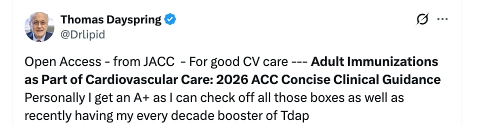
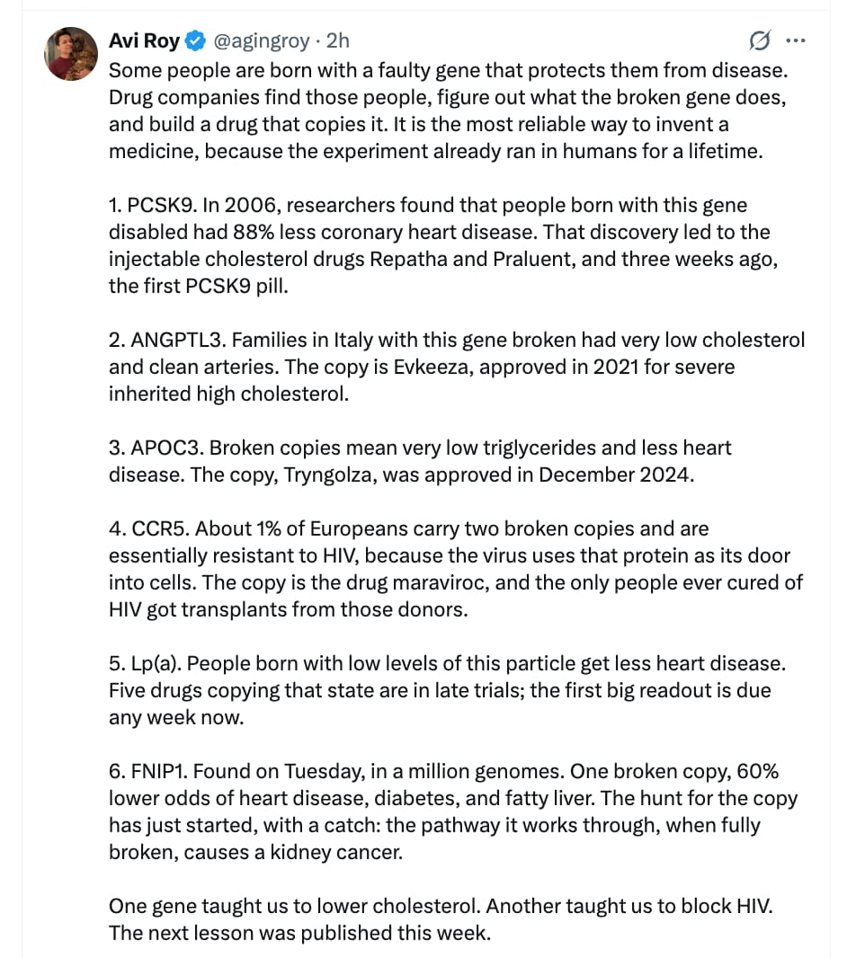
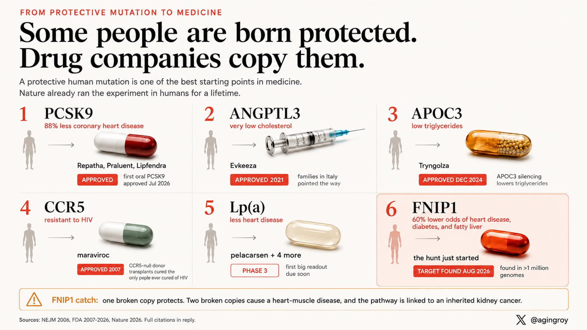
Lose the belly fat to live longer and healthier:
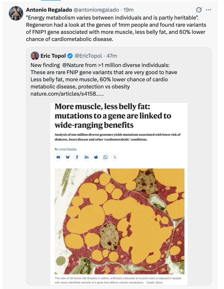
Source Papers:
Lose the belly fat to live longer and healthier:
Source Papers:
Full paper here:
The higher rate of rapid progression of atherosclerosis in healthy keto dieters with high lipids aside (KETO-CTA vs. NATURE-CT).
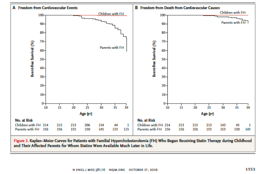
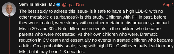
Children with FH under statin treatments vs. their parents with FH. Are people going to pretend the parents as children were “metabolically unhealthy”?
I’m here to share the results of my statin experiment and to ask my super smart friends for advice on what my next steps might be.
As some of you know, I used to be statin intolerant and went years with nothing for my lipids.
I’m currently on Repatha, ezetimibe, colchicine, and bempedoic acid. I don’t technically need more, but I’m high risk, and I’d still like my lipids to be lower to make up for all the time I wasn’t on anything.
After using some very old Livalo a couple of months ago with success, on 7/10/26 I started taking Livalo 1 mg per day. Unlike in the olden days, this time I feel fine and it no longer makes me feel sofa-bound.
Now on to potential myopathy… GAH
Oddly, I almost never get sore when I workout. Over this past month, I’m getting more and more sore. I can’t be sure it’s related, but it’s looking that way.
My first thought was, well, I’m more concerned with my heart than being pain free, so hurting during workouts is a fine trade off…… BUT, it’s now to the point that I can’t lift as much weight, and I simply don’t have muscle to spare (osteoperios and tiny)
Before I go off Livalo to see if that is truly the cause, I wanted to mention that Opus told me colchicine has the potential to make Livalo’s effective dose higher. Apparently combining it with bempodoic acid might also be a contributing cause.
If you were me, would you go off Livalo for a month to see if it was a coincidence or not? If it winds up being causal, would you then just add back, perhaps, 1mg 2x per week?
Or, is the statin potentially more beneficial than colchicine and/or bempodoic acid on someone with already good lipids? If so, instead of touching Livalo yet, would you’d pause each of those for a month to see if it’s the combo creating the issue?
If my pre-Livalo lack of soreness from working out is helped by colchicine, pausing it while being on Livalo might not tell me much. For all I know it’s the LDN though… not sure.
Open to any suggestions!!
There is potential of pitavastatin interaction with colchicine. Dropping colchicine should be looked at in the context of overall inflammatory biomarkers, hsCRP, IL-6, GlycA etc. - if those are low, then dropping colchicine might be an option, if not, I’d keep it. In other words, if given a choice I’d rather take pitavastatin and drop colchicine as long as my inflammatory markers are low, if not, I’d keep the colchicine and drop the pitavastatin given that you are controlling your lipids pretty well already. If you have myalgia, my suspicion would fall on pitavastatin (Livalo), because while the effect of pitavastatin on muscles is much less than other statins, it’s still a statin and there are reports (including from users on this site). You might just be very sensitive to statins, and in your place, if pitavastatin is giving you problems, I’d drop it - there are other drugs coming down the pike which you can get on once they’re available. I love statins, pitavastatin especially, but they are not for everybody, and if you are getting such serious side effects as you describe, I would drop them without hesitation. Remember, the goal is to safely drop your ApoB, the goal is not to simply be on a statin, and you can drop your ApoB/LDL with other classes of drugs. I’m not a doctor and this is not medical advice etc. just my thoughts as a lay student of lipids and CVD.
You should replace the statin with equivalent compound if you get myopathy even if in the majority of cases it’s the nocebo effect. First it’s a real effect even if it’s not from the statin, so you can’t wish it away, and second I think it increases your risk of the very rare incidence of rhabdo which is dangerous. Colchicine has many interactions some which are dangerous, so it’s something you should go over with a doctor carefully, and search for drug interactions in the literature/websites.
Obicetrapib is available in Europe soon, which you might be able to order in some way if you’re comfortable with not waiting for outcome trials, but if you do, do check for drug interactions for novel compounds!
Is there any statin you haven’t tried? There could still be one that works for you and doesn’t have drug interactions.
Some with Statin Associated Myopathy benefit from CoQ10 supplementation (200mg/d)
For several reasons, this study may not be relevant in this case.
(1)The statins examined did not include pitavastatin, therefore the relevance is in question.
From the study:
“Depending on the inclusion criteria of each RCT, different types of statins were administered (Simvastatin, Atorvastatin, Rosuvastatin, Lovastatin, Pravastatin, Fluvastatin), ranging from a dosage of 10 mg/d to 80 mg/d.”
(2)The causative mechanism of statin associated myalgia that is examined in this study is based on statins depleting CoQ10 levels:
From the study:
“Several causative mechanisms are discussed for the development of SAMS,(9) but a statin-induced reduction of Coenzyme Q10 (CoQ10) levels is of particular interest.(13) Statins (3-Hydroxy-3-methylglutaryl CoA reductase inhibitors) inhibit the endogenous synthesis of cholesterol by inhibiting the rate-limiting enzyme of the mevalonate pathway.(14) Thus, statins intervene very early in the mevalonate metabolic pathway and inhibit the formation of intermediary products including geranylgeranyl pyrophosphate. This isoprenoid is essential for the endogenous synthesis of CoQ10.(15) As CoQ10 is only supplied in small quantities with food and is predominantly synthesised endogenously,(16,17) statin intake can lead to reduced CoQ10 levels.(17)”
Re: (2) Pitavastatin uniquely among statins does not deplete serum levels of CoQ10, (I posted studies showing this elswhere in this site) therefore the mechanism examined by this study is irrelevant in the case of pitavastatin.
No expert by any measure but I would drop colchicine for reasons others have indicated (might make statin sides worse) plus I’ve read some bad stories about it especially if taken for longer periods. Then after you’ve waited for couple weeks and assessed the situation, I would drop PITA (if you really have to). Even though you’re saying you’re high risk you’re already doing a lot especially now that you are doing Rpatha in addition to EZe and BAcid. Don’t overdo it just because you think you’re high risk. Three lipid lowering meds should be more than enough for you.
From personal experience, most medications you take together with pitavastatin cause increased side effects. Take it isolated in the evening.
Thank you all for taking the time to provide excellent feedback.
I decided to go off livalo to see if the pain subsides. If it does, I can then re-evaluate options when I’m sure that was the cause (weighing another trial against BA and colchicine etc, or just closing the chapter).
Because it’s been getting worse, and the mention of rhabdo, I thought stopping to figure it out seemed like the conservative move.
My pilates instructor and my PT have individually been making comments at my lower ability to go hard due to pain, so whatever is going on is real.
And yessss, I want to get Obicetrapib the day my hot little hands can get a hold of it!!!
FYI, I take ubiquinol a few days per week
I’ll also reopen the can of worms better known as colchicine.
Thanks again everyone!!!
I noticed no one has mentioned the potential root cause of your myalgia yet. Colchicine isn’t the culprit here. You likely carry a malignant hyperthermia susceptibility gene, which leads to dysfunction in your ryanodine receptors (RyR). Pitavastatin contains a fluorophenyl structure, which can form a triad that targets these ryanodine receptors.
Note that this mechanism assumes your creatine kinase (CK) levels are NOT elevated. If your lab results come back showing normal CK, then this is almost certainly the issue. Switching to simvastatin or pravastatin should directly resolve the problem.
muscle-related adverse reactions signals were meaningful when the two drugs were combined in the order of colchicine combined with fluvastatin (ROR 187.38, 95% CL 96.68-363.17; IC 6.99 95% CL 1.65-5.68); colchicine combined with simvastatin in 135 cases (ROR 30.08. 95% CL 25.25-35.85; IC 4.80 95% CL 3.96-5.12); and colchicine combined with rosuvastatin (ROR 25.73, 95% CL 20.16-32.83; IC 4.59 95% CL 3.38-4.98) versus colchicine combined with atorvastatin (ROR 25.73, 95% CL 22.33-29.66; IC 4.59 95% CL 3.97-4.91) with almost identical signal intensity, followed by colchicine combined with pravastatin (ROR 13.67, 95% CL 9.17-20.37; IC 3.73 95% CL 1.87-4.47), whereas no signals were generated for lovastatin or pitavastatin.
You can measure creatinine kinase as well, I remembered now I did this before/after taking a statin, Gil Carvalho did the same.
Not sure what the guidelines are.
If you notice mild muscle pain after you start to take a statin, contact your healthcare professional. You may have a blood test to measure levels of an enzyme called creatinine kinase. This test can diagnose rhabdomyolysis or milder forms of muscle pain.
edit: I saw Kawaii posted about this now as well
Mogging
This is what peak performance looks like
What is interesting is that rosuvastatin and pravastatin not only cause myalgia in me but also make me feel like I’ve been hit by a car or something. Pitavastatin and atorvastatin only cause myalga in my feet when I’ve been taking them at too high doses or too low without break or in combination with other medications.

In this National Lipid Association presentation, lipidologists Dr. Peter H. Jones and Dr. Thomas Dayspring dismantle the long-standing clinical misconception of High-Density Lipoprotein Cholesterol (HDL-C) as the “good cholesterol” and establish a modern, physiologically rigorous framework for evaluating high-density lipoproteins (HDLs). Historically, observational epidemiology demonstrated an inverse correlation between plasma HDL-C levels and atherosclerotic cardiovascular disease (ASCVD). However, extensive randomized controlled trials (RCTs) evaluating HDL-C-raising pharmacotherapies—including niacin, fibrates, and early cholesteryl ester transfer protein (CETP) inhibitors—consistently failed to demonstrate reductions in major adverse cardiovascular events (MACE). Furthermore, Mendelian randomization studies confirmed that genetic variations raising plasma HDL-C do not confer causal cardiovascular protection (Voight et al., 2012).
HDLs represent 90% of all circulating lipoprotein particles in plasma, yet they carry a minor fraction of overall plasma lipids compared to ApoB-containing “dump truck” lipoproteins (LDL, VLDL, IDL). Instead of acting primarily as cholesterol transporters, HDLs are heterogeneous, pleiotropic platforms carrying over 200 distinct proteomic and lipidomic species. They participate in innate immunity, antioxidant defenses, endothelial maintenance, and anti-thrombotic activity. Plasma HDL-C tests measure solely the mass of cholesterol contained within HDL particles, completely failing to reflect HDL functionality or cellular cholesterol efflux capacity.
The dynamic lifecycle of HDL involves complex lipid remodeling. Apolipoprotein A-1 (ApoA-1) undergoes phospholipidation and esterification via Lecithin-Cholesterol Acyltransferase (LCAT) to form mature, spherical HDLs. Crucially, 60% of HDL-C originates directly from hepatic lipidation and 20–30% from enterocytes, while macrophage/arterial plaque efflux accounts for less than 10%. Furthermore, CETP-mediated lipid exchange between HDL and ApoB particles, combined with renal catabolism of shrunken, triglyceride-rich HDLs, drives particle clearance. Novel, potent CETP inhibitors like obicetrapib significantly reduce ApoB and LDL-C while generating massive HDL1 particles (up to 18 nm). These large particles cause Nuclear Magnetic Resonance (NMR) spectral overlap, creating false-positive elevations in small LDL particle counts. Ultimately, clinical lipid management must abandon HDL-C treatment targets, focusing strictly on ApoB and non-HDL-C reduction, alongside lifestyle optimization to preserve endogenous HDL particle functionality.
Produced by Gemini 1.5 Pro