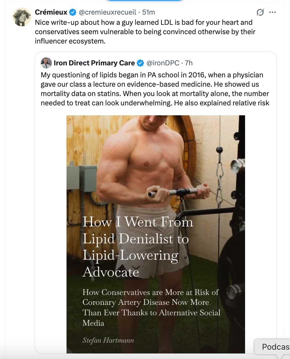
Full writeup here:
4 Likes
I mean, I’m happy he turned around and found a more evidence based path, and respect for being able to publicly change his mind, so I’m not going to criticize him, but… I can’t help but be dismayed how he fell into the denialist camp in the first place. Seriously - this is your health, your one and only body - this is not like arguing which oil is best for your car engine, if wrong, you can always get another car. If this was some unclear case, I could understand - but the evidence was there all along, all you had to do is read the damn papers and look at the bloody evidence! Like, before you decide to take or not any medication, don’t you do tons of research to make sure you understand all the risk/benefit and applicability scenarios?? It takes me months/years, to get on any med, or even switch between classes of statins from atorvastatin to pitavastatin. And regardless of how much I learn, I guess I’m just not bold enough to then start “teaching” whatever I think is the truth, become an “influencer” or whatnot - participating on a site like this is as far as I’ll go. Yet there he was, actually teaching that wrong-headed stuff that he gleaned from dogdy doctors, misinformation and a complete lack of basic analytical skills. If you ever step up to actually teaching people, I imagine you’d move heaven and earth before you dared to open your mouth, because it’s a huge responsibility when you are dealing with life and death and health issues. I don’t think I would ever feel I’ve studied enough to be so bold. I’m content to implement my research in my own life, and at best share opinions on a board like rapamycin.news., frequently with the disclaimer and caveat YMMV.
Almost all the papers he cites were there before he fell into the denialist camp. He could have read them. Why is he listening to anyone telling him what to do with his health based on someone’s “authority” - is that part of the conservative tendency to worship authority? It can’t even be that, because the consensus of establishment was firmly in the lipid and statin corner. Perhaps it’s another tendency - to follow charismatic personalities, to believ that some person is the “holder of truth”, and you then rely on their word without questioning. I don’t even know why we need to bring politics into this - as he did - because it doesn’t matter if you are a lefty or rightwinger, you can read a study just as well - politics is not an excuse! You have a brain, regardless of your politics, use it.
The problem is in not carefully looking at the evidence in the first place. Unless he really learned his lesson, he could fall into another dogma again just as easily.
4 Likes
Another look, not new, but worth revisiting. The STAREE trial was discussed in this thread above back at the end of August.
Atorvastatin, Cardiovascular Events, and Disability-free Survival in Older Adults
https://www.nejm.org/doi/full/10.1056/NEJMoa2607314
"RESULTS
A total of 9971 participants were enrolled: 4984 were assigned to receive atorvastatin and 4987 to receive placebo. The mean (±SD) age of the participants was 74.7±4.5 years, and 51.9% were women. After a median of 5.9 years, a primary cardiovascular event had occurred in 297 participants (10.9 events per 1000 person-years) in the atorvastatin group and in 412 participants (15.5 events per 1000 person-years) in the placebo group (hazard ratio, 0.70; 95% confidence interval [CI], 0.61 to 0.82; P<0.001). Death from any cause, dementia, or persistent physical disability occurred in 637 participants (21.6 events per 1000 person-years) in the atorvastatin group and in 676 participants (23.0 events per 1000 person-years) in the placebo group (hazard ratio, 0.94; 95% CI, 0.84 to 1.05; P=0.25). Serious adverse events occurred in 131 participants (2.7%) in the atorvastatin group and in 129 (2.7%) in the placebo group, with musculoskeletal, hepatobiliary, and diabetes-related adverse events occurring more commonly in the atorvastatin group.
CONCLUSIONS
Treatment with atorvastatin led to a lower risk of major cardiovascular events than placebo at a median of 5.9 years but did not result in longer disability-free survival among community-dwelling older adults without clinical cardiovascular disease.(Funded by the National Health and Medical Research Council and others; STAREE ClinicalTrials.gov number, NCT02099123.)"
3 Likes
Serial Changes in Epicardial Adipose Tissue Attenuation Identify Residual Atherosclerotic Risk Independent of LDL Lowering
https://www.jacc.org/doi/10.1016/j.jcmg.2026.05.014
This study is about what drives coronary plaque progression, even after LDL is lowered. Participants were stratified into three target LDL cholesterol groups, and more than half of the participants in every single group experienced an increase in noncalcified plaque percent atheroma volume. The LDL groups were:
- LDL <1.4 mmol/L (<55 mg/dL): Plaque progression was observed in 52.2% of participants (12 out of 23).
- LDL 1.4–1.8 mmol/L (55–70 mg/dL): Plaque progression was observed in 50.0% of participants (22 out of 44).
- LDL >1.8 mmol/L (>70 mg/dL): Plaque progression was observed in 57.1% of participants (133 out of 233)
What drives plaque progression:
- Adipose tissue composition and inflammatory activity , rather than total fat volume , drive ongoing noncalcified plaque remodeling
- Epicardial adipose tissue (EAT) attenuation serves as a marker for residual inflammatory risk, driving ongoing atherosclerotic plaque progression even when LDL cholesterol goals are met.
- Regional epicardial fat inflammation likely influences coronary plaques via localized pericoronary adipose tissue (PCAT) signaling pathways
So even when patients achieve aggressive lipid targets (<1.4 mmol/L or <55 mg/dL), standard lipid-lowering therapy alone does not fully arrest plaque progression. Ongoing noncalcified plaque remodeling in these patients was driven by non-lipid pathways—specifically residual inflammatory activity captured by changes in epicardial and pericoronary adipose tissue attenuation.
1 Like
Well, as I’ve posted before there are tons of studies addressing this - here’s a few (I’ll cross-link in the other thread).
SGLT2 inhibitors are associated with reductions in epicardial adipose tissue volume and thickness: a meta-analysis
https://www.nature.com/articles/s41366-026-02110-6
Empagliflozin Suppresses the Differentiation/Maturation of Human Epicardial Preadipocytes and Improves Paracrine Secretome Profile
The article:
Empagliflozin significantly reduced epicardial adipose tissue, subcutaneous adipose tissue, myocardial extracellular volume, matrix volume, cardiomyocyte volume, aortic stiffness and several inflammatory biomarkers, compared to placebo in nondiabetic patients with HFrEF.
And the above referenced in full:
Mechanistic Insights of Empagliflozin in Nondiabetic Patients With HFrEF: From the EMPA-TROPISM Study
https://www.jacc.org/doi/10.1016/j.jchf.2021.04.014
Effect of empagliflozin on ectopic fat stores and myocardial energetics in type 2 diabetes: the EMPACEF study
Effect of SGLT2-Inhibitors on Epicardial Adipose Tissue: A Meta-Analysis
https://www.mdpi.com/2073-4409/10/8/2150
High released lactate by epicardial fat from coronary artery disease patients is reduced by dapagliflozin treatment
https://pubmed.ncbi.nlm.nih.gov/31783199/
Effects of Empagliflozin on Carotid Intima-Media Thickness and Epicardial Fat Tissue Volume in Patients with Type-2 Diabetes Mellitus
3 Likes
Yep, both SLGT2i and GLP1s reduce epicardial fat volume and inflamation
3 Likes
It is actually very easy to fall into that trap. For the longest time, I used to get influenced by that space and didn’t know any better. I just didn’t see any “evidence based” doctors with any sort of platform so it was mostly those people running free. I was taking about 30-40 supplements and no statin thinking it was bad. I never fell for the LDL is good trap though because I would see the number clearly go up if I started eating worse and knew that eating worse wasn’t healthy.
Once the more evidence based folks starting actually addressing each of their points and debunking them with evidence, I started to realize there was another side to the story. I credit Layne Norton with really being one of the firsts to do this and now it’s become widespread (Gil and Stanfield come to mind).
I give this guy a lot of credit for admitting his wrongs. Sure, it’s easy to say he should have known better in the first place but at least he arrived in the right spot in the end. Hopefully, it encourages others to admit their wrongs as well.
I will also add that most of my friends tend to be more conservative so I hear them say a lot of crazy things when it comes to health. I am more independent and aligned with them on other topics in general, but they are taking every peptide you can imagine, NAD injections, megadosing vitamin d, no statins, think seed oils are the worst thing ever, etc. I’ll help them if they ask but it is a waste of time to correct them all.
5 Likes
Aren’t we living in interesting times that what you think about science is commonly dependent on your political beliefs.
PS, I always admire you for admitting your past mistakes and making a better path forward for yourself.
3 Likes
People tend to be tribal in their thinking. Hence even policy viewpoints can depend on the tribe that people associate with rather than people associating with a political tribe because they agree with the policies.
People tend to disassociate with a tribe when the leaders of the tribe do things that they dislike normally because it causes them problems (or people close to them).
Sadly, but not surprisingly necessarily, this goes as far as scientific views.
4 Likes
Science is a buzzword. Evidence is not.
4 Likes
All I can say is that when my father wasn’t lowering his LDL, his arterial plaque scores kept going up at a normal level.
When he started LLT with statins and BA and Ezetemibe, his arterial plaque froze in it’s tracks. There are the influencers and then there’s the data. I’ll go with the data every time.
If you want to halt arteriosclerosis, lower your ApoB and LDL below 55.
10 Likes
We do know that ezetimibe inhibits dietary cholesterol uptake in the intestine and disposes of it fecally. So ezetimibe lowers LDL-C lipid levels in the blood. But what happens when you give ezetimibe to vegetarians who get very little dietary cholesterol? Does it mean since ezetimibe doesn’t have any dietary cholesterol to inhibit the absorption of, it therefore will not lower lipid blood levels? That is the question explored in this study - an oldie (2006!), but goodie - and I like this study a lot, it was very well designed and quite decisive in its findings, which we can have a high degree of confidence in. The bottom line, regardless of your dietary cholesterol intake, ezetimibe works as intended.
The lipid-lowering effect of ezetimibe in pure vegetarians
https://www.jlr.org/article/S0022-2275(20)43275-4/fulltext
" Results of previous studies have shown that ezetimibe (10 mg/day) reduces LDL cholesterol in patients with mild hypercholesterolemia on a normal-cholesterol diet (dietary intake of 200–500 mg/day) by 16–22%. However, the LDL cholesterol-lowering effect of ezetimibe in subjects with an extremely low dietary cholesterol intake (vegetarians) has not been studied. We conducted a randomized, double-blind, placebo-controlled, two-phase crossover study in 18 healthy pure vegetarians to assess the effect of ezetimibe (10 mg/day) on plasma lipids, cholesterol absorption, and its synthesis. Treatment periods lasted 2 weeks each, with an intervening 2 week washout period. Fractional cholesterol absorption was determined using the continuous dual stable isotope feeding method. Mean dietary cholesterol intake in the pure vegetarians was extremely low and averaged 29.4 ± 16.8 and 31.4 ± 14.4 mg/day during the placebo and ezetimibe administration phases, respectively. Fractional cholesterol absorption during the placebo phase was 48.2 ± 8.2% and was decreased by 58% during ezetimibe treatment to 20.2 ± 6.2% (P < 0.001). This change in intestinal cholesterol absorption was followed by a significant reduction in LDL cholesterol of 17.3%. In individuals with extremely low dietary cholesterol intake, treatment with ezetimibe (10 mg/day) leads to a significant reduction of cholesterol absorption and a clinically relevant decrease of plasma LDL cholesterol, comparable to that of subjects with a normal dietary cholesterol intake. Thus, the lipid-lowering effect of ezetimibe is mediated mainly through a reduction of the absorption of endogenous (biliary) cholesterol."
Meanwhile for those worried about oxidized dietary cholesterol, they can rest easy that ezetimibe works as intended as another old ezetimibe study demonstrates.
Ezetimibe inhibits the incorporation of dietary oxidized cholesterol into lipoproteins
https://www.jlr.org/article/S0022-2275(20)43392-9/fulltext
“Oxidized cholesterol is present in significant quantities in the typical Western diet. When ingested, oxidized cholesterol is absorbed by the small intestine and incorporated into both chylomicrons and LDL, resulting in LDL that is more susceptible to further oxidation. Feeding studies in animal models and epidemiological studies in humans have suggested that oxidized cholesterol in the diet increases the development of atherosclerosis. In this study, we determined the effect of ezetimibe, a drug that inhibits small intestinal absorption of cholesterol, on the levels of oxidized cholesterol in the serum after a test meal containing oxidized cholesterol. We demonstrate that ezetimibe, 10 mg per day for 1 month, markedly reduced the levels (50% decrease) of oxidized cholesterol in the serum after feeding a test meal containing either α-epoxy cholesterol or 7-keto cholesterol, two of the predominant oxidized cholesterols found in the diet. Moreover, the decrease in oxidized cholesterol in the serum was attributable to a decrease in the incorporation of dietary oxidized cholesterol into both chylomicrons and LDL. Because there was no decrease in postprandial triglyceride levels, we conclude that this decrease in oxidized cholesterol levels in the serum is attributable to decreased absorption and not to enhanced clearance. Whether this decrease in oxidized cholesterol absorption prevents or delays the development of atherosclerosis remains to be determined.”
3 Likes
As the resident vegan, I’ll share that I put off taking EZ for a minute thinking there was nothing for it to do… but lo and behold, my numbers went down after starting… so, my n=1 shows the study was correct 
I then discovered most of the cholesterol EZ blocks comes from your own bile, not your food.
5 Likes
Yes, by the time I learned about ezetimibe, that fact was already well-known, probably from studies like the one mentioned above.
2 Likes
2024 study with graphs of plaque on CTA over time. Average TPV (Total Plaque Volume) appears to increase linearly from year/age 45.
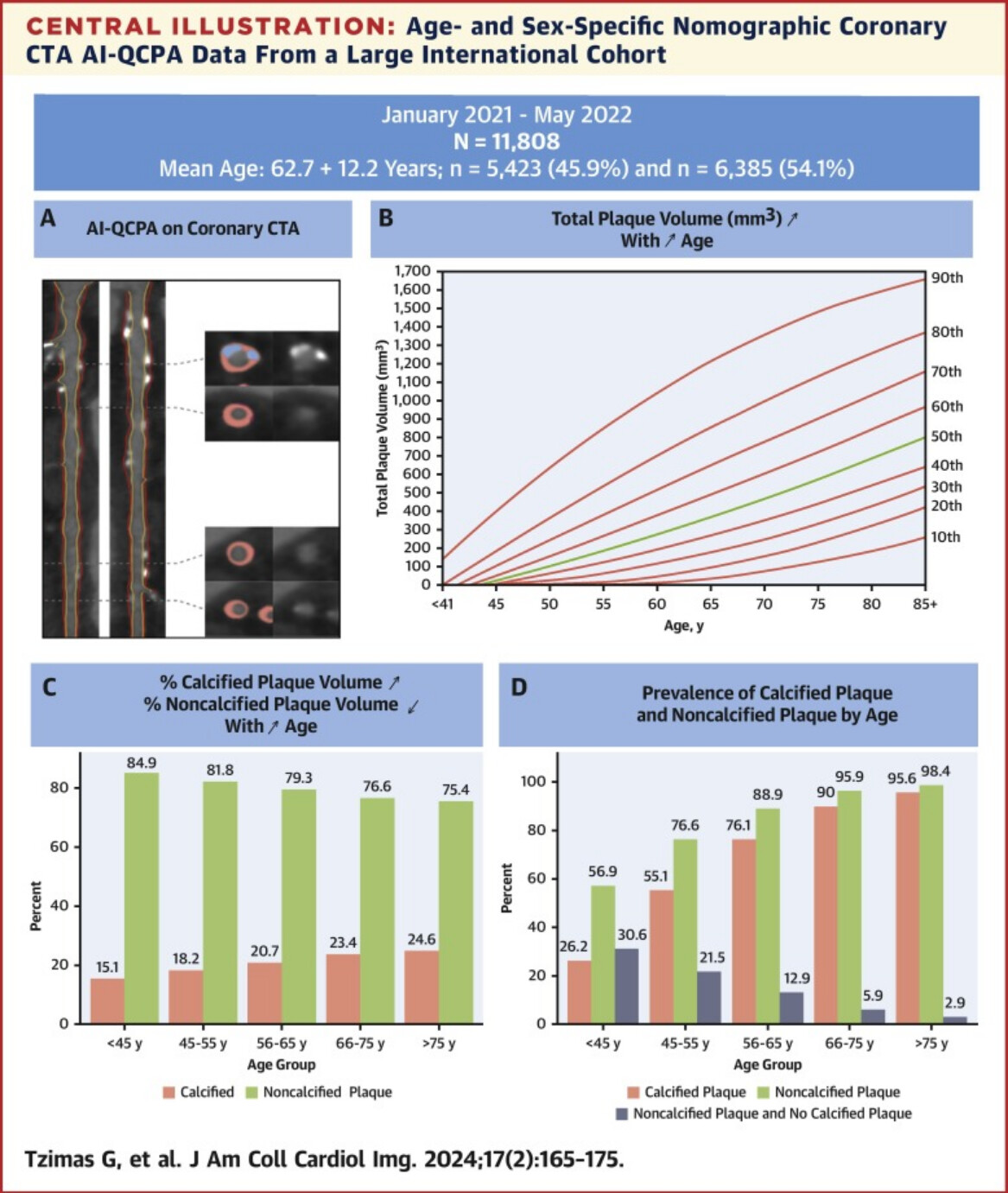
https://www.sciencedirect.com/science/article/pii/S1936878X23002346
2 Likes
OK, but did you know that the same thing applies to plant sterols  ? I would guess it’s logical. Undoubtedly it is a good thing in case of sitosterolemia. But is it also good in the absence of sitosterolemia, given that some have proposed - controversially - plant sterols as beneficial in CVD? Inquiring minds want to know. Any medication deserves a thorough establishment of all effects. A couple of papers (the EZE had a tiny # of subjects):
Ezetimibe Reduces Plant Sterol Accumulation and Favorably Increases Platelet Count in Sitosterolemia
A general look at phytosterols (review):
Phytosterols and Cardiovascular Disease
2 Likes
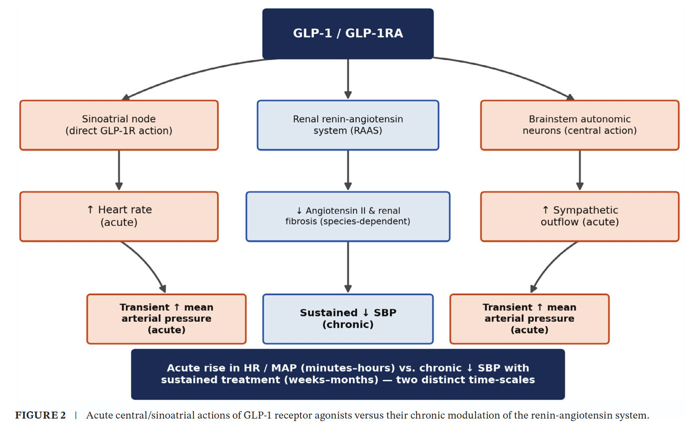
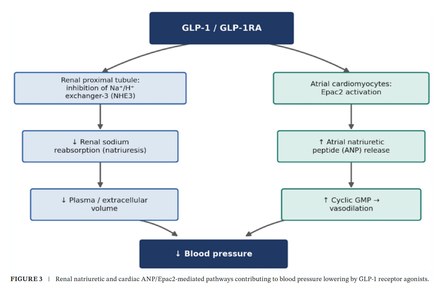
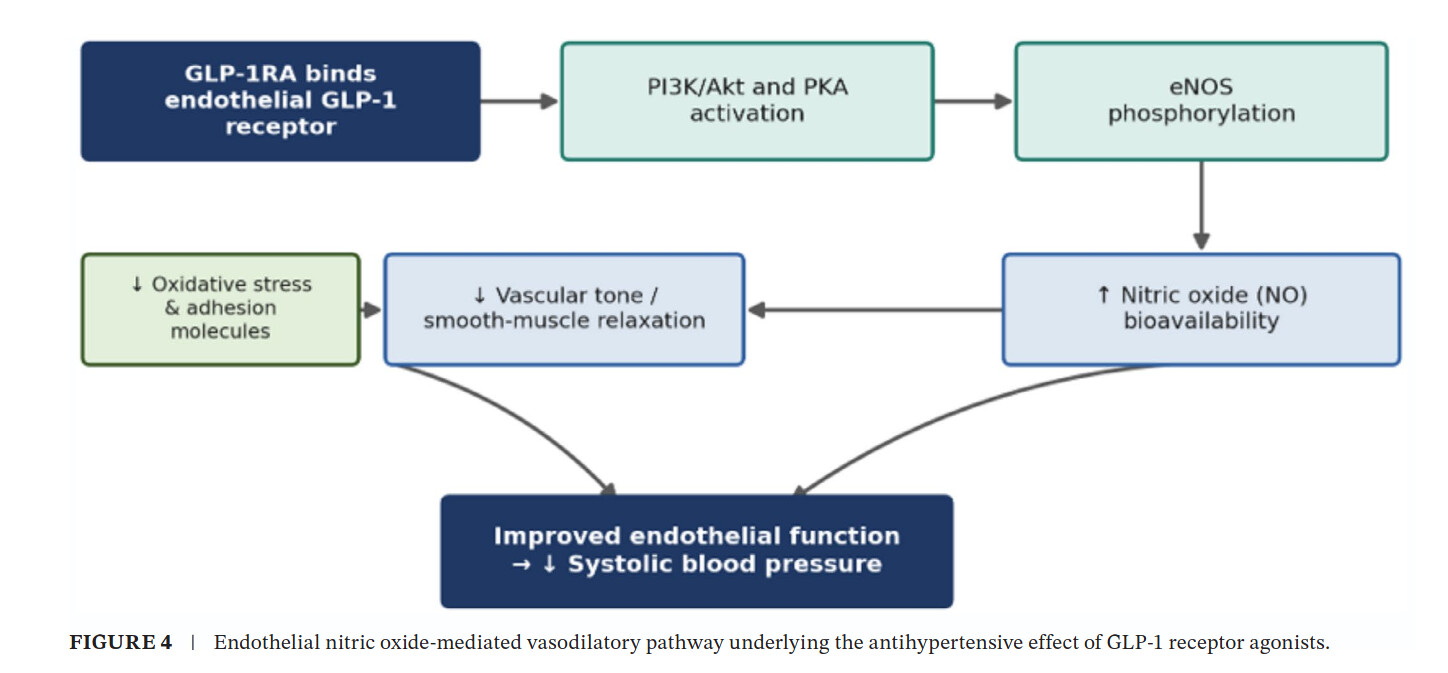
Glucagon-Like Peptide-1 Agonists and Blood Pressure Regulation: Molecular Mechanisms and Cardiovascular Implications
https://bpspubs.onlinelibrary.wiley.com/doi/epdf/10.1002/prp2.70329
AI summary:
1 Like
Let us keep in mind that rapamycin appears to polarize M1/M2 in the opposite direction from GLP-1RA. And in a contest between the two, rapamycin takes precedence as it’s upstream of where GLP-1RA work here and affects the very mechanism of polarization leaving GLP-1RA nothing to work with. If you are taking both, be aware that it is rapamycin that will be dominant. In this context pulsed administration of rapamycin becomes interesting, as one could speculate that in the trough of zero, GLP-1RA could step in to again re-balance M1 and M2.
However, the interplay of GLP-1 and rapamycin - for that matter other anti-diabetics too, is quite complex and frequently counter-intuitive. When I research a drug for inclusion into my stack, I spend a huge amount of time and effort trying to map out the possible interactions, because that is really where the rubber hits the road. It makes little sense to look at the actions of a drug on paper - such as the quote above “promotes M1 to M2 macrophage polarization, reduces pro-inflammatory cytokines” - if the other drugs in your stack completely abolish that effect, because you are counting on an effect that will be absent. You must then decide how to balance out these drugs by whatever means - dose, protocol, timing, or even inclusion.
Rapamycin and anti-diabetic drugs including GLP-1RA have a complex interplay in glucose control too. I have frequently noted in many threads that rapamycin definitely can be deleterious to pancreatic beta cells. This is a serious issue I have spent a lot of time on as someone who both takes rapamycin and has been pre-diabetic (for more than a decade).
Below is a paper that is super helpful in illuminating some of these issues, including the rapamycin and GLP-1 interplay, M1/M2 and other issues discussed. I didn’t post it before as it doesn’t fit neatly into any of the threads - rapamycin, glucose control, GLP-1 and anti-diabetics, pancreatic health. But because it focuses so much on the inflammatory aspect, I guess I’ll post it in this thread, even though it doesn’t really fit perfectly. I would strongly urge anyone interested in understanding these issues to read the whole paper to really get a good grounding in the underlying physiology. Enjoy!
mTOR: A double-edged sword for diabetes
https://jlb.onlinelibrary.wiley.com/doi/full/10.1002/JLB.3MR0317-095RR
5 Likes
Thank you for sharing—this is a very interesting paper.
The effect of rapamycin on β-cells and insulin resistance is a topic I’m particularly interested in. It provides insights that complement other papers I have read regarding its impact on calcium uptake.
I asked Opus 5.5 (extra high mode) to reconcile these papers, specifically focusing on my context: glucose dysregulation, pulsed rather than chronic rapamycin, and non-T1DM.
Here it is:
Short answer: the review doesn’t contradict the calcium idea, but it doesn’t support it either. It is about how diabetes develops (immune cells and islet growth) and rests almost entirely on rodent and cell-culture work. The calcium paper you’re remembering (Lombardi 2017) is real. Its “calcium” finding is best read as one side of a mitochondrial defect in how β-cells link glucose metabolism to insulin release. A second group described the same defect from the Krebs-cycle side. The bigger issue for you is different: nothing yet shows that a β-cell mechanism causes your ~1-week CGM rise. And your protocol has a built-in confound that could produce the same signal without rapamycin. I also need to retract several things I told you in May.
1. Which parts of the review apply to you
| Review section | Relevance | Why |
|---|---|---|
| Immune half (NK, CD8, Th1/Th17, M1 cells destroying islets) | None | This describes autoimmune or inflammatory β-cell injury. Your GAD65, IA-2 and ZnT8 antibodies were all negative (2026-07-22) and hs-CRP is 0.44. Your NK count of 490 is a blood finding, not islet infiltration. |
| β-cell growth and mass (mTORC1 → cyclin D2) | Negligible | This is slow mass adaptation in rodents. It can’t produce a reversible 1-week signal. |
| mTORC2 → β-cell toxicity (Barlow 2012, ref 122) | Possible, but needs prolonged exposure | Rapamycin inhibited mTORC1 acutely, but the toxicity tracked mTORC2 dissociation and loss of PKB (Akt) activity. Knocking down RICTOR to inactivate mTORC2 selectively reproduced the toxicity of chronic rapamycin. (C) |
| Liver glucose production (Houde 2010, ref 53) | A real candidate | In rats given 2 mg/kg/day for 15 days, the liver switched on its glucose-production (gluconeogenic) program and caused severe glucose intolerance, even though hepatic insulin signalling to Akt was preserved. Chronic dosing again, and apparently not via mTORC2. (C) |
The review also states that inhibiting mTOR promotes insulin resistance. That is the chronic picture. The one acute human study points the other way. With amino acids infused to switch on mTOR, rapamycin raised insulin-stimulated glucose uptake by 17% at meal-like insulin levels, with no effect at low insulin. (B: one clamp study in healthy volunteers.) You eat 1.6 g/kg of protein and take leucine during runs. An acute pulse is therefore more likely to help muscle insulin action than hurt it. Peripheral insulin resistance is the least likely acute driver for you.
2. What the calcium paper actually showed
Lombardi 2017 tested rat β-cell lines plus mouse and human islets at 25 nM for 24 h:
- Glucose-stimulated insulin secretion fell; viability was unchanged; secretion in response to leucine and glutamine was impaired while the response to KCl (direct depolarisation) was intact; oxygen consumption fell; ER Ca²⁺ stores were depleted with increased leak; mitochondrial Ca²⁺ uptake fell; and all three IP3R isoforms were upregulated, which the authors proposed as the mechanism.
- The intact KCl response matters. The insulin-release machinery itself works; the lesion sits upstream, in metabolism.
Shimodahira 2010 (cited in the review) used rat islets for 24 h:
- ATP fell, glucose oxidation fell, and α-ketoglutarate dehydrogenase (α-KGDH) activity fell, while the respiratory chain downstream of complex II was unaffected.
These two findings likely describe one lesion. α-KGDH is one of the mitochondrial enzymes that calcium switches on. So less mitochondrial Ca²⁺ uptake (Lombardi) and lower α-KGDH activity (Shimodahira) are plausibly the same defect seen from opposite ends. Calcium is a node in that loop, not necessarily its root. (C: this link is my synthesis; neither paper tested it.)
What the paper does not show:
- Which mTOR complex is involved. It was never tested. The closest data (Barlow, also in human islets) point to mTORC2 under prolonged exposure.
- How long the effect lasts. There was no washout experiment, so nothing tells us about recovery after the drug clears.
-
Whether the dose matches yours. The 25 nM figure comes from Desai 2003, which measured portal-vein drug levels in islet-transplant recipients. That is continuous dosing, sampled where the islets were infused.
- The label gives a blood-to-plasma ratio of 36 ± 18, meaning the drug sits mostly in blood cells, with about 92% of plasma drug bound to protein.
- So free drug ≈ whole blood ÷ 36 × 0.08 ≈ 0.2% of the whole-blood level. A whole-blood 23 ng/mL (= 25 nM) works out to about 0.05 nM free.
- Culture medium has no red cells and far less protein, so most of the 25 nM in a dish reaches the cells.
- Caveats: red-cell binding saturates, so the free fraction at your peak is higher than this. Rapamycin also accumulates inside cells bound to FKBP12.
- Net: the cell-culture magnitude is an upper bound for your pulse, not an estimate. (C)
Sources:
- Lombardi et al., Sci Rep 2017 — Sirolimus depletes intracellular Ca²⁺ stores in β-cells
- Shimodahira et al., J Endocrinol 2010 — Rapamycin impairs metabolism-secretion coupling
- Barlow et al., Diabetologia 2012 — Rapamycin islet toxicity via mTORC2
- Houde et al., Diabetes 2010 — Chronic rapamycin upregulates hepatic gluconeogenesis
- Krebs et al., Diabetes 2007 — mTOR regulates nutrient-sensitive glucose uptake in man
- Arriola Apelo et al., Aging Cell 2016 — Intermittent rapamycin and glucose homeostasis
- Rapamune (sirolimus) FDA label — distribution
- Burstein — Insulin sensitivity in trained athletes upon cessation of training (published Diabetes 1985)
- Bajpeyi et al., J Appl Physiol 2009 — Persistence of insulin sensitivity during training cessation
1 Like